
Cartilages of larynx easily understandable way with images and university important points
cartilages of larynx anatomy diagram thyroid cricoid arytenoid epiglottis
larynx cartilage anatomy
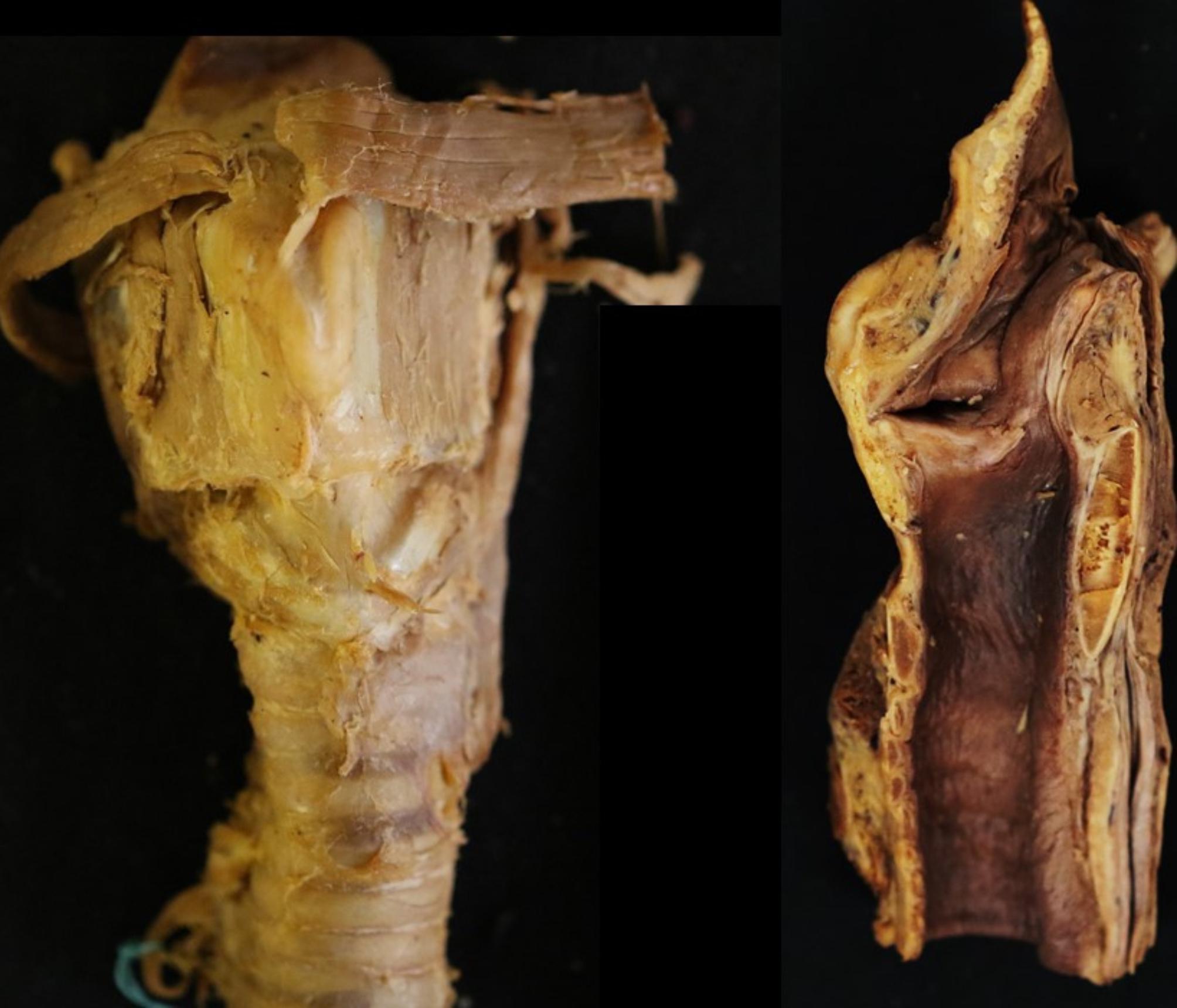
This composite clinical photograph displays a dissected human larynx, providing a detailed anatomical study of the perilaryngeal structures. The left panel shows a frontal (anterior) view of the larynx and upper trachea. Key visible structures include the thyroid cartilage, cricoid cartilage, and the intervening cricothyroid ligament. Striated muscle fibers and connective tissues are seen superiorly near the hyoid region, while the trachea is identifiable inferiorly by its characteristic horizontal cartilaginous rings. The right panel presents a midsagittal section, exposing the internal laryngeal cavity. This view highlights the contrast between the external yellowish connective tissues and the darker, brownish mucosal lining of the airway. Anatomical landmarks such as the epiglottis, laryngeal vestibule, and the posterior plate of the cricoid cartilage are visible in cross-section. These images are typically used in surgical anatomy or otolaryngology to study the morphometry of the cricothyroid ligament and laryngeal dimensions for procedures like cricothyrotomy or endotracheal intubation training.
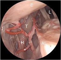
This diagnostic clinical image, likely obtained via endoscopic laryngoscopy, illustrates the complex vascular anatomy of the left larynx. The photograph focuses on the branching patterns of the superior laryngeal artery (SLA) in relation to key endolaryngeal structures. Visible landmarks include the epiglottis (E) superiorly, the laryngeal lumen (L), the left arytenoid region (AL), and the thyroid cartilage (T). A specific branch of the SLA is highlighted, demonstrating a caudal and lateral course marked with an asterisk (*). This vessel is seen traversing the paraglottic space and exiting the larynx in close proximity to the lower horn (H) of the thyroid cartilage. The image serves as an educational tool for identifying surgical landmarks and understanding the varied arterial supply to the laryngeal mucosa and intrinsic muscles, which is critical for phonosurgery and oncologic resections to avoid intraoperative hemorrhage.
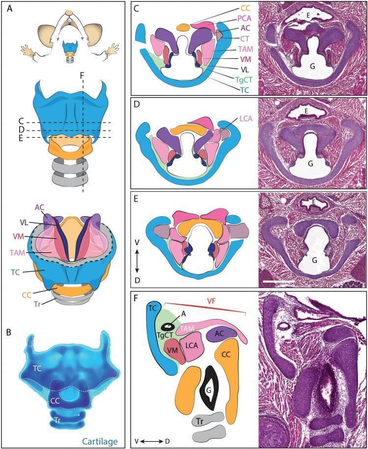
This multi-panel educational figure illustrates the comparative anatomy of the laryngeal apparatus in a mouse model, relevant to mammalian developmental biology. Panel A provides a schematic ventral view of the larynx, designating sectional planes for horizontal (C-E) and sagittal (F) orientations. Panel B shows a whole-mount adult larynx stained with Alcian Blue to highlight cartilaginous structures, including the thyroid cartilage (TC), cricoid cartilage (CC), and tracheal rings (Tr). Panels C, D, and E display Hematoxylin and Eosin (H&E) stained horizontal sections of an E18.5 embryonic larynx paired with corresponding anatomical diagrams. These sections demonstrate the spatial relationship between the esophagus (E), glottis (G), and laryngeal muscles such as the thyroarytenoid (TAM) and vocalis (VM). Panel F presents a midline sagittal H&E section with a detailed diagram identifying the vocal fold (VF) complex, including the vocal ligament (VL), lateral cricoarytenoid muscle (LCA), and thyroglottal connective tissue (TgCT). The figure utilizes standard dorso-ventral (D-V) axes to orient the development and arrangement of the laryngeal skeleton and soft tissue components.
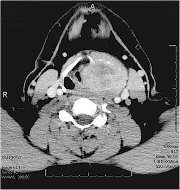
Axial contrast-enhanced CT scan of the neck at the level of the larynx. The image demonstrates a large, heterogeneously enhancing mass measuring approximately 5 x 5.7 cm, centered in the left laryngeal region. Key findings include the complete destruction of the left thyroid cartilage and medial extension of the tumor, which significantly compresses the laryngeal ventricle and narrows the airway at the level of the vocal cords. The mass exhibits central areas of decreased attenuation (suggestive of necrosis or cystic components) and early punctate calcifications. Disruption of normal anatomy is evident through the lateral displacement and compression of the left carotid sheath and sternocleidomastoid muscle. The lesion extends superiorly toward the hyoid bone, obliterating the left pyriform sinus. This diagnostic image illustrates the local invasiveness typical of primary laryngeal bone or cartilage tumors, such as chondrosarcoma or giant cell tumor of the larynx.
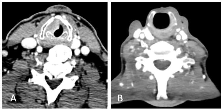
Two side-by-side axial contrast-enhanced CT scans of the neck at the level of the larynx, demonstrating preoperative and postoperative anatomy for laryngeal carcinoma. Image (A) shows the preoperative state with an asymmetric soft tissue mass involving the left vocal fold, causing narrowing of the glottic airway. The thyroid cartilage appears intact but adjacent to the primary lesion. Image (B) displays the postoperative anatomy six months after surgical intervention (likely a partial laryngectomy or supracricoid laryngectomy). Notably, the thyroid cartilage has been removed, resulting in a reconstructed neolarynx with a patent, wider airway compared to the preoperative scan. Both images clearly visualize the cervical vertebrae, posterior neck musculature, and carotid sheath vasculature. This comparison illustrates the radical alteration of laryngeal architecture following oncological resection and surgical reconstruction for throat cancer.
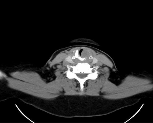
This diagnostic image is an axial contrast-enhanced CT scan of the neck at the level of the larynx. The image demonstrates a significant soft-tissue nodular lesion originating from the left glottic and subglottic region. A hallmark of this pathology is the visible partial destruction and infiltration of the left thyroid cartilage, along with suspected involvement of the cricoid ring and ipsilateral arytenoid cartilage. The mass results in substantial narrowing and displacement of the laryngeal airway lumen toward the right. Adjacent soft tissue structures show bilateral cervical lymph node hyperplasia. This visual evidence is characteristic of an invasive laryngeal neoplasm, such as adenoid cystic carcinoma, highlighting the clinical significance of CT in assessing cartilage invasion and airway compromise for surgical planning. The scan is a key educational resource for identifying laryngeal anatomy, cartilage landmarks, and signs of malignant infiltration in head and neck oncology.
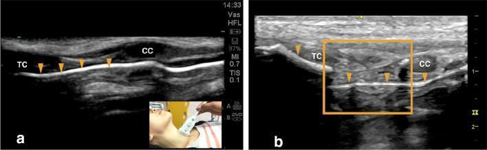
This diagnostic comparison image consists of two high-frequency airway ultrasound scans (a and b) in a longitudinal orientation, illustrating normal versus traumatic sonoanatomy of the larynx. Image 'a' demonstrates a normal airway with a continuous, linear hyperechoic interface (arrowheads) representing the intact air-mucosal interface. Key anatomical structures identified include the hypoechoic thyroid cartilage (TC) and cricoid cartilage (CC). Image 'b' displays pathological findings following blunt neck trauma. The air-mucosal interface is notably disrupted (arrowhead), indicating loss of mucosal integrity. A highlighted region (orange box) reveals a fracture of the cricoid cartilage and the development of an endolaryngeal hematoma, characterized by heterogenous echogenicity within the soft tissues. These images serve as an educational resource for medical professionals and students in emergency medicine and radiology to differentiate normal laryngeal anatomy from significant laryngeal trauma, such as cartilage fractures and internal soft tissue injuries, using point-of-care ultrasound (POCUS).
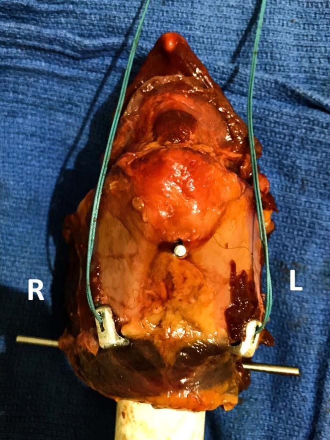
This clinical photograph displays a surgical setup on a harvested larynx to demonstrate the application of custom laryngeal clamps used in laryngoplasty. Two U-shaped stainless steel clamps are positioned bilaterally on the caudoventral margins of the thyroid cartilage laminae, labeled 'R' and 'L' for right and left. Each clamp consists of a short dorsal arm and a long ventral arm, which wrap around the caudal edge of the cartilage. A thick green polyester suture is integrated into each construct. The suture path involves threading through dual holes in the dorsal arm of the clamp, passing through a pre-drilled hole in the thyroid cartilage, and finally through a slit on the clamp's dorsal arm. This arrangement is designed to reinforce the anchorage point on the cartilage, distributing mechanical load to prevent cartilage tearing during laryngoplasty maneuvers. The setup is mounted on a mechanical base for biomechanical testing, illustrating the structural relationship between the prosthetic implants, the suture, and the cartilaginous anatomy.
laryngeal cartilages posterior view thyroid cricoid arytenoid corniculate cuneiform epiglottis labeled diagram
Cartilages of the Larynx — Simplified with Diagrams
Quick Overview
| Type | Cartilages | Number |
|---|---|---|
| Unpaired (single) | Thyroid, Cricoid, Epiglottis | 3 |
| Paired | Arytenoid, Corniculate, Cuneiform | 3 x 2 = 6 |
1. Thyroid Cartilage — the "shield"
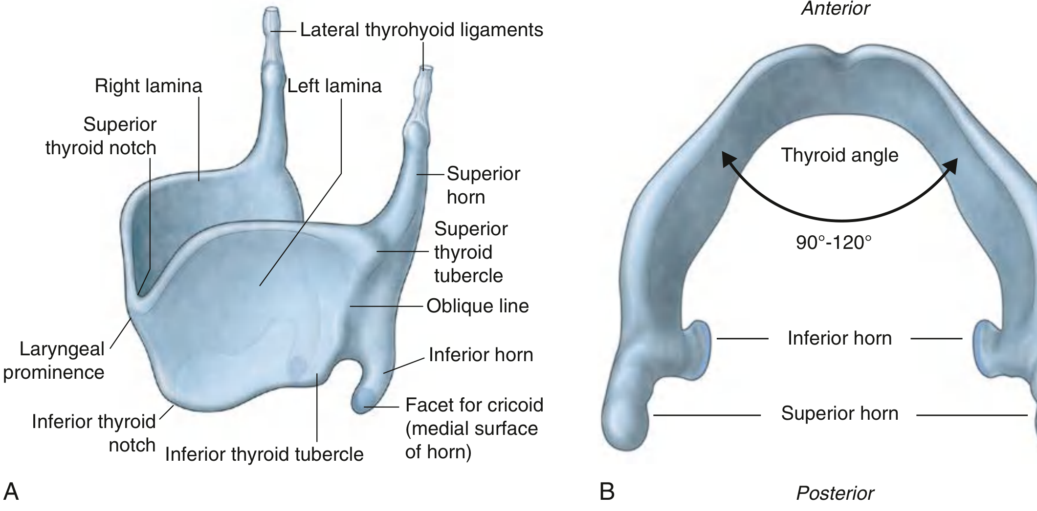
- Angle of fusion: 90° in males, 120° in females → prominence more visible in men (forensic sex determination point)
- Has superior horn (connects to hyoid via lateral thyrohyoid ligament) and inferior horn (articulates with cricoid → cricothyroid joint)
- Oblique line on lateral surface = attachment for sternothyroid, thyrohyoid, inferior constrictor
- Type: Hyaline cartilage → ossifies with age (relevant to radiograph/CT interpretation and forensic age estimation)
2. Cricoid Cartilage — the "signet ring"
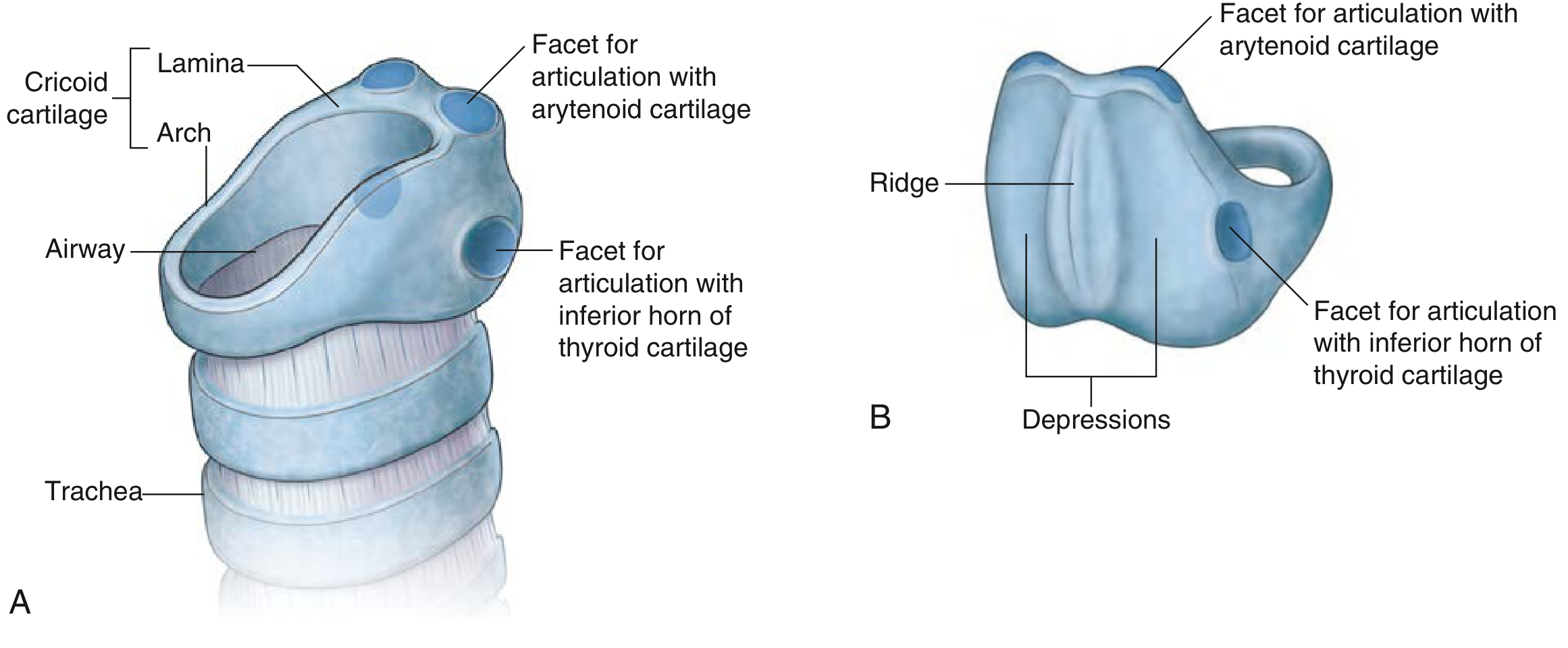
- Only complete cartilaginous ring in the entire respiratory tract — clinically vital in cricothyrotomy (emergency airway) and pediatric subglottic narrowing
- Broad lamina posteriorly, narrow arch anteriorly
- Articulates with thyroid cartilage (cricothyroid joint) and arytenoid cartilage (cricoarytenoid joint)
- Type: Hyaline → ossifies
3. Epiglottis — the "leaf-shaped flap"
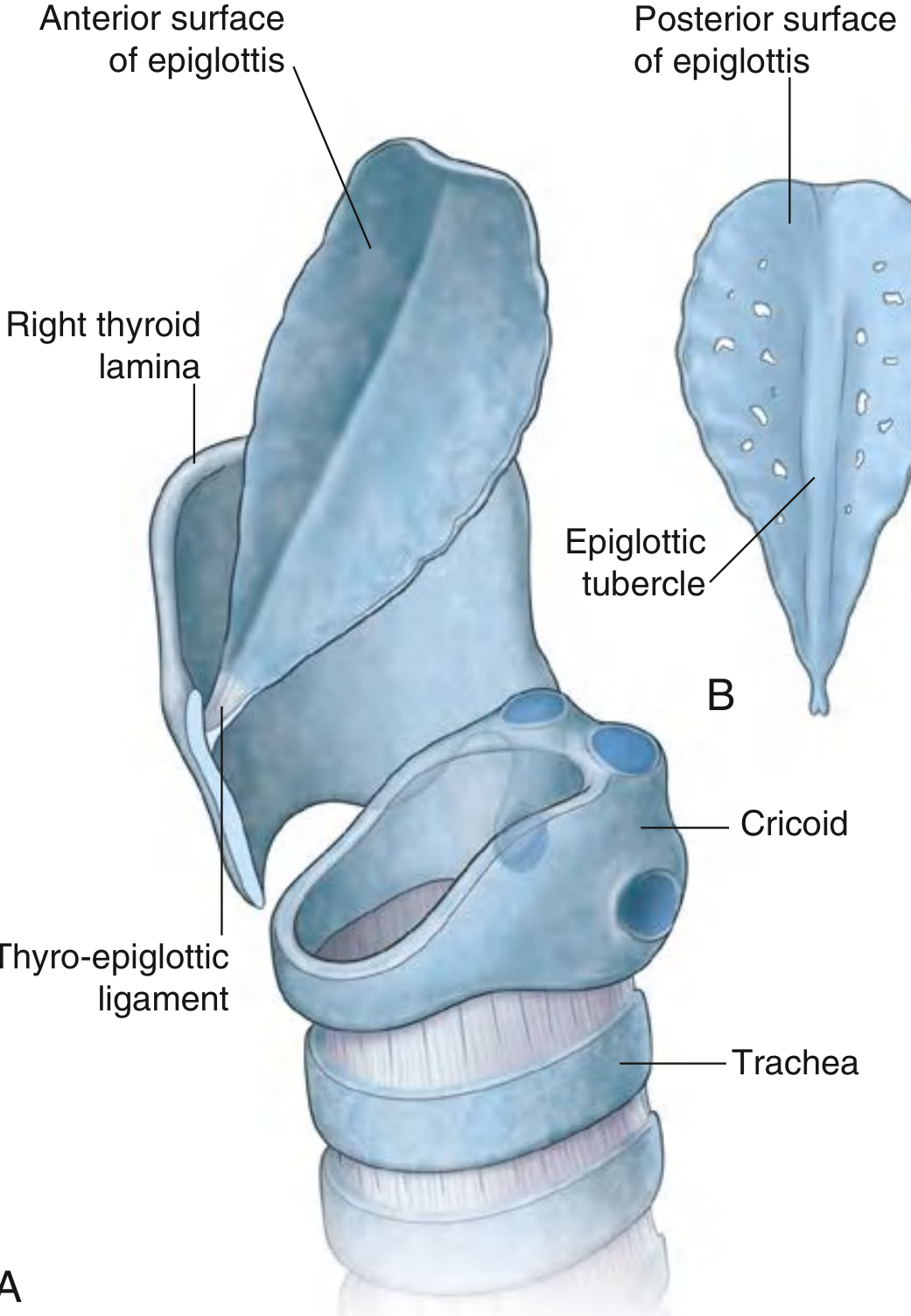
- Leaf-shaped, attached by its stalk to the back of the thyroid cartilage via the thyroepiglottic ligament
- Also attached to the hyoid bone by the hyoepiglottic ligament
- Guards the laryngeal inlet during swallowing
- Type: Elastic cartilage → does NOT ossify, also does not undergo malignant change (important distinguishing exam point vs thyroid/cricoid/arytenoid)
4. Arytenoid Cartilages — the "pyramids" (most important functionally)
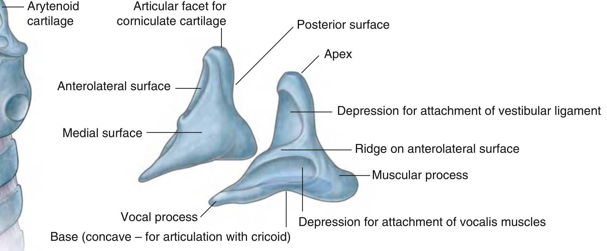
- Pyramid-shaped, paired; base sits on cricoid lamina (cricoarytenoid joint)
- Vocal process (anterior angle) → attachment of the vocal ligament
- Muscular process (lateral angle) → attachment of posterior and lateral cricoarytenoid muscles
- Apex articulates with the corniculate cartilage
- Base and body = hyaline (can ossify); vocal process and apex = elastic cartilage — a classic "trick" MCQ point
5. Corniculate and Cuneiform Cartilages — the "small helpers"
- Corniculate (cartilage of Santorini): tiny cones sitting on the apex of each arytenoid, project into the aryepiglottic fold
- Cuneiform (cartilage of Wrisberg): small rod-shaped cartilages lying within the aryepiglottic fold, anterior to the corniculate cartilages
- Both are elastic cartilage, purely supportive, no muscle attachments
Joints of the Larynx (frequently asked)
| Joint | Type | Movement | Functional effect |
|---|---|---|---|
| Cricothyroid joint | Synovial | Thyroid tilts forward/down on cricoid | Tenses/lengthens vocal ligaments (pitch control) |
| Cricoarytenoid joint | Synovial | Arytenoids slide + rotate | Abducts/adducts vocal cords (opens/closes glottis) |
- Posterior cricoarytenoid (PCA) muscle = only abductor of the vocal cords — clinical relevance: bilateral recurrent laryngeal nerve palsy paralyzes PCA bilaterally → unopposed adductors → stridor/airway obstruction, a classic viva question.
University Exam Cheat-Sheet
| Point | Answer |
|---|---|
| Total cartilages | 9 (3 unpaired + 3 paired) |
| Hyaline cartilages (ossify with age) | Thyroid, Cricoid, Arytenoid (except vocal process/apex) |
| Elastic cartilages (never ossify) | Epiglottis, Corniculate, Cuneiform, apex & vocal process of arytenoid |
| Only complete cartilage ring | Cricoid |
| Largest cartilage | Thyroid |
| Landmark for cricothyrotomy | Cricothyroid membrane, between thyroid and cricoid cartilage |
| Only vocal cord abductor | Posterior cricoarytenoid muscle |
| Cartilage prone to malignant change | Hyaline ones (thyroid, cricoid), not the elastic epiglottis |
| Sex difference used forensically | Thyroid angle: 90° (male) vs 120° (female) |
| Nerve supply above vocal cords | Internal laryngeal nerve (sensory) |
| Nerve supply below vocal cords + all intrinsic muscles (except cricothyroid) | Recurrent laryngeal nerve |
| Motor to cricothyroid | External laryngeal nerve |
Ca larynx all points easy to remember with easy understandable with explaining images according to mainly dhingara Give all university points structurally
carcinoma larynx glottic supraglottic vocal cord tumor laryngoscopy
vocal cord leukoplakia growth larynx endoscopic view
laryngeal carcinoma treatment
Carcinoma Larynx — Complete University-Ready Notes
1. Epidemiology & Etiology
| Risk factor | Detail |
|---|---|
| Sex | Male : Female = 7-10 : 1 (declining as female smoking rises) |
| Age | 5th-6th decade |
| Smoking | Single most important factor - dose-related |
| Alcohol | Synergistic with smoking (multiplicative, not additive, risk) |
| Others | Wood dust, asbestos, nickel, mustard gas exposure, radiation, GERD/LPR, HPV (mainly supraglottic), Plummer-Vinson syndrome (post-cricoid) |
- Leukoplakia
- Chronic hyperplastic laryngitis
- Pachydermia laryngis
- Laryngeal papilloma (in adults - risk of malignant change; juvenile papilloma does NOT turn malignant)
2. Site Classification (Most Important Section)
| Region | Boundaries | Approx. Incidence |
|---|---|---|
| Supraglottic | Epiglottis, aryepiglottic fold, false cords, ventricle | ~30-35% |
| Glottic | True vocal cords, anterior & posterior commissure | ~55-60% (most common) |
| Subglottic | Below true cords to lower border of cricoid | ~5% (rarest) |
- Glottis has a poor lymphatic supply → early glottic cancer rarely metastasizes → best prognosis, presents early (hoarseness)
- Supraglottis has a rich, bilateral lymphatic network → early nodal metastasis, often presents late with a neck node
- Subglottis has sparse but bilateral lymphatics draining to pretracheal/paratracheal nodes → silent until late (stridor/dyspnea) → worst prognosis
3. Pathology
- 95% Squamous cell carcinoma (well to moderately differentiated most common)
- Rare types: verrucous carcinoma, adenocarcinoma, chondrosarcoma (from cartilage), sarcoma
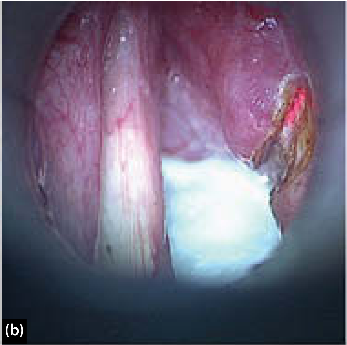
4. Spread (Table 14.1 style - very commonly asked)
| Primary site | Spreads to |
|---|---|
| Supraglottic | Pre-epiglottic space → anterior commissure; false cords/mucosa → paraglottic space → true cords |
| Glottic (anterior cord) | Anterior commissure → contralateral cord |
| Glottic (posterior cord) | Thyroid cartilage & cricothyroid membrane → arytenoid/cricothyroid joint |
| Subglottic | Trachea, thyroid gland, cervical esophagus |
5. Clinical Features (High-Yield "Red Flag" List)
| Site | Earliest symptom | Later symptoms |
|---|---|---|
| Glottic | Hoarseness of voice (any hoarseness >3 weeks = mandatory laryngoscopy) | Stridor, dyspnea, hemoptysis |
| Supraglottic | Vague throat discomfort, foreign body sensation, referred otalgia | Dysphagia, muffled "hot potato" voice, neck node (often first sign) |
| Subglottic | Usually silent | Stridor, dyspnea (presents late as airway is already narrow) |
6. TNM Staging (simplified)
| Stage | T | Feature |
|---|---|---|
| T1 | Confined to site of origin, normal cord mobility | T1a: one cord; T1b: both cords |
| T2 | Extension to adjacent subsite / impaired cord mobility | |
| T3 | Cord fixation, or invasion of paraglottic space/inner cortex of thyroid cartilage | |
| T4a | Through thyroid cartilage, or invades tissues beyond larynx (trachea, soft tissues of neck, thyroid, esophagus) | |
| T4b | Invades prevertebral space, mediastinum, or encases carotid artery |
7. Investigations
- Indirect laryngoscopy (mirror exam) - first line, OPD
- Videolaryngoscopy / stroboscopy - assesses cord mobility & mucosal wave
- Direct laryngoscopy with biopsy - gold standard for diagnosis
- CT/MRI neck - assesses cartilage invasion, paraglottic/pre-epiglottic space, nodal disease (CT better for cartilage, MRI better soft tissue contrast)
- Chest X-ray/CT thorax - to rule out second primary or metastasis (lung is the commonest distant metastatic site and also site of synchronous second primary from the same smoking field)
- PET-CT - for staging/recurrence in advanced disease
8. Treatment (Stage-wise - classic exam table)
| Stage | Treatment |
|---|---|
| Early (T1-T2 N0) | Radiotherapy OR transoral laser microsurgery (TLM) / partial laryngectomy - similar cure rates, voice-preservation preferred |
| Locally advanced (T3-T4, node positive) | Larynx preservation protocol: concurrent chemoradiation, OR total laryngectomy + neck dissection + adjuvant radiotherapy |
| Extensive cartilage invasion / non-organ-preservable | Total laryngectomy is preferred (chemoradiation alone gives <50% control in this group) |
| Metastatic | Palliative chemotherapy/radiotherapy |
9. Prognosis (5-year survival, exam numbers)
| Site | Approx. 5-yr survival (early stage) |
|---|---|
| Glottic | Best prognosis (~90% for T1) |
| Supraglottic | Intermediate |
| Subglottic | Worst (rare, presents late) |
Quick Revision Mnemonic Box
- 9 laryngeal cartilages framework you already know underpins all of the above sites.
- "Hoarseness = glottic, Neck node = supraglottic, Silent stridor = subglottic."
- Cricoid = only complete ring → subglottic ca threatens airway earliest despite silent presentation.
- Smoking + alcohol = synergistic, not just additive.
- Any hoarseness > 3 weeks in a smoker → direct laryngoscopy + biopsy, no excuses.