
Gram staining and zn staining procedure
Reading File
Now let me search the medical library for both staining procedures simultaneously.
Finding Sources
Finding Sources
Good results found. Let me now read the detailed content from the best sources for both staining procedures simultaneously.
Reading File
Reading File
Let me get the ZN staining detailed procedure and the staining diagram.
Reading File
Finding Sources
Let me now fetch the staining diagram and also get the Gram stain step-by-step from Jawetz.
Loading Image
Reading File
I now have all the content needed for a thorough answer. Let me compile everything.
Gram Staining and Ziehl-Neelsen (ZN) Staining
GRAM STAIN
Background
The differential staining procedure was described in 1884 by the Danish physician Hans Christian Gram. It remains one of the most useful procedures in microbiology and medicine.
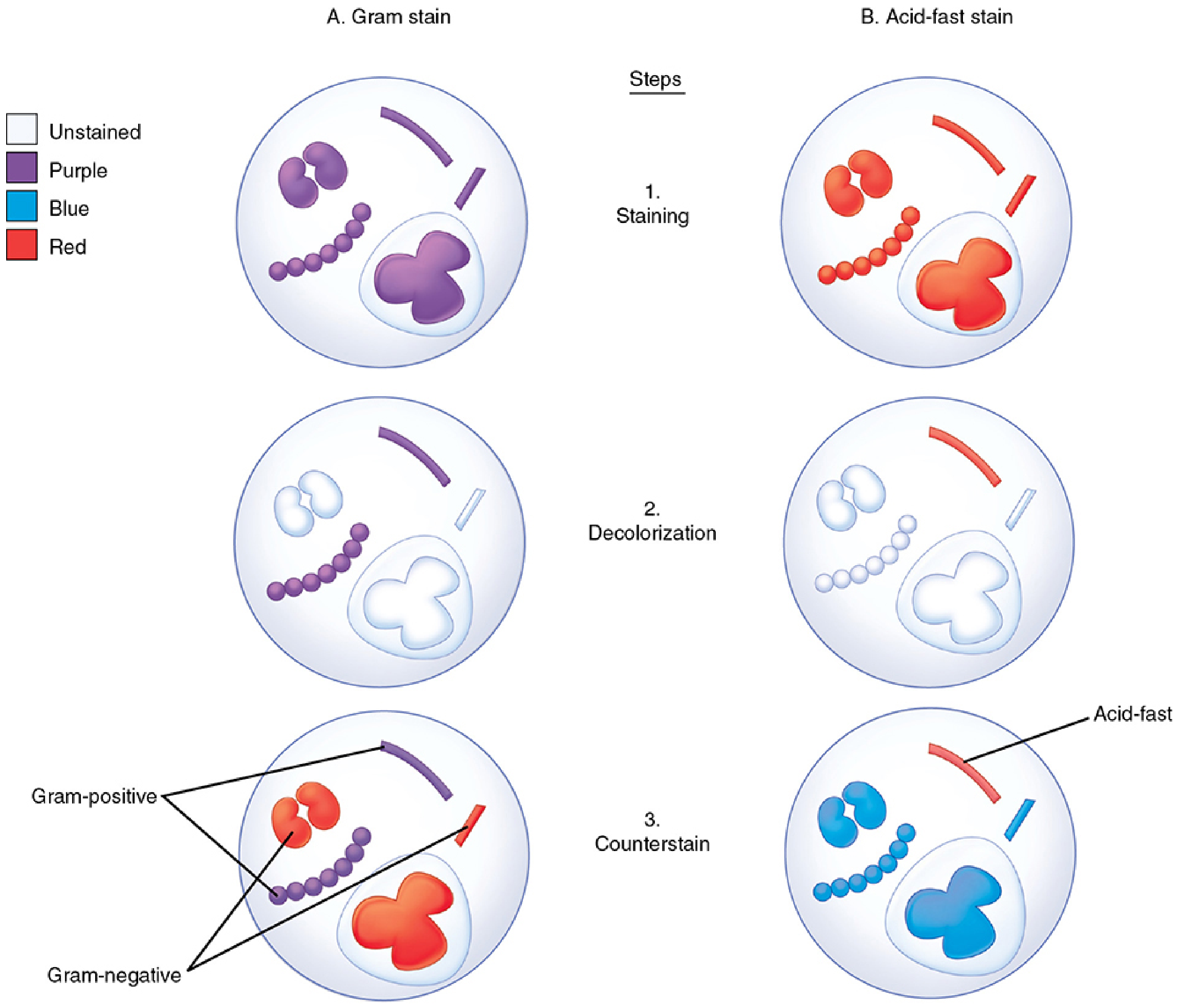
The diagram above shows all three steps of both the Gram stain (left) and acid-fast stain (right) side by side.
Reagents Required
- Crystal violet - primary stain (basic dye)
- Gram's iodine (iodine in potassium iodide) - mordant
- Acetone-alcohol (or acetone or 95% alcohol) - decolorizer
- Safranin (red dye) - counterstain
Step-by-Step Procedure
| Step | Action | Reagent | Time | Result |
|---|---|---|---|---|
| 1. | Prepare smear, air dry, heat-fix | - | - | Bacteria fixed to slide |
| 2. | Primary stain | Crystal violet | 1 min | All bacteria stain purple |
| 3. | Wash with water | Water | Brief | Excess dye removed |
| 4. | Apply mordant | Gram's iodine (KI) | 1 min | Crystal violet-iodine complex forms in all bacteria |
| 5. | Wash with water | Water | Brief | - |
| 6. | Decolorize | Acetone-alcohol | 10-30 sec | Gram-negative cells lose color; Gram-positive retain purple complex |
| 7. | Wash immediately with water | Water | Brief | Stop decolorization |
| 8. | Counterstain | Safranin | 1 min | Decolorized cells take up red counterstain |
| 9. | Wash, blot dry, examine | - | - | - |
Mechanism
- Iodine acts as a mordant - it forms insoluble purple complexes (crystal violet-iodine) with ribonuclear protein inside cells
- On decolorization with acetone-alcohol:
- Gram-positive organisms: thick peptidoglycan cell wall contracts and traps the dye complex → remain purple
- Gram-negative organisms: thin peptidoglycan + outer lipopolysaccharide membrane dissolves in solvent → dye washes out → cells decolorized → pick up safranin counterstain → appear pink/red
Note: An intact cell wall is essential for a positive Gram reaction. Old, dead, or organisms damaged by antibiotics may appear Gram-negative even if they are intrinsically Gram-positive.
Results Interpretation
| Result | Color | Examples |
|---|---|---|
| Gram-positive | Purple/violet | Staphylococcus, Streptococcus, Clostridium, Bacillus |
| Gram-negative | Pink/red | E. coli, Klebsiella, Pseudomonas, Neisseria |
| Background (leukocytes, debris) | Red | - |
ZIEHL-NEELSEN (ZN) / ACID-FAST STAIN
Background
Acid-fastness is a property of mycobacteria (e.g., M. tuberculosis, M. leprae) and some related organisms (e.g., Nocardia - weakly acid-fast). These organisms stain poorly with ordinary dyes due to their high lipid (mycolic acid) content in the cell wall. However, once stained, they resist decolorization with strong mineral acid in alcohol - hence the term "acid-fast."
Reagents Required
- Carbol-fuchsin (basic fuchsin + phenol in alcohol-water) - primary stain
- 1% H₂SO₄ in 95% alcohol (acid-alcohol) - decolorizer
- Methylene blue (or malachite green) - counterstain
Step-by-Step Procedure (Ziehl-Neelsen Hot Method)
| Step | Action | Reagent | Condition | Result |
|---|---|---|---|---|
| 1. | Prepare smear, air dry, heat-fix | - | - | Specimen fixed |
| 2. | Flood slide with primary stain | Carbol-fuchsin | Heat gently until steam rises (do NOT boil), keep steaming for 3-5 minutes | All bacteria stain red |
| 3. | Cool and wash | Water | - | - |
| 4. | Decolorize | Acid-alcohol (3% HCl in 95% ethanol) | Until no more red color runs off (~2 min) | Non-acid-fast organisms lose red color |
| 5. | Wash with water | Water | Brief | - |
| 6. | Counterstain | Methylene blue | 1 min | Decolorized cells take up blue color |
| 7. | Wash, blot dry, examine | - | - | Oil immersion |
The heating step is the key feature of the Ziehl-Neelsen method that allows carbol-fuchsin to penetrate the waxy mycobacterial cell wall.
Mechanism
- Mycobacteria have a very high mycolic acid content in the cell wall
- Normally, carbol-fuchsin cannot penetrate this waxy wall → heating helps drive the stain through
- Once inside, the dye binds to mycolic acids and forms stable complexes
- When decolorized with strong acid-alcohol, non-acid-fast organisms lose the stain
- Acid-fast organisms (mycobacteria) retain carbol-fuchsin due to their waxy wall → appear red
- All other organisms take up the methylene blue counterstain → appear blue
Results Interpretation
| Result | Color | Organism |
|---|---|---|
| Acid-fast (AFB positive) | Bright red/pink rods | Mycobacterium tuberculosis, M. leprae, M. avium |
| Non-acid-fast | Blue | Most other bacteria, WBCs |
| Weakly acid-fast | Red (with weak acid decolorizer) | Nocardia, Cryptosporidium oocysts |
Variants of Acid-Fast Stain
| Method | Heating | Decolorizer | Use |
|---|---|---|---|
| Ziehl-Neelsen (hot method) | Yes - steam | Acid-alcohol (3% HCl) | Classic AFB stain |
| Kinyoun (cold method) | No | Same acid-alcohol (higher phenol in carbol-fuchsin) | Convenience |
| Fluorochrome (auramine-rhodamine) | No | Acid-alcohol | Most sensitive; screening method of choice in most labs |
| Modified acid-fast (Fite stain) | No | Weaker acid | Nocardia, M. leprae (partially acid-fast) |
Side-by-Side Comparison
| Feature | Gram Stain | ZN/Acid-Fast Stain |
|---|---|---|
| Invented by | Hans Christian Gram (1884) | Franz Ziehl & Friedrich Neelsen (1880s) |
| Primary stain | Crystal violet | Carbol-fuchsin |
| Mordant | Gram's iodine | Heat (or higher phenol) |
| Decolorizer | Acetone-alcohol | Acid-alcohol (3% HCl) |
| Counterstain | Safranin (red) | Methylene blue |
| Positive result | Purple | Red/pink |
| Negative result | Pink/red | Blue |
| Organisms detected | Most bacteria | Mycobacteria, acid-fast organisms |
| Key principle | Cell wall permeability to dye-iodine complexes | Mycolic acid resists acid decolorization |
Sources: Sherris & Ryan's Medical Microbiology, 8th Ed. | Jawetz, Melnick & Adelberg's Medical Microbiology, 28th Ed. | Medical Microbiology, 9th Ed.
What is acid fastness
Acid fastness is the property of certain bacteria (mainly mycobacteria) to resist decolorization with strong acid-alcohol after being stained with carbol-fuchsin. This is due to the high mycolic acid content in their cell walls, which makes the dye-organism bond very stable.
Searching sources...
Finding Sources
Reading File
Reading File
Reading File
Loading Image
Let me also fetch the mycobacterial cell wall diagram for a complete visual explanation.
Loading Image
Excellent - now I have everything needed for a complete, well-illustrated answer.
Acid Fastness
Definition
Acid fastness is the property of certain microorganisms to resist decolorization by strong acid-alcohol after being stained with carbol-fuchsin. Organisms possessing this property are called Acid-Fast Bacilli (AFB).
In simpler terms: these bacteria are very hard to stain initially, but once stained, they hold onto the dye so tightly that even the most powerful decolorizing agents (concentrated acid + alcohol) cannot wash it out.
Structural Basis - Why Are Mycobacteria Acid-Fast?
The key is the uniquely high lipid content of the mycobacterial cell wall - over 60% of the total cell wall mass is lipid.
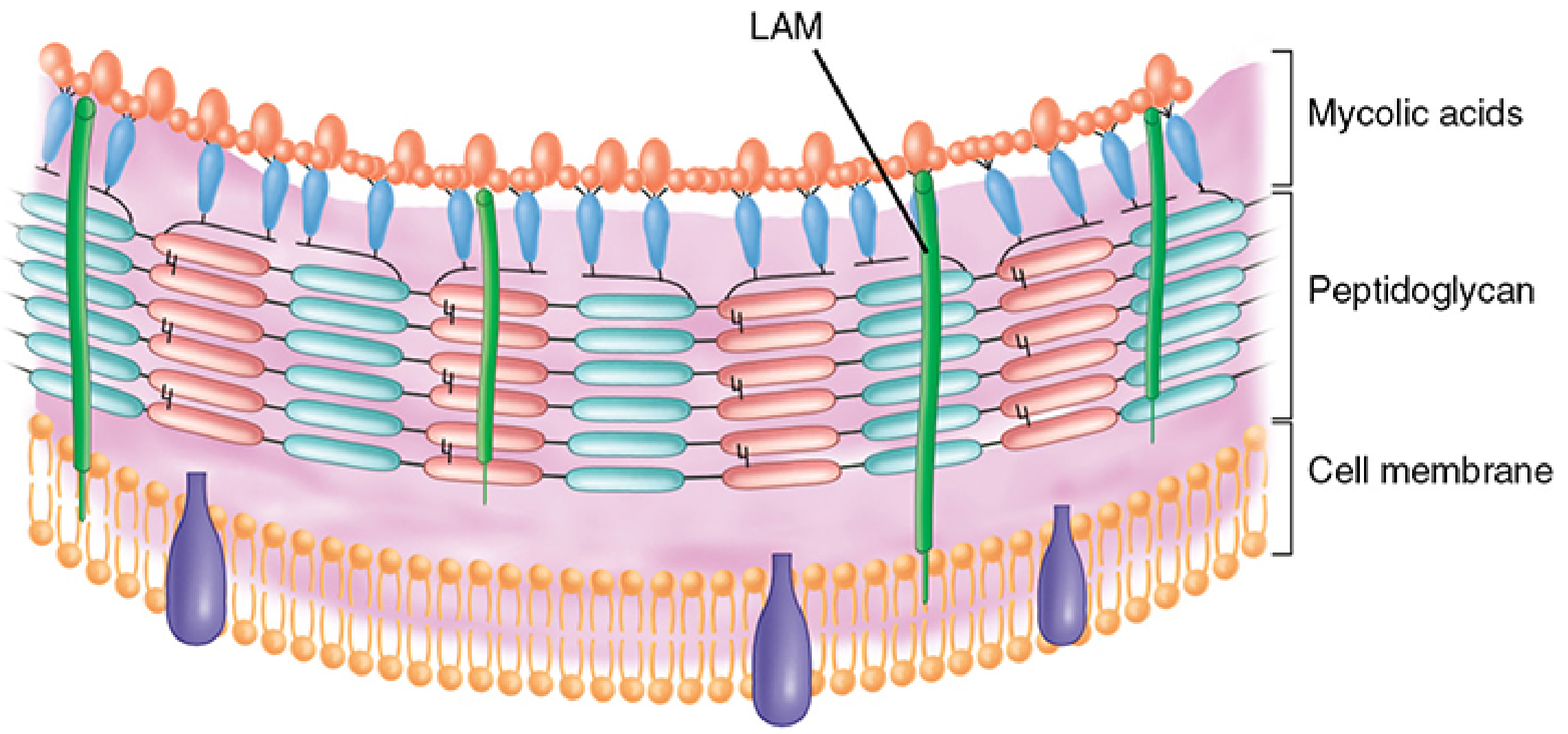
Mycobacterial cell wall: mycolic acids form a dense, waxy outer layer linked to arabinogalactan and peptidoglycan beneath.
The wall contains:
| Component | Role in Acid Fastness |
|---|---|
| Mycolic acids | Long-chain, branched fatty acids (C₇₈-C₉₀); form a dense, waxy hydrophobic outer layer - the PRIMARY determinant of acid fastness |
| Lipoarabinomannan (LAM) | A lipid-polysaccharide complex from plasma membrane to surface; structurally analogous to LPS of Gram-negatives |
| Arabinogalactan | Links mycolic acids to underlying peptidoglycan |
| Peptidoglycan | Inner structural layer |
This "waxy coat" makes the cell wall:
- Impenetrable under ordinary staining conditions
- Hydrophobic - resists water-based stains
- Hardy - resistant to many antibiotics and disinfectants
- Very low cell wall permeability overall
Mechanism of Staining and Retention
| Phase | What Happens |
|---|---|
| Staining | Carbol-fuchsin + heat forces the dye through the waxy wall. Fuchsin binds to mycolic acids inside the wall. |
| Decolorization | Acid-alcohol applied - non-acid-fast organisms lose the dye easily. Mycobacteria retain fuchsin because the waxy mycolic acid layer traps it. |
| Counterstaining | Methylene blue stains all decolorized (non-AFB) elements blue. AFB stand out as red/pink rods on a blue background. |
Critically, acid fastness depends on both the integrity of the cell wall and the presence of lipids. Removing lipids with hot acid, or disrupting the cell wall by sonication, destroys acid fastness.
Microscopic Appearance of AFB
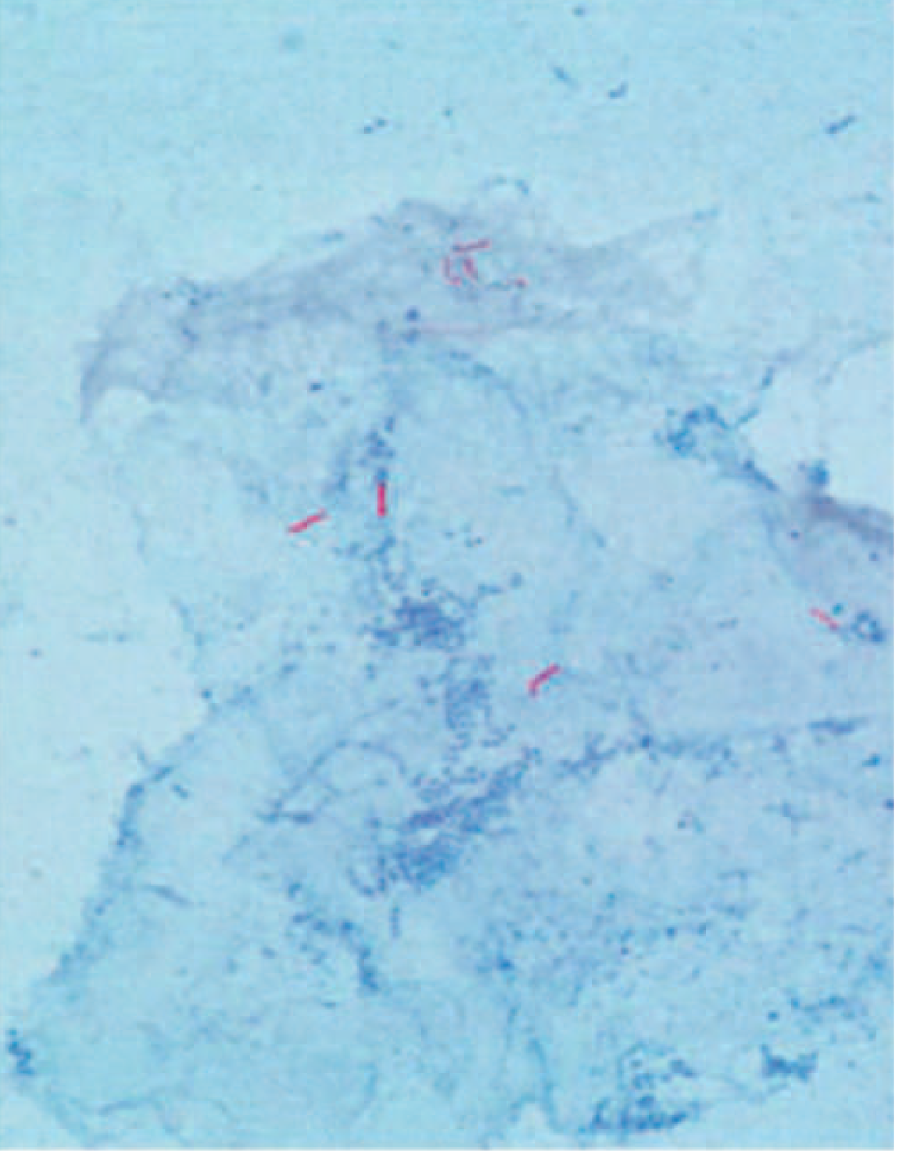
Acid-fast smear showing M. tuberculosis: bright red/pink bacilli against a blue background (methylene blue counterstain). From Harrison's Principles of Internal Medicine, 22nd Ed.
Organisms Showing Acid Fastness
| Category | Examples |
|---|---|
| Fully acid-fast | Mycobacterium tuberculosis, M. bovis, M. leprae, M. avium |
| Weakly acid-fast (partial) | Nocardia spp., Rhodococcus spp., Legionella micdadei |
| Protozoa | Cryptosporidium oocysts, Isospora (now Cystoisospora) |
Weakly acid-fast organisms like Nocardia resist decolorization only with dilute acid (not the strong acid-alcohol used for mycobacteria). This is why a modified acid-fast stain (Fite stain) with a weaker decolorizer is used for them.
Relation to the Length of Mycolic Acids
The degree of acid fastness correlates with the chain length of mycolic acids:
- Mycobacteria: C₇₈-C₉₀ (longest) → strongly acid-fast
- Nocardia: C₅₀-C₆₂ → weakly acid-fast
- Corynebacterium: C₂₂-C₃₆ → not acid-fast (too short to confer resistance)
Clinical Significance
Acid fastness is diagnostically important because it allows rapid presumptive identification of mycobacteria directly from patient specimens (sputum, CSF, tissue) before culture results are available. A positive AFB smear in the right clinical context is strong evidence for mycobacterial infection and justifies starting anti-tubercular therapy.
The same waxy cell wall that causes acid fastness also explains why mycobacteria:
- Are naturally resistant to many antibiotics
- Survive intracellularly within macrophages
- Require long treatment durations (months to years)
Sources: Sherris & Ryan's Medical Microbiology, 8th Ed. | Jawetz, Melnick & Adelberg's Medical Microbiology, 28th Ed. | Harrison's Principles of Internal Medicine, 22nd Ed. | Medical Microbiology, 9th Ed.
This is a shared conversation. Sign in to Orris to start your own chat.