
Deep fasica in abdomen
deep fascia abdominal wall layers anatomy diagram
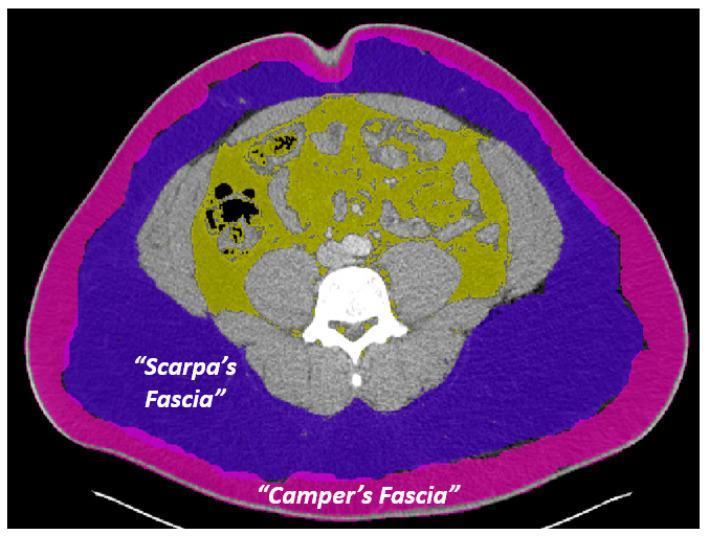
This diagnostic image is an axial computed tomography (CT) scan of the abdomen at the level of the umbilicus, utilizing color-coded overlays to delineate superficial and deep adipose compartments. The image serves as an anatomical diagram for differentiating abdominal wall layers. The outermost layer, highlighted with a pink overlay, represents Camper's fascia (superficial subcutaneous adipose tissue), appearing as a contiguous fatty layer immediately deep to the skin. Internal to this is the purple overlay designating Scarpa's fascia (deep subcutaneous adipose tissue), which is separated from the deeper visceral fat by the abdominal musculature. The yellow overlay identifies the visceral fat (intra-abdominal fat), which is distributed irregularly within the peritoneal cavity, interspersed between and surrounding internal organs and the vertebral body. This visualization emphasizes the spatial relationship between subcutaneous and visceral adipose depots, illustrating clinical concepts related to body composition analysis, metabolic health, and surgical anatomy of the abdominal wall.
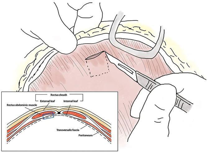
This medical illustration depicts the surgical harvesting of an autologous rectus sheath graft, combining a primary surgical field diagram and an anatomical cross-section. The main illustration shows a surgeon's hands performing a coeliotomy, using a scalpel to incise the internal leaf of the rectus sheath. A dashed square indicates the planned 1 x 1 cm graft harvest site. Retractors are shown maintaining the abdominal incision. The bottom-left inset provides a schematic cross-sectional view of the abdominal wall layers, labeled from superficial to deep: external leaf of the rectus sheath, rectus abdominis muscle, internal leaf of the rectus sheath, transversalis fascia, and peritoneum. A blue highlighted box in the cross-section correlates with the surgical site, specifically identifying the internal leaf and underlying peritoneum as the tissue layers being harvested. This visual serves to educate on surgical anatomy and the technique for obtaining autologous tissue for reconstructive procedures, such as gallbladder or common bile duct repair.
Deep Fascia of the Abdomen
Layers of the Abdominal Wall (Superficial to Deep)
- Skin
- Subcutaneous tissue
- Superficial fascia (Camper's + Scarpa's)
- Deep fascia (investing fascia of muscles)
- Muscles (external oblique, internal oblique, transversus abdominis; rectus abdominis medially)
- Transversalis fascia (the deepest layer of true deep fascia)
- Preperitoneal (extraperitoneal) fat and areolar tissue
- Parietal peritoneum
Cross-sectional anatomy
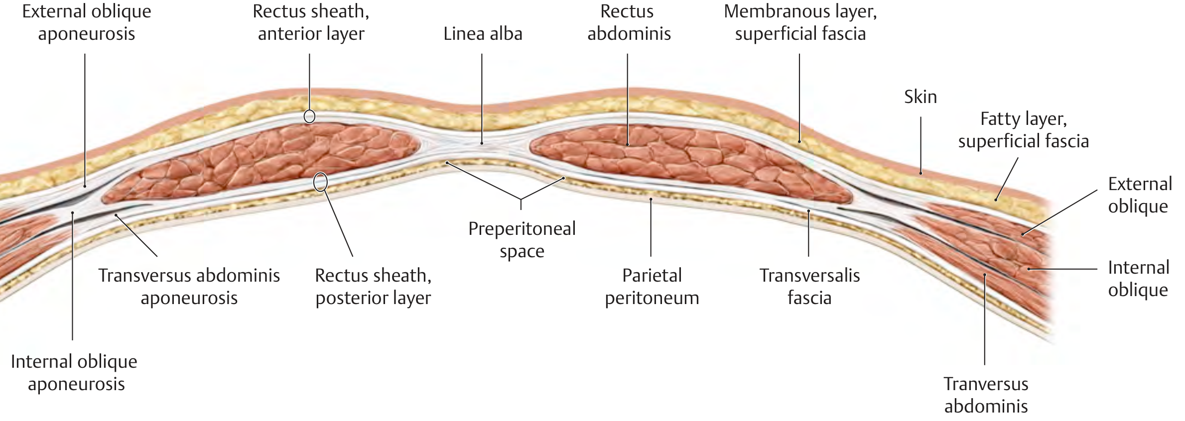
1. Superficial Fascia (technically not "deep" but relevant for contrast)
| Layer | Old Name | New TA Name | Key Features |
|---|---|---|---|
| Superficial (fatty) layer | Camper's fascia | Fatty layer of abdomen | Continuous with superficial thigh fascia; extends to scrotum/labia majora; contains superficial epigastric vessels |
| Deep (membranous) layer | Scarpa's fascia | Membranous layer of abdomen | Compressed fibrous components of superficial fascia; continuous with Colles' fascia in perineum; forms fundiform ligament of penis |
- Scarpa's fascia attaches to the fascia lata of the thigh about a finger-breadth below the inguinal ligament, limiting spread of fluid (e.g., urine extravasation in urethral rupture) inferiorly.
- (Mulholland and Greenfield's Surgery, p. 3635)
2. Investing Deep Fascia of the Muscles (Fascia Innominata)
- The fascia covering the external oblique is sometimes called the fascia innominata. It is bound inferiorly to the inguinal ligament and pubis, then continues onto the thigh to blend with the fascia lata, sealing the space inferior to the inguinal ligament.
- This creates an important surgical boundary: the myopectineal orifice of Fruchaud, which includes Hesselbach's (inguinal) triangle superiorly - the weakest part of the groin.
- (Mulholland and Greenfield's Surgery, p. 3635)
3. Transversalis Fascia - The True Deep Fascia of the Abdominal Cavity
Structure
- Lines the internal surface of the transversus abdominis (and the posterior rectus sheath)
- Crosses the midline anteriorly, associating with the transversalis fascia of the opposite side
- Continuous superiorly with the fascia on the inferior surface of the diaphragm
- Continuous posteriorly with the deep fascia covering posterior abdominal wall muscles (iliac fascia, psoas fascia) and attaches to the thoracolumbar fascia
- After attaching to the iliac crest, it blends inferiorly into the parietal pelvic (endopelvic) fascia
"There is therefore a continuous layer of deep fascia surrounding the abdominal cavity that is thick in some areas, thin in others, attached or free, and participates in the formation of specialized structures." - Gray's Anatomy for Students, p. 343
Bilaminar Debate
- Anterior lamina - more uniform, adherent to the deep surface of the transversus abdominis and rectus abdominis
- Posterior lamina - variable in density, interspersed with adipose tissue; often called the preperitoneal fascia. The inferior epigastric vessels run between/within this layer.
- (Mulholland and Greenfield's Surgery, p. 3648)
Derivatives and Analogs
| Structure | Relationship to Transversalis Fascia |
|---|---|
| Internal spermatic fascia | Transversalis fascia evaginates at the deep inguinal ring to form this covering of the spermatic cord |
| Iliopubic tract | Thickened band of transversalis fascia at the zone of transition between iliac and transversus abdominis muscles; runs parallel to the inguinal ligament; key landmark in laparoscopic hernia repair |
| Iliopectineal arch | Separates the femoral nerve/iliac muscle from femoral vessels |
| Femoral sheath | Formed partly by transversalis fascia anteriorly and iliac fascia posteriorly |
| Anterior wall of femoral canal | Contributes to femoral sheath |
4. Rectus Sheath
- Above the arcuate line (halfway between umbilicus and pubis): both anterior and posterior sheaths are present
- Anterior layer: external oblique aponeurosis + anterior half of internal oblique aponeurosis
- Posterior layer: posterior half of internal oblique aponeurosis + transversus abdominis aponeurosis + transversalis fascia
- Below the arcuate line: all three aponeuroses pass anterior to the rectus abdominis; the posterior sheath consists only of transversalis fascia
Clinical Relevance
| Condition | Fascial Basis |
|---|---|
| Urine extravasation (urethral rupture) | Limited by Scarpa's fascia inferiorly (attaches to fascia lata); spreads into abdominal wall, scrotum, and perineum |
| Inguinal hernia | Weakness in transversalis fascia at the deep inguinal ring or Hesselbach's triangle |
| Herniation spread | Deep to Scarpa's fascia (fascial spaces communicate) |
| Laparoscopic hernia repair | Iliopubic tract is a critical landmark; mesh placed in preperitoneal space between transversalis fascia layers |
| Diastasis recti | Progressive stretching of the linea alba (not a true hernia - no fascial defect); midline bulge |
| Port site closure | Deep fascia (transversalis fascia) must be closed under direct vision to prevent trocar-site hernia |
- Gray's Anatomy for Students, p. 343 - Transversalis fascia as a continuous deep fascial layer
- THIEME Atlas of General Anatomy - Abdominal wall cross-sectional layers and nomenclature
- Sabiston Textbook of Surgery, p. 1647 - Surgical anatomy of anterior abdominal wall
- Mulholland and Greenfield's Surgery, pp. 3632-3649 - Superficial fascia, Scarpa's fascia, transversalis fascia derivatives