
Wydm by effector cells in cns
Effector cells in the CNS release cytokines, cytotoxic molecules, and other mediators to carry out immune responses. The main ones are: T cells (CD8+ release perforin/granzymes and IFN-γ), microglia (TNF-α, IL-1β, reactive oxygen species), astrocytes (IL-6, chemokines), and NK cells (perforin/granzymes).
Searching sources...
Reading File
Finding Sources
Finding Sources
Reading File
Reading File
Finding Sources
Reading File
Finding Sources
In central nervous system
The question "what do effector cells release in the CNS" refers to the mediators secreted by immune effector cells (T cells, microglia, macrophages, NK cells) within CNS tissue during inflammation or immune attack.
Searching sources...
Finding Sources
Finding Sources
Finding Sources
Finding Sources
Reading File
Finding Sources
Finding Sources
Finding Sources
Finding Sources
Here is a consolidated answer on what effector cells release in the CNS:
What Effector Cells Release in the CNS
The CNS is an immune-privileged site, but during inflammation (e.g., multiple sclerosis, viral encephalitis, autoimmune disease), activated effector cells infiltrate or are resident there and release a range of soluble mediators.
1. T Cells (CD4+ and CD8+)
CD4+ Th1 cells are the dominant effectors in most CNS autoimmune conditions. They release:
| Mediator | Effect in CNS |
|---|---|
| IFN-γ (interferon-gamma) | Activates microglia/macrophages; upregulates MHC II on APCs |
| TNF-β (lymphotoxin) | Direct cytotoxic injury to oligodendrocytes and axons |
| TNF-α | Promotes demyelination, oligodendrocyte death |
| IL-2 | T cell autocrine growth factor; sustains effector response |
CD4+ Th17 cells also infiltrate the CNS and release:
- IL-17 - drives neutrophil recruitment, disrupts blood-brain barrier
- IL-22 - inflammatory, barrier disruption
CD8+ cytotoxic T cells (CTLs) kill target cells by releasing:
- Perforin - forms pores in target cell membranes
- Granzymes (A and B) - serine proteases that trigger apoptosis in target cells (neurons, oligodendrocytes)
- IFN-γ - antiviral and pro-inflammatory
Key textbook quote: "Activated T cells and the microglia-macrophages can contribute to tissue injury via non-antigen-restricted mechanisms. Each of these cell types releases an array of soluble factors... Cytokines characteristic of T cells include interleukin 2 (IL-2), interferon gamma (IFN-γ), and tumor necrosis factor β (TNF-β; lymphotoxin)."
- Bradley and Daroff's Neurology in Clinical Practice
2. Microglia / Macrophages (Resident + Recruited)
These are the major innate effector cells of the CNS. When activated ("M1 state"), they release:
| Mediator | Effect |
|---|---|
| TNF-α | Oligodendrocyte death, demyelination, BBB disruption |
| IL-1β | Pro-inflammatory, promotes T cell activation, fever |
| IL-6 | Drives acute-phase response; promotes Th17 differentiation |
| IL-12 | Drives Th1 polarization of infiltrating T cells |
| Reactive oxygen species (ROS) | Oxidative damage to myelin and axons |
| Nitric oxide (NO) | Produced via iNOS; blocks axonal conduction, causes demyelination |
| Proteases (MMP-9) | Degrade extracellular matrix; facilitate immune cell entry through BBB |
| Complement proteins | Opsonization and lysis of target cells |
3. Astrocytes (Reactive Astrogliosis)
In the reactive state, astrocytes release:
- IL-6, IL-1β - amplify inflammatory signals
- Chemokines (CXCL10, CCL2) - recruit T cells and monocytes into CNS
- Glutamate (excess) - excitotoxicity and neuronal death
- VEGF - promotes vascular permeability
4. NK Cells
NK cells can enter the inflamed CNS and release:
- Perforin + Granzymes - kill infected or stressed neurons
- IFN-γ - activates macrophages and microglia
- TNF-α - cytotoxic effects
Summary Table
| Cell Type | Key Mediators Released |
|---|---|
| CD4+ Th1 | IFN-γ, TNF-α/β, IL-2 |
| CD4+ Th17 | IL-17, IL-22 |
| CD8+ CTL | Perforin, Granzymes, IFN-γ |
| Microglia/Macrophage | TNF-α, IL-1β, IL-6, IL-12, ROS, NO, MMP-9 |
| Astrocyte | IL-6, IL-1β, Chemokines, Glutamate |
| NK Cell | Perforin, Granzymes, IFN-γ, TNF-α |
Clinical relevance: In MS, the dominant pathway is myelin-specific Th1/Th17 cells entering the CNS and releasing IFN-γ and IL-17, which activate microglia/macrophages, leading to demyelination through TNF-α, ROS, and NO. This is the rationale for therapies targeting IFN-β, TNF blockade, and B-cell depletion (rituximab, ocrelizumab).
Pregangilonic sympathetic outflow detailed
Preganglionic sympathetic fibers originate from the intermediolateral cell column (IML) of the spinal cord at T1-L2 (thoracolumbar outflow). They are myelinated (B fibers), short, and synapse in paravertebral or prevertebral ganglia using acetylcholine on nicotinic receptors.
Searching sources...
Finding Sources
Finding Sources
Good - multiple excellent sources found. Let me read the most detailed ones in parallel.
Reading File
Reading File
Reading File
Reading File
Reading File
Reading File
Excellent - enough detail gathered. Let me now compile the full answer.
Loading Image
Here is a detailed breakdown of preganglionic sympathetic outflow:
Preganglionic Sympathetic Outflow - Detailed
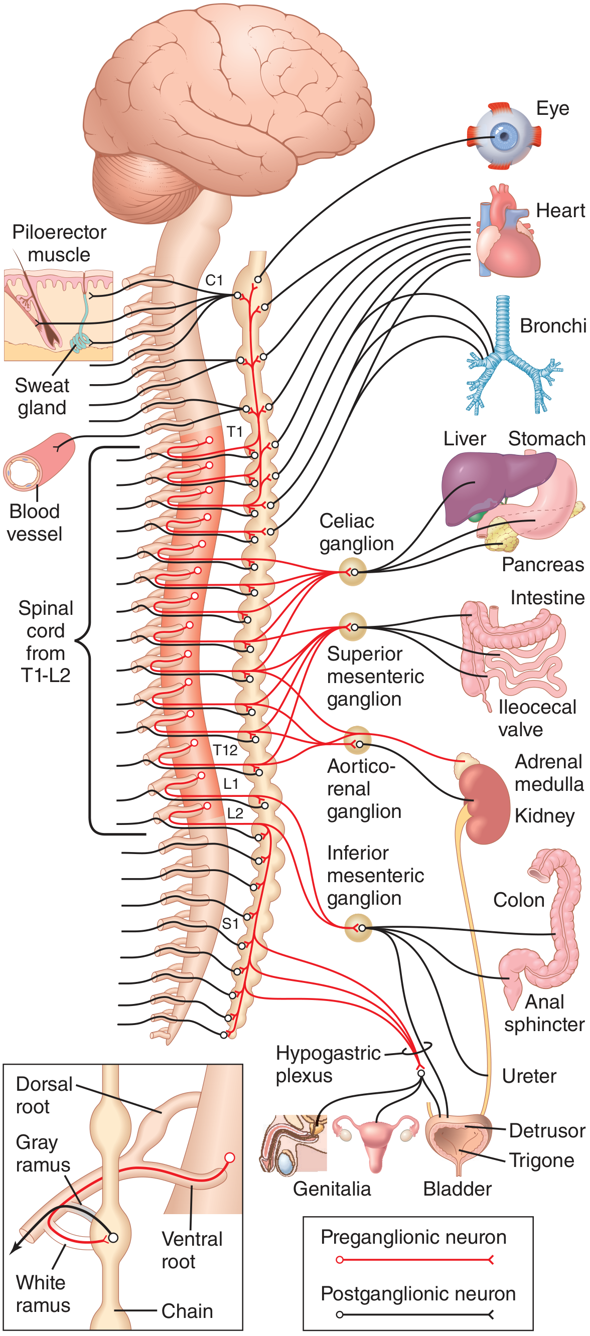
1. Origin - Cell Bodies
- Located in the intermediolateral cell column (IML) / lateral horn of the spinal cord gray matter
- Spans segments T1 to L2 (some texts say T1-L3)
- This is why the sympathetic system is called the thoracolumbar outflow
- White rami communicantes exist only at T1-L2 levels
2. Fiber Characteristics
| Property | Detail |
|---|---|
| Myelination | Lightly myelinated (B fibers) |
| Color | White (contribute to white rami communicantes) |
| Length | Short (ganglia lie close to spinal cord) |
| Neurotransmitter | Acetylcholine (ACh) |
| Receptor on postganglionic | Nicotinic (N2) receptors |
| Divergence | Each preganglionic neuron branches onto ~200 postganglionic neurons - allowing widespread body-wide effects |
3. Path from Spinal Cord to Ganglion
Step-by-step route:
- Cell body in IML of lateral horn (T1-L2)
- Axon exits via the ventral (anterior) root
- Travels briefly in the spinal nerve
- Diverges into the white ramus communicans (myelinated, so white)
- Enters the paravertebral sympathetic chain ganglion
4. Three Fates After Entering the Sympathetic Chain
Once inside a paravertebral ganglion, the preganglionic fiber has three options:
| Fate | Description |
|---|---|
| 1. Synapse at same level | Synapses with postganglionic neuron in the ganglion it entered |
| 2. Ascend or descend in the chain | Travels up or down to synapse in a ganglion at a different vertebral level |
| 3. Pass through without synapsing | Exits the chain, travels via splanchnic nerves to synapse in a prevertebral ganglion in the abdomen |
"After entering a paravertebral ganglion, a preganglionic sympathetic axon may (1) synapse within that segmental paravertebral ganglion, (2) travel up or down the sympathetic chain to synapse within a neighboring paravertebral ganglion, or (3) pass through the chain and then through one of the sympathetic nerves radiating outward from the chain, finally synapsing in a peripheral sympathetic (prevertebral) ganglion."
- Medical Physiology (Boron & Boulpaep)
5. Segmental Distribution of Preganglionic Outflow
| Spinal Level | Target Region |
|---|---|
| T1 | Head (ascends to superior cervical ganglion) |
| T2 | Neck |
| T3-T6 | Thorax (heart, lungs, bronchi) |
| T7-T11 | Abdomen (stomach, liver, pancreas, small intestine) |
| T12, L1, L2 | Legs and pelvic organs |
The distribution is partly determined by the embryological origin of the organ. For example, the heart receives fibers from the neck region of the sympathetic chain because the heart originally developed in the neck of the embryo before descending into the thorax.
- Guyton & Hall Textbook of Medical Physiology
6. Ganglia Where Preganglionic Fibers Synapse
A. Paravertebral (Sympathetic Chain) Ganglia
- 22-23 ganglia arranged bilaterally along the vertebral column from cervical to coccyx
- Key named ganglia:
| Ganglion | Formed From | Supplies |
|---|---|---|
| Superior cervical ganglion | Fusion of C1-C4 | Head and neck (pupil dilator, eyelid elevator, sweat glands of face) |
| Middle cervical ganglion | C5-C6 | Heart, thyroid |
| Stellate (cervicothoracic) ganglion | C7-C8 + T1 fused | Heart, lungs, upper limb |
| Thoracic ganglia (T2-T12) | Individual | Thoracic wall, intercostals |
| Lumbar ganglia | L1-L2 | Lower limbs |
| Coccygeal (ganglion impar) | Right + left chains merge | Perineum |
B. Prevertebral (Collateral) Ganglia
- Lie anterior to the aorta, near the roots of major visceral arteries
- Preganglionic fibers reach them via splanchnic nerves (bypass the chain without synapsing):
| Splanchnic Nerve | Spinal Origin | Synapse In |
|---|---|---|
| Greater splanchnic | T5-T9 | Celiac ganglion |
| Lesser splanchnic | T10-T11 | Superior mesenteric + aorticorenal ganglia |
| Least splanchnic | T12 | Aorticorenal ganglion |
| Lumbar splanchnic | L1-L2 | Inferior mesenteric ganglion / hypogastric plexus |
7. Special Case: Adrenal Medulla
- Preganglionic fibers from T5-T11 travel via greater splanchnic nerves all the way to the adrenal medulla without synapsing
- The chromaffin cells of the adrenal medulla are modified postganglionic neurons (embryologically derived from neural crest)
- These cells secrete epinephrine (80%) and norepinephrine (20%) directly into the bloodstream in response to ACh from preganglionic fibers
- This is why the adrenal medulla acts like a "sympathetic ganglion released into the blood"
"Preganglionic sympathetic nerve fibers pass without synapsing, all the way from the intermediolateral horn cells, through the sympathetic chains, through the splanchnic nerves, into the adrenal medullae. These secretory cells embryologically are derived from nervous tissue and are actually postganglionic neurons."
- Guyton & Hall
8. White vs. Gray Rami Communicantes
| Feature | White Ramus Communicans | Gray Ramus Communicans |
|---|---|---|
| Fiber type | Myelinated preganglionic | Unmyelinated postganglionic |
| Color | White | Gray |
| Direction | Spinal nerve → sympathetic chain | Sympathetic chain → spinal nerve |
| Levels present | T1-L2 only | All spinal cord levels (C2 to coccyx) |
| Function | Carries preganglionic fibers into ganglia | Carries postganglionic fibers back to spinal nerves |
Summary Flowchart
IML lateral horn (T1-L2)
       ↓
Ventral root → Spinal nerve → White ramus communicans
       ↓
Sympathetic chain ganglion
  ├─ Synapse here → postganglionic via gray ramus → body wall, skin, blood vessels, sweat glands
  ├─ Ascend/Descend chain → synapse in cervical/lumbar ganglia → head, neck, legs
  └─ Pass through (no synapse) → splanchnic nerves → prevertebral ganglia → abdominal/pelvic viscera
                                                   ↓ (special)
                                           Adrenal medulla → epinephrine/norepinephrine released
Sources: Guyton & Hall Textbook of Medical Physiology; Medical Physiology (Boron & Boulpaep); Gray's Anatomy for Students; Goodman & Gilman's Pharmacological Basis of Therapeutics
Breif me about sympathetic trunks
The sympathetic trunks are two paravertebral chains of ganglia running from the base of the skull to the coccyx, connected by internodal rami. They contain 22-23 ganglia on each side and allow preganglionic fibers from T1-L2 to reach all spinal levels.
Searching sources...
Finding Sources
Finding Sources
Reading File
Reading File
Reading File
Reading File
Finding Sources
Loading Image
Finding Sources
Reading File
Here is a concise but thorough brief on the sympathetic trunks:
Sympathetic Trunks - Brief
Overview
The sympathetic trunks (also called sympathetic chains) are two bilateral paravertebral nerve cords running alongside the vertebral column on each side. They are the highway that allows preganglionic fibers emerging only from T1-L2 to reach every spinal level in the body - from head down to the coccyx.
"On each side, a paravertebral sympathetic trunk extends from the base of the skull to the inferior end of the vertebral column where the two trunks converge anteriorly to the coccyx at the ganglion impar."
- Gray's Anatomy for Students
Structure
| Feature | Detail |
|---|---|
| Number | Two (one on each side of vertebral column) |
| Extent | Base of skull → coccyx |
| Ganglia | ~22-23 paravertebral ganglia on each side |
| Termination | Both trunks merge anteriorly to coccyx at the ganglion impar |
| Position | Anterior to transverse processes / anterolateral to vertebral bodies |
| Connections to spinal nerves | Via white rami (T1-L2 only) and gray rami (all levels) |
Regional Parts and Their Ganglia
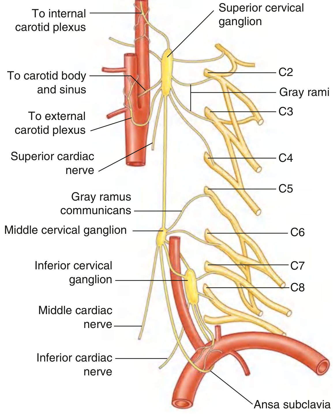
1. Cervical Sympathetic Trunk
- No white rami here (no direct preganglionic input from the cord at cervical levels)
- Receives ascending preganglionic fibers from upper thoracic cord (T1-T3)
- Has 3 ganglia:
| Ganglion | Level | Branches / Supplies |
|---|---|---|
| Superior cervical ganglion | C1-C2 (largest ganglion in the trunk) | Internal/external carotid plexuses, gray rami to C1-C4, pharynx, superior cardiac nerve |
| Middle cervical ganglion | C6 (may be absent) | Gray rami to C5-C6, middle cardiac nerve, thyroid |
| Inferior cervical ganglion | C7 (usually fuses with T1 → stellate/cervicothoracic ganglion) | Gray rami to C7-T1, vertebral artery plexus, inferior cardiac nerve, subclavian loop (ansa subclavia) |
The stellate ganglion (cervicothoracic ganglion) is clinically important - blockade here causes Horner's syndrome (ptosis, miosis, anhidrosis, enophthalmos).
2. Thoracic Sympathetic Trunk
- ~12 ganglia (1st thoracic usually fused into stellate ganglion)
- Has both white and gray rami communicantes at each level
- Position: anterior to necks of ribs superiorly, shifting to costovertebral joints in mid-thorax, then anterolateral to vertebral bodies inferiorly
- Key contribution: gives rise to the splanchnic nerves (preganglionic fibers bypass the chain without synapsing here):
| Nerve | Origin | Destination |
|---|---|---|
| Greater splanchnic | T5-T9 | Celiac ganglion |
| Lesser splanchnic | T10-T11 | Superior mesenteric + aorticorenal ganglia |
| Least splanchnic | T12 | Aorticorenal ganglion |
- Also contributes postganglionic fibers to the cardiac plexus, pulmonary plexus, aortic plexus, esophageal plexus
3. Lumbar Sympathetic Trunk
- ~4 ganglia
- Has white rami only from L1-L2 (end of thoracolumbar outflow)
- Gray rami to all lumbar spinal nerves
- Gives lumbar splanchnic nerves → inferior mesenteric ganglion and hypogastric plexus
- Lies on the anterolateral surface of lumbar vertebral bodies, medial to psoas major
4. Sacral Sympathetic Trunk
- ~4-5 ganglia on the anterior surface of the sacrum, medial to the anterior sacral foramina
- No white rami at sacral levels (no preganglionic input directly here)
- Gray rami to all sacral spinal nerves
- Contributes to the hypogastric plexus and innervation of pelvic organs
5. Ganglion Impar (Coccygeal Ganglion)
- Single unpaired ganglion where the two trunks merge in front of the coccyx
- The terminal point of the entire sympathetic chain
Rami Communicantes - Summary
| Ramus | Fiber Type | Direction | Present At |
|---|---|---|---|
| White ramus communicans | Myelinated preganglionic | Spinal nerve → trunk | T1-L2 only |
| Gray ramus communicans | Unmyelinated postganglionic | Trunk → spinal nerve | All levels (C2 to coccyx) |
Four Pathways for Preganglionic Fibers Once in the Trunk
- Synapse in the ganglion at the same level they entered
- Ascend or descend the trunk and synapse in a ganglion at a different level
- Pass through the trunk and exit as splanchnic nerves to synapse in prevertebral ganglia
- Pass through without synapsing to reach the adrenal medulla directly
Clinical Relevance
| Condition | Mechanism |
|---|---|
| Horner's syndrome | Lesion of cervical trunk / superior cervical ganglion (T1 preganglionic or the ganglion itself) → loss of sympathetics to the eye and face |
| Raynaud's phenomenon | Excessive sympathetic vasoconstriction via the chain → sympathectomy (surgical or chemical) at T2-T3 relieves symptoms |
| Hyperhidrosis | Overactive T2-T3 sympathetic trunk to sweat glands → treated by thoracoscopic sympathectomy |
| Stellate ganglion block | Used for pain, arrhythmias, PTSD, and sympathetically-maintained pain syndromes |
Sources: Gray's Anatomy for Students; Neuroanatomy through Clinical Cases (Blumenfeld); Fischer's Mastery of Surgery
how increase in sympethetic activity affect body in brief also in detail tell me how it effects 1.piloerection 2. in respiration which is about lungs 3.in cardiovascular system 4. renal blood flow 5. skeletal muscle 6.cornary blood flow 7.cerebreal affects 8.heart especially about sa node in heart 9.git 10. urinary bladder 11.liver cells 12.insulin 13. sodium potassium atpase in cell keep these notes about each oragan or cell stated here very brief and detail with annotting about receptor presence and their working about sympathetic outflow
Sympathetic activation (fight-or-flight) releases norepinephrine from postganglionic fibers and epinephrine from the adrenal medulla, acting on α and β adrenergic receptors throughout the body to prepare for emergency response.
Searching sources...
Finding Sources
Finding Sources
Finding Sources
Finding Sources
Finding Sources
Finding Sources
Reading File
Finding Sources
Reading File
Finding Sources
Reading File
Finding Sources
Finding Sources
Here is the complete receptor-annotated detailed breakdown of all 13 systems:
Increased Sympathetic Activity: Effects on the Body
Overall mechanism: Postganglionic sympathetic fibers release norepinephrine (NE) onto target organs. The adrenal medulla releases epinephrine (Epi, 80%) + NE (20%) into blood. These act on α1, α2, β1, β2, β3 adrenergic receptors. α receptors prefer NE; β receptors prefer Epi.
"Stimulation of the β1-receptor causes positive inotropic and chronotropic effects on the heart, increased renin secretion in the kidney, and lipolysis in adipocytes, as well as bronchodilation and vasodilation in skeletal muscle. The α1 subtype mediates vascular and smooth muscle contraction causing vasoconstriction and increased blood pressure."
- Goldman-Cecil Medicine
1. Piloerection
Brief: Sympathetic stimulation causes hair to stand up ("goosebumps").
Receptor: α1-adrenergic
Mechanism:
- Postganglionic sympathetic fibers innervate the arrector pili muscle (smooth muscle attached to hair follicle)
- NE binds α1 receptors → Gq protein → PLC activation → IP3 + DAG → ↑intracellular Ca²⁺ → smooth muscle contraction
- Contraction pulls the hair follicle upright → piloerection
- In animals: traps air for warmth and makes animal appear larger (threat display)
- In humans: vestigial reflex, produces goosebumps (cutis anserina)
Fiber type: Postganglionic sympathetic, unmyelinated (C fibers), via gray rami → skin branches
2. Respiration - Lungs (Bronchial Effects)
Brief: Sympathetic activity causes bronchodilation and reduces secretions.
Receptor: β2-adrenergic (dominant on bronchial smooth muscle) + α1 (on bronchial vessels)
Mechanism:
- Epinephrine (from adrenal medulla) and NE bind β2 receptors on bronchial smooth muscle
- β2 → Gs → ↑cAMP → PKA activation → phosphorylates myosin light chain kinase (MLCK) → inactivates it → smooth muscle relaxation → bronchodilation
- Simultaneously, PKA also opens K⁺ channels → hyperpolarization → further relaxation
- Bronchial glands: α1 stimulation → ↓mucus secretion (reduced secretions)
- Pulmonary vasculature: α1 → mild vasoconstriction (diverts blood to better-ventilated areas)
- Net result: ↑airway caliber → ↓airway resistance → ↑airflow to meet increased O2 demand during fight-or-flight
Clinical use: β2 agonists (salbutamol/albuterol) mimic this for asthma treatment
3. Cardiovascular System (General)
Brief: ↑heart rate, ↑contractility, ↑blood pressure, peripheral vasoconstriction.
Receptors: β1 (heart), α1 (vessels), β2 (some vascular beds)
| Effect | Receptor | Mechanism |
|---|---|---|
| ↑Heart rate (chronotropy) | β1 | ↑cAMP → PKA → ↑If (funny current) in SA node → faster spontaneous depolarization |
| ↑Contractility (inotropy) | β1 | ↑cAMP → PKA → phosphorylates L-type Ca²⁺ channels + troponin I → ↑Ca²⁺ influx + faster relaxation |
| ↑Conduction velocity | β1 | ↑cAMP in AV node → faster conduction |
| Vasoconstriction (skin, gut, kidney) | α1 | Gq → PLC → ↑Ca²⁺ → MLCK → smooth muscle contraction |
| Vasodilation (skeletal muscle) | β2 | Gs → ↑cAMP → smooth muscle relaxation |
| ↑Blood pressure | α1 + β1 | ↑peripheral resistance + ↑cardiac output |
4. Renal Blood Flow
Brief: Sympathetic activation decreases renal blood flow via vasoconstriction.
Receptors: α1 (afferent arteriole, predominant) + β1 (juxtaglomerular cells)
Mechanism:
- NE → α1 on afferent arterioles → vasoconstriction → ↓glomerular capillary pressure → ↓GFR and ↓renal blood flow
- Simultaneously, NE → β1 on juxtaglomerular (JG) cells → ↑cAMP → ↑renin release → activates RAAS → AngII → further vasoconstriction + aldosterone → Na⁺/water retention → ↑blood pressure
- α1 also acts on efferent arteriole but less potently than afferent
- Net result: ↓renal perfusion (blood diverted to heart, muscles, brain) + ↑renin → longer-term BP support
- Severe sympathetic activation (e.g., shock) can cause acute tubular necrosis from ischemia
Note: Dopamine (DA1 receptors on renal vasculature) causes vasodilation - used therapeutically in renal protection.
5. Skeletal Muscle Blood Flow
Brief: Sympathetic activity causes vasodilation in skeletal muscle (via epinephrine/β2), but vasoconstriction via NE/α1 at rest.
Receptors: β2 (metabolic vasodilation, Epi-mediated) + α1 (NE-mediated vasoconstriction at rest)
Mechanism:
- At rest: NE dominates → α1 → mild vasoconstriction (vascular tone maintenance)
- During fight-or-flight: adrenal Epi floods blood → β2 receptors on skeletal muscle arterioles → Gs → ↑cAMP → vasodilation → ↑blood flow to muscles
- Cholinergic sympathetic fibers (unique to skeletal muscle) also release ACh → muscarinic → vasodilation (anticipatory, before exercise begins)
- Local metabolic factors (↑CO2, ↓O2, adenosine) also override sympathetic tone during active exercise
- Net: ↑blood flow to contracting muscle, ↑O2 delivery, ↑glucose delivery
6. Coronary Blood Flow
Brief: Net effect = vasodilation (despite some α1-mediated constriction), primarily driven by metabolic demand.
Receptors: β2 (vasodilation) + α1 (vasoconstriction) + DA1 (dopaminergic vasodilation)
Mechanism:
- Coronary vessels have both α1 and β2 receptors
- Direct sympathetic NE → α1 → mild coronary vasoconstriction
- BUT: ↑heart rate + ↑contractility (β1 effects) → ↑myocardial O2 consumption → local metabolic vasodilators (adenosine, CO2, H⁺, K⁺) released → dominant vasodilation overrides α1 constriction
- Epi via β2 also contributes directly to vasodilation
- DA1 receptors on coronary vasculature: dopamine → vasodilation (exploited in cardiogenic shock treatment)
- Net: coronary blood flow increases during sympathetic activation to meet increased cardiac demand
7. Cerebral Effects
Brief: Cerebral circulation is relatively protected from sympathetic vasoconstriction; autoregulation dominates.
Receptors: α1 (present but weak effect) + β2 (present)
Mechanism:
- Cerebral arterioles do have α1 adrenergic receptors but they are far less responsive than peripheral vessels
- Cerebral autoregulation strongly overrides sympathetic tone - maintains constant blood flow across MAP 60-150 mmHg
- Systemic ↑BP (from sympathetic activation) may slightly reduce cerebral perfusion via myogenic response
- CNS arousal effects: sympathetic activation → release of NE from locus coeruleus → ↑alertness, attention, arousal
- ↑Glucose availability to brain (via hepatic glycogenolysis)
- Blood-brain barrier limits access of circulating catecholamines
- In severe hypertension (e.g., pheochromocytoma crisis): autoregulation fails → hypertensive encephalopathy
8. Heart - SA Node (Specifically)
Brief: Sympathetic activation increases the firing rate of the SA node → tachycardia.
Receptor: β1-adrenergic (primary) + β2 (minor)
Detailed Mechanism at SA Node:
- NE/Epi binds β1 receptor on SA nodal cells
- β1 → Gs protein → ↑adenylyl cyclase → ↑cAMP
- ↑cAMP → directly binds and opens HCN channels (If, "funny current") - these carry inward Na⁺ current during diastolic depolarization
- Faster If current → more rapid spontaneous diastolic depolarization (phase 4)
- ↑cAMP also → PKA → phosphorylates L-type Ca²⁺ channels (ICaL) → ↑Ca²⁺ entry → faster upstroke (phase 0) and lower threshold
- PKA also phosphorylates ryanodine receptors (RyR2) in SR → ↑Ca²⁺ sparks → further acceleration
- Result: shorter cycle length → ↑heart rate (positive chronotropy)
Other cardiac effects via β1:
- AV node: ↑conduction velocity (positive dromotropy)
- Ventricular muscle: ↑contractility (positive inotropy) via ↑Ca²⁺ influx
- ↑Relaxation rate (positive lusitropy) via PKA phosphorylation of phospholamban → ↑SERCA pump activity
9. Gastrointestinal Tract (GIT)
Brief: Sympathetic activation inhibits GIT activity - reduces motility, secretions; contracts sphincters.
Receptors: α1, α2 (dominant inhibitory) + β2 (smooth muscle relaxation)
| Effect | Receptor | Mechanism |
|---|---|---|
| ↓Motility (peristalsis) | α2 (presynaptic) | NE → α2 on myenteric plexus neurons → ↓ACh release → ↓peristalsis |
| ↓Motility (direct) | β2 | ↑cAMP → smooth muscle relaxation → ↓gut movement |
| ↑Sphincter tone | α1 | ↑Ca²⁺ → smooth muscle contraction → closed sphincters (esophageal, pyloric, ileocecal, internal anal) |
| ↓Secretions | α2 | ↓cAMP in secretory cells → ↓gastric acid, ↓intestinal secretions |
| ↓Splanchnic blood flow | α1 | Vasoconstriction → blood diverted to muscles/heart |
Net result: GIT is effectively "shut down" during fight-or-flight. Blood is diverted away. Digestion pauses.
10. Urinary Bladder
Brief: Sympathetic activation promotes urine retention - relaxes detrusor, contracts internal sphincter.
Receptors: β2/β3 (detrusor muscle) + α1 (internal urethral sphincter + bladder neck)
Mechanism:
- Detrusor muscle (bladder wall): NE/Epi → β2 and β3 → Gs → ↑cAMP → smooth muscle relaxation → bladder fills, does not contract → urine retained
- Internal urethral sphincter (bladder neck): NE → α1 → Gq → ↑Ca²⁺ → smooth muscle contraction → sphincter closes → urine cannot escape
- Trigone and base: α1 → contraction reinforces sphincter closure
- Net: bladder capacity ↑, micturition inhibited - logical during fight-or-flight (you don't urinate when fleeing)
- Clinical relevance: α1 blockers (tamsulosin, prazosin) relax the internal sphincter → used in BPH to aid urination. β3 agonists (mirabegron) relax detrusor → used in overactive bladder.
11. Liver Cells (Hepatocytes)
Brief: Sympathetic activation triggers glycogenolysis and gluconeogenesis in the liver → ↑blood glucose.
Receptors: α1 (dominant in liver) + β2 (minor)
Mechanism:
- NE/Epi → α1 on hepatocytes → Gq → PLC → ↑IP3 → ↑Ca²⁺ from ER + ↑DAG → PKC activation
- ↑Ca²⁺ → activates phosphorylase kinase → activates glycogen phosphorylase → breaks down glycogen → glucose-1-phosphate → glucose released into blood
- Epi → β2 (minor contribution) → Gs → ↑cAMP → PKA → also activates glycogen phosphorylase and inhibits glycogen synthase
- ↑Gluconeogenesis: PKA → ↑expression of PEPCK and other gluconeogenic enzymes
- Net: rapid ↑blood glucose to fuel muscles and brain during stress
Summary of signaling: NE → α1 → IP3/Ca²⁺/PKC → glycogenolysis; Epi → β2 → cAMP/PKA → glycogenolysis + gluconeogenesis
12. Insulin (Pancreatic β-Cells)
Brief: Sympathetic activation inhibits insulin secretion and promotes glucagon secretion.
Receptors: α2 (inhibitory, dominant on β-cells) + β2 (stimulatory, minor)
Mechanism:
- NE → α2 receptors on pancreatic β-cells → Gi protein → ↓adenylyl cyclase → ↓cAMP → ↓PKA activity → ↓exocytosis of insulin granules → ↓insulin secretion
- α2 also → opens K⁺ channels → hyperpolarization → prevents Ca²⁺ entry → further ↓insulin release
- Glucagon (α-cells): β2 → Gs → ↑cAMP → ↑glucagon secretion (counteracts hypoglycemia)
- Net metabolic effect: ↓insulin + ↑glucagon → ↑blood glucose (from liver) + ↑lipolysis (from fat)
- This ensures glucose stays available for brain and muscles during emergency
Clinical relevance: Non-selective β-blockers (propranolol) block β2 on β-cells → can mask hypoglycemia and impair recovery from low blood sugar (dangerous in diabetics).
13. Na⁺/K⁺-ATPase (Sodium-Potassium Pump) in Cells
Brief: Sympathetic activation (especially via β2) stimulates Na⁺/K⁺-ATPase → drives K⁺ into cells → can cause hypokalemia.
Receptor: β2-adrenergic
Mechanism:
- Epi/NE → β2 receptors on cell membranes → Gs → ↑cAMP → PKA activation
- PKA phosphorylates the Na⁺/K⁺-ATPase α-subunit → ↑pump activity
- Pump moves 3 Na⁺ out and 2 K⁺ in per cycle → net K⁺ uptake into cells
- This occurs in skeletal muscle, liver, heart cells
- Result: serum K⁺ falls (hypokalemia) - transient but significant
- Also occurs via β2 → ↑insulin release (minor) which also stimulates Na⁺/K⁺-ATPase
Physiological purpose:
- Maintains high intracellular K⁺ (needed for resting membrane potential)
- During exercise/stress, K⁺ leaks out of contracting muscles; β2-driven pump activity counteracts excessive hyperkalemia
Clinical relevance:
- Stress-induced hypokalemia (e.g., post-MI surge in catecholamines → ↓K⁺ → arrhythmias)
- β2 agonists (salbutamol) used to treat hyperkalemia by driving K⁺ into cells
- Non-selective β-blockers can blunt this pump stimulation → relative K⁺ retention
Master Summary Table
| System | Receptor | NE/Epi | Effect |
|---|---|---|---|
| Piloerection | α1 | NE | Arrector pili contracts → hair erect |
| Lungs (bronchi) | β2 | Epi | Bronchodilation, ↓secretions |
| Cardiovascular | β1 (heart), α1 (vessels) | NE+Epi | ↑HR, ↑BP, ↑contractility, vasoconstriction |
| Renal blood flow | α1 + β1 | NE | ↓RBF, ↑renin, ↓GFR |
| Skeletal muscle BF | β2 (Epi) / α1 (NE) | Epi dominant | Vasodilation (Epi) |
| Coronary BF | β2 + metabolic | Epi | Net vasodilation (metabolic demand wins) |
| Cerebral | Autoregulation dominant | - | Minimal direct effect; ↑arousal |
| SA node (heart) | β1 | NE+Epi | ↑If → ↑HR (tachycardia) |
| GIT | α2 (presynaptic), β2, α1 | NE | ↓Motility, ↑sphincter tone, ↓secretions |
| Urinary bladder | β2/β3 (detrusor), α1 (sphincter) | NE | Detrusor relaxes, sphincter contracts → retention |
| Liver | α1 (dominant), β2 | NE+Epi | Glycogenolysis → ↑blood glucose |
| Insulin (β-cells) | α2 (inhibitory) | NE | ↓Insulin, ↑glucagon |
| Na⁺/K⁺-ATPase | β2 | Epi | ↑Pump activity → K⁺ into cells → hypokalemia |
Sources: Goldman-Cecil Medicine; Medical Physiology (Boron & Boulpaep); Goodman & Gilman's Pharmacology; Neuroanatomy through Clinical Cases (Blumenfeld)
This is a shared conversation. Sign in to Orris to start your own chat.