
Secretion of bicarbonate ions digestive physiology To get full marks in mbbs first year physiology university exams
bicarbonate secretion pancreatic duct mechanism diagram
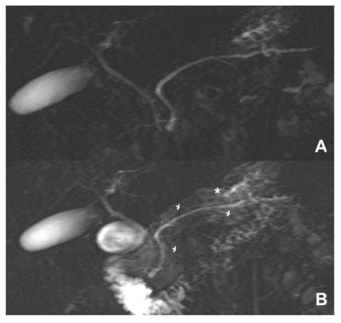
This Comparison Chart consists of two side-by-side (vertical) Diagnostic Images (A and B) showing Magnetic Resonance Cholangiopancreatography (MRCP) findings in a case of Sphincter of Oddi dysfunction (SOD). Image A presents a baseline MRCP where the gallbladder and primary biliary tree are visible, but the pancreatic parenchyma and small ductal side branches are poorly delineated. Image B displays a Secretin-stimulated MRCP (S-MRCP) of the same anatomical region. Key visual features in Image B include diffuse pancreatic parenchymal enhancement (indicated by an asterisk), which appears as a cloud-like increase in signal intensity throughout the gland. Additionally, small arrowheads highlight improved visualization of the Main Pancreatic Duct (MPD) and its side branches due to secretin-induced bicarbonate secretion. The contrast between A and B demonstrates the functional utility of S-MRCP in assessing pancreatic exocrine reserve and identifying subtle abnormalities in ductal dynamics, which are diagnostic indicators for recurrent acute pancreatitis or chronic pancreatitis secondary to SOD.
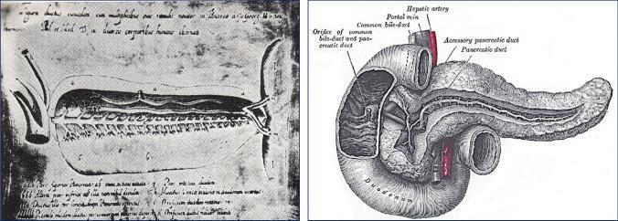
A side-by-side comparison of historical anatomical illustrations of the pancreatic-duodenal region. The left image is a 1642 copperplate engraving by Johann Georg Wirsüng, representing the earliest anatomical drawing of the main pancreatic duct (Wirsüng’s canal) within a lobulated pancreas. The right image is a more detailed 1918 illustration from Gray’s Anatomy, showcasing the complex relationship between the pancreas, duodenum, and biliary system. Key labeled structures in the modern diagram include the head, body, and tail of the pancreas, the main pancreatic duct, and the accessory pancreatic duct (duct of Santorini). The duodenum is shown encircling the pancreatic head, with a windowed view revealing the orifice of the common bile duct and pancreatic duct at the major duodenal papilla. Associated vasculature, including the hepatic artery and portal vein, are also depicted. This comparison illustrates the historical progression of anatomical knowledge regarding exocrine pancreatic structures and their clinical relevance to gastrointestinal physiology.
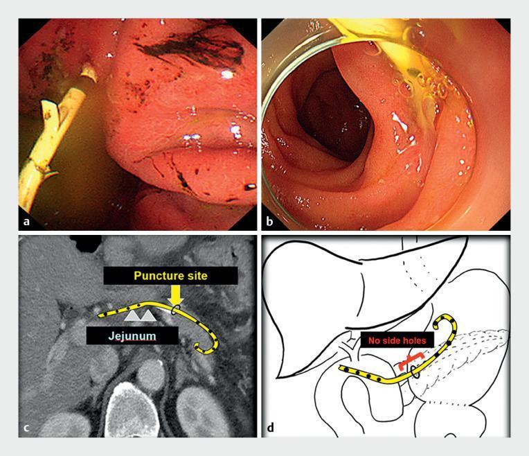
This composite educational graphic illustrates a case of endoscopic ultrasound-guided pancreatic duct drainage (EUS-PDD) and a subsequent complication. Panels (a) and (b) present endoscopic views showing a plastic pancreatic stent within the gastrointestinal tract. In (a), the gastric side view reveals the stent with side holes visible outside the duct, surrounded by erythematous, irritated mucosa with minor dark spots. In (b), the jejunal side view shows the stent emerging through healthy-appearing mucosa. Panel (c) is a coronal CT scan with a superimposed illustration showing the pigtail stent traversing from the jejunum into the pancreatic parenchyma. The puncture site is highlighted, demonstrating the curved path of the perforated stent. Panel (d) provides a schematic anatomical diagram illustrating the malposition of the stent; it highlights that the drainage holes are located outside the pancreatic duct near the anastomotic site, explaining the mechanism of ductal occlusion and subsequent acute pancreatitis. This set serves as a teaching tool for interventional gastroenterology regarding stent placement accuracy and troubleshooting in surgically altered anatomy.
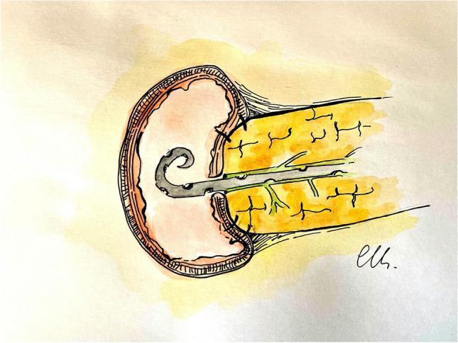
This medical illustration depicts a completed Robotic-assisted minimally invasive Pancreaticojejunostomy (RmPJ) anastomosis. The diagram showcases the surgical union between the transected pancreas (yellow structure) and the jejunum (pink circular structure). A central pancreatic duct is visible with a grey drainage stent in place, ensuring ductal patency. The anastomosis is constructed using an invaginating technique, specifically an outer anterior layer where the jejunal seromuscular wall is rolled over the pancreatic parenchyma. Visible dark sutures are placed at regular intervals along the anterior and posterior margins, demonstrating a continuous running barbed suture technique. This surgical approach is commonly utilized in pancreaticoduodenectomy (Whipple procedure) to manage the pancreatic remnant and mitigate the risk of postoperative pancreatic fistula. The illustration highlights key anatomical relationships including the pancreatic capsule, parenchyma, and the layered wall of the small intestine during reconstructive abdominal surgery.
Secretion of Bicarbonate Ions - Digestive Physiology (MBBS First Year)
Overview
1. PANCREATIC BICARBONATE SECRETION (Most Important)
Site of Secretion
- Bicarbonate and water are secreted by the epithelial cells of the ductules and ducts of the pancreas (NOT the acini - the acini secrete enzymes).
Composition
- HCO3- concentration can rise to up to 145 mEq/L - approximately 5 times the plasma concentration.
- The final pH of pancreatic juice averages 8.0 (alkaline).
- Total pancreatic juice secreted per day: approximately 1 litre.
Cellular Mechanism of HCO3- Secretion
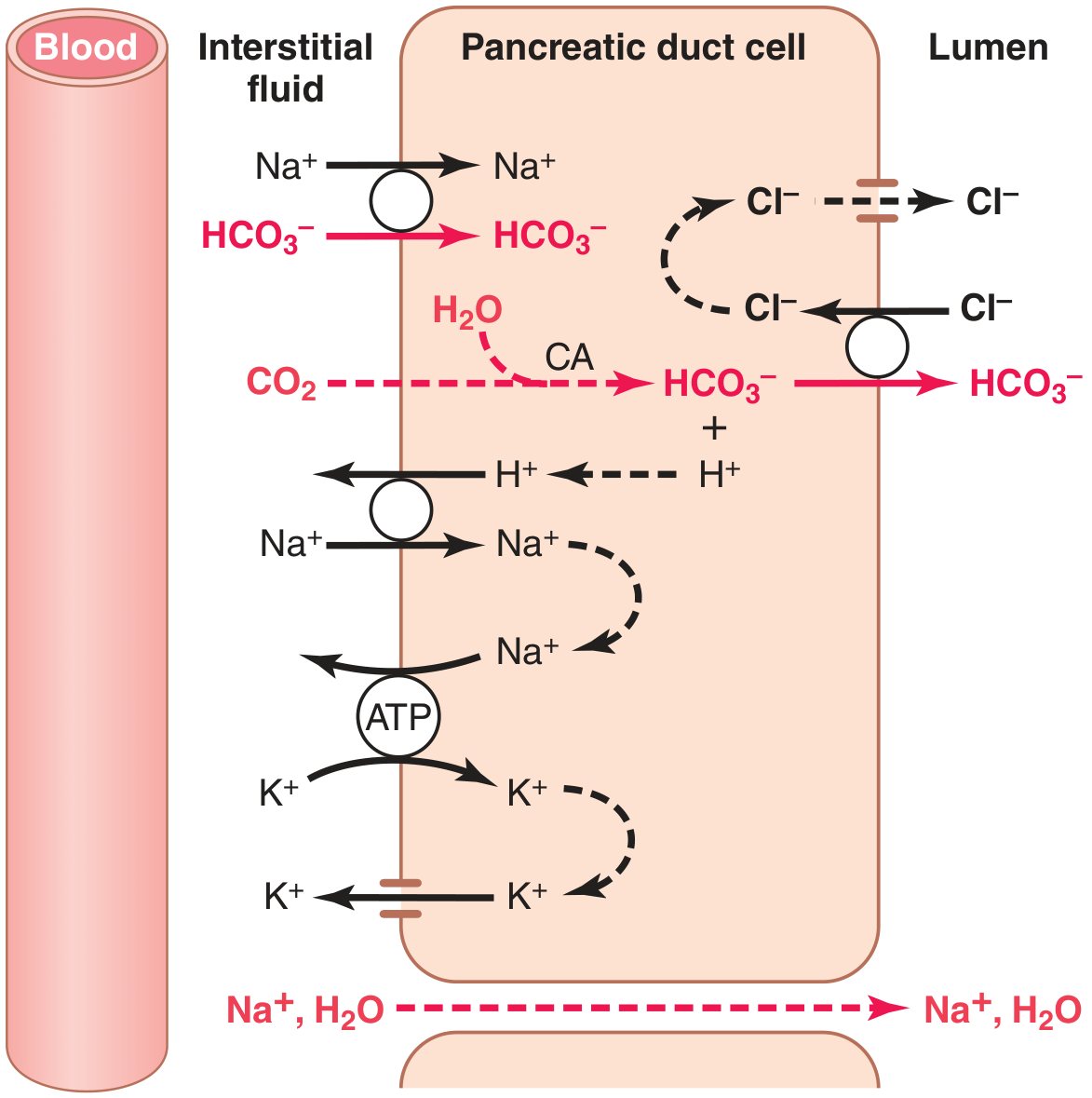
- CO2 diffuses from the blood into the duct cell.
- Under the influence of carbonic anhydrase (CA), CO2 + H2O → H2CO3 (carbonic acid).
- H2CO3 dissociates → HCO3- + H+.
- Additional HCO3- also enters the cell across the basolateral membrane by co-transport with Na+.
- HCO3- is exchanged for Cl- by secondary active transport across the luminal (apical) membrane → HCO3- enters the duct lumen.
- The Cl- that enters the cell is recycled back into the lumen via special Cl- channels (CFTR).
- The H+ formed intracellularly is exchanged for Na+ across the basolateral membrane (H+/Na+ exchange).
- Na+ also enters via the basolateral Na+/HCO3- co-transporter and is then transported into the duct lumen.
- The negative voltage of the lumen pulls Na+ through tight junctions.
- The resulting osmotic gradient drives water into the duct lumen → forming an isosmotic sodium bicarbonate solution.
Key enzyme to remember: Carbonic anhydrase - essential for intracellular HCO3- generation.
2. REGULATION OF PANCREATIC HCO3- SECRETION
Three Basic Stimuli:
| Stimulus | Source | Primary Effect |
|---|---|---|
| Secretin | S cells of duodenum & jejunum | Stimulates copious HCO3- and water secretion from duct cells |
| Acetylcholine (ACh) | Vagal and enteric nerve endings | Mainly stimulates enzyme secretion from acini; minor fluid effect |
| CCK (Cholecystokinin) | I cells of duodenum & upper jejunum | Mainly stimulates enzyme secretion from acini; minor fluid effect |
Secretin - The KEY Hormone for HCO3- Secretion
- Secretin is a 27-amino acid polypeptide (mol. wt. ~3400).
- It is stored in inactive form as prosecretin in S cells of the duodenal and jejunal mucosa.
- Trigger: Acid chyme with a pH < 4.5 to 5.0 entering the duodenum - especially HCl from the stomach.
- Effect: Secretin is released into the blood → acts on pancreatic duct cells → large quantities of HCO3-rich fluid (up to 145 mEq/L HCO3-, low Cl-).
Phases of Pancreatic Secretion:
| Phase | Mechanism | % of Total Enzyme Secretion |
|---|---|---|
| Cephalic | Vagal (ACh) | ~20% |
| Gastric | Vagal (ACh) continued | 5-10% |
| Intestinal | Secretin (HCO3-) + CCK (enzymes) | 70-80% |
Potentiation:
- When ACh + CCK + Secretin all act simultaneously, total secretion is far greater than the sum of individual effects (multiplicative/potentiation effect).
3. FUNCTIONS OF PANCREATIC HCO3-
-
Neutralizes HCl from the stomach:
- HCl + NaHCO3 → NaCl + H2CO3 → NaCl + CO2 + H2O
- CO2 is absorbed into the blood and expired via lungs.
- This protects the duodenal mucosa (which cannot withstand acid gastric juice) from peptic ulceration.
-
Provides optimal pH for pancreatic enzyme activity - pancreatic enzymes work best at pH 7.0-8.0 (slightly alkaline/neutral).
-
Inactivates pepsin - pepsin is inactivated at neutral/alkaline pH, preventing protein digestion in the duodenum.
4. OTHER SOURCES OF BICARBONATE IN THE DUODENUM
Brunner's Glands (Glands of Brunner)
- Located in the submucosa of the duodenum (especially the first few centimeters).
- Secrete a bicarbonate-rich alkaline mucus.
- Stimulated by secretin, vagal stimulation, and local irritation.
- Function: Neutralize gastric chyme, lubricate the mucosa, secrete EGF (epidermal growth factor).
Bile (from Liver)
- Bile secreted into the duodenum via the common bile duct also contains bicarbonate.
- Contributes to the overall alkalinization of duodenal contents.
- Secretin stimulates bicarbonate and water secretion from the biliary ductal epithelium.
Gastric Mucosal HCO3-
- Surface epithelial cells of the stomach secrete a small amount of HCO3-.
- This forms the pre-epithelial alkaline layer (the mucus-bicarbonate barrier) that protects the gastric mucosa from its own HCl.
- For each H+ secreted by the parietal cell luminally, one HCO3- exits the parietal cell across the basolateral membrane → alkalinizes submucosal blood → taken up by surface cells → secreted onto the mucosal surface.
- Stimulated by prostaglandin E2 (PGE2) - this is why NSAIDs (which block PG synthesis) reduce gastric mucosal protection.
5. CLINICAL RELEVANCE (for MBBS exams)
| Condition | Relevance to HCO3- |
|---|---|
| Peptic Ulcer Disease | Failure of HCO3- secretion from pancreas/Brunner's glands → acid damage |
| Cystic Fibrosis | Mutant CFTR (Cl- channel) → impaired Cl- recycling → impaired HCO3- secretion → thick pancreatic secretions |
| NSAID use | Inhibits PGE2 → reduces gastric mucosal HCO3- → mucosal damage |
| Secretinoma / Zollinger-Ellison | Massive acid load overwhelms HCO3- buffering → multiple ulcers |
| Pancreatitis | Reduced HCO3- secretion capacity |
EXAM SUMMARY TABLE
| Source | Cell/Location | Stimulus | HCO3- Concentration | Function |
|---|---|---|---|---|
| Pancreas | Duct/ductule cells | Secretin (primary), ACh, CCK | Up to 145 mEq/L | Neutralize HCl, optimal enzyme pH |
| Brunner's glands | Duodenal submucosa | Secretin, vagus | Alkaline | Neutralize chyme, protect mucosa |
| Bile | Biliary duct epithelium | Secretin | Moderate | Neutralize duodenal acid |
| Gastric mucosa | Surface epithelial cells | PGE2 | Low | Pre-epithelial mucosal defense |
QUICK RECALL POINTS FOR EXAM
- HCO3- is secreted by duct cells, NOT acinar cells of the pancreas.
- Secretin is the key hormone for HCO3- secretion - triggered by acid (pH <4.5-5.0) in the duodenum.
- Carbonic anhydrase is the key enzyme in the mechanism.
- Maximum HCO3- concentration = 145 mEq/L (~5x plasma).
- HCO3-/Cl- exchanger on the luminal side + CFTR (Cl- recycling channel) = the key transport proteins.
- Final pH of pancreatic juice = 8.0.
- Neutralization reaction: HCl + NaHCO3 → NaCl + H2CO3 → NaCl + CO2 + H2O.
- Brunner's glands are in the submucosa (not mucosa) of the duodenum.
- Gastric HCO3- secretion is stimulated by PGE2 and inhibited by NSAIDs.
- All three stimuli (Secretin + CCK + ACh) show potentiation - their combined effect exceeds the sum of individual effects.