
Bronchial Asthma in pregnancy 10 marks answer
Bronchial Asthma in Pregnancy
Definition and Prevalence
Physiological Background in Pregnancy
Effect of Pregnancy on Asthma (the "Rule of Thirds")
- ~1/3 improve
- ~1/3 worsen
- ~1/3 remain unchanged
- Pregnancy-associated changes in sex hormone levels
- Psychosocial stress
- Nonadherence to medications
- Higher prevalence of GERD
- Nasal congestion
- Altered immunity
- Respiratory infections
Effect of Asthma on Pregnancy (Maternal and Fetal Complications)
- Preeclampsia (increased risk)
- Gestational diabetes
- Ante- and postpartum hemorrhage
- Anemia
- Depression
- Preterm labour
- Preterm birth (spontaneous and induced)
- Small for gestational age (SGA)
- Low birthweight (<2500 g)
- Congenital anomalies (increased risk)
- Perinatal mortality (in severe, uncontrolled cases)
Diagnosis in Pregnancy
- Most patients have a pre-existing history of asthma
- Differential diagnosis: Dyspnea of pregnancy (no wheezing/cough/obstruction), pulmonary embolism, pulmonary edema, COPD, laryngeal dysfunction, hyperventilation
- Confirmed by: FEV1 reduced with at least 12% or 200 mL improvement after inhaled albuterol
- Methacholine challenge is contraindicated in pregnancy
- Exhaled nitric oxide (FeNO) is significantly elevated in pregnant asthmatics vs. healthy pregnant controls - useful marker
- Skin testing for allergies is not recommended (risk of systemic reactions); specific IgE blood testing is preferred
| Severity | Symptoms | Nighttime | FEV1/PEFR |
|---|---|---|---|
| Intermittent | ≤2 days/week | ≤2×/month | ≥80% |
| Mild Persistent | >2 days/week, not daily | 3-4×/month | ≥80% |
| Moderate Persistent | Daily | >1×/week | 60%-80% |
| Severe Persistent | Throughout the day | Often 7×/week | <60% |
Management
- Minimal or no maternal symptoms day or night
- No limitations of activities
- Maintenance of normal or near-normal pulmonary function
- Minimal use of rescue bronchodilators
1. Objective Assessment and Monitoring
- Spirometry is the preferred method; PEFR is an acceptable alternative
- Normal PEFR in pregnancy: 380-550 L/min
- PEFR zones: Green >80%, Yellow 50-80%, Red <50% of personal best
- Patients with moderate-severe asthma should have daily PEFR monitoring
- FEV1 <80% predicted is associated with preterm birth and low birthweight
2. Trigger Avoidance
- Avoid allergens: house dust mites, animal dander, cockroach antigens, mold, pollens
- Avoid tobacco smoke (active and passive)
- Control GERD
- Avoid NSAIDs/aspirin and beta-blockers in sensitive patients
- Exercise-induced asthma: pre-treat with albuterol inhaler 10-15 minutes before exercise
3. Patient Education
- Action plans (green/yellow/red zones)
- Proper inhaler technique
- Emphasis that medication risks are lower than the risk of uncontrolled asthma
- Symptom monitoring and self-management
4. Pharmacologic Therapy
| Severity | Treatment |
|---|---|
| Intermittent | Short-acting β2-agonist (SABA: albuterol) PRN |
| Mild Persistent | Low-dose inhaled corticosteroid (ICS: budesonide preferred) + SABA |
| Moderate Persistent | Low-medium dose ICS + long-acting β2-agonist (LABA: salmeterol) or increase ICS dose |
| Severe Persistent | High-dose ICS + LABA ± oral corticosteroids |
- Albuterol (salbutamol): Drug of choice for acute relief; well-established safety profile
- Budesonide (ICS): Preferred ICS in pregnancy due to most safety data; ICS is the cornerstone of treatment for ALL levels of persistent asthma in pregnancy
- Salmeterol/formoterol (LABA): Safe in combination with ICS; do not use as monotherapy
- Theophylline: Acceptable in moderate-severe asthma; monitor serum levels (therapeutic 5-12 mcg/mL); crosses placenta - can cause neonatal irritability/tachycardia
- Systemic corticosteroids (oral/IV): Benefits outweigh risks in acute exacerbations; associated with cleft palate (first trimester exposure), gestational diabetes, preeclampsia when used long-term
- Montelukast (LTRA): Use with caution (FDA warning - possible association with congenital anomalies); may be needed if asthma cannot be controlled otherwise
- Ipratropium bromide: Safe; added to albuterol in acute exacerbations
- Biologic agents (omalizumab, etc.): Limited fetal safety data; as IgG molecules, expected to cross placenta in 2nd trimester; use only when clearly needed
Management of Acute Exacerbation in Pregnancy
- Supplemental O2 to maintain SpO2 >95%
- Nebulized albuterol 2.5 mg every 20 minutes for 3 doses
- Fetal monitoring (CTG if viable)
| Response | Criteria | Action |
|---|---|---|
| Good | FEV1/PEFR ≥70%, sustained 60 min | Discharge home; oral steroids if needed |
| Incomplete | FEV1/PEFR 50-70%, mild-moderate symptoms | Add ipratropium 0.5 mg, systemic corticosteroids 40-80 mg/day; consider terbutaline 0.25 mg SC; consider IV magnesium sulfate 2g over 20 min |
| Poor | FEV1/PEFR <50%, PCO2 >42 mmHg, confusion | Admit to ICU; intubation if required |
Fetal Surveillance
- Serial ultrasound for fetal growth in moderate-severe or poorly controlled asthma
- Antepartum fetal surveillance (NST/biophysical profile) starting at 32 weeks for moderate-severe asthma
- After recovery from a severe exacerbation
Labor and Delivery
- Asthma attacks are uncommon during labor (high endogenous cortisol and prostaglandins E2 may be protective)
- Stress-dose corticosteroids should be given to patients on prolonged systemic steroids during labor
- Regional anesthesia is preferred (reduces minute ventilation, O2 consumption, prevents hyperinflation)
- If general anesthesia required: ketamine and halogenated agents are preferred (bronchodilatory)
- Safe oxytocics: Oxytocin and prostaglandin E2
- Avoid: Prostaglandin F2-alpha (carboprost), morphine, meperidine, ergometrine - all may trigger bronchospasm
Preconceptional Counseling
- Consult pulmonologist/allergist/MFM specialist before conception
- Baseline pulmonary function testing
- Optimize asthma control before conception
- Stop smoking; avoid triggers
- Continue asthma medications during pregnancy
- Reassure: well-controlled asthma = excellent pregnancy outcomes
Summary
Pulmonary tuberculosis in pregnancy 10 marks answer
Pulmonary Tuberculosis in Pregnancy
Introduction and Epidemiology
Effect of Pregnancy on TB
- Fatigue and malaise of early TB can be confused with symptoms of normal pregnancy
- Physiological dyspnoea in pregnancy may mask respiratory symptoms
- A high index of suspicion is essential
Effects of TB on Pregnancy (Complications)
- Severe preeclampsia and eclampsia
- Placenta previa
- Postpartum hemorrhage
- Sepsis
- Anemia
- Higher morbidity and mortality (if untreated)
- TB-positive women have an 80% increased rate of pregnancy complications compared to TB-negative women
- Intrauterine growth restriction (IUGR)
- Preterm birth (spontaneous and induced)
- Low birthweight
- Perinatal mortality
- Congenital tuberculosis (rare but documented)
Routes of Fetal Transmission (Congenital TB)
- Hematogenous spread through the placenta (most common)
- Contiguous spread from endometrial tuberculosis
- Aspiration of TB-infected amniotic fluid
High-Risk Factors for TB in Pregnancy (Box 58.1)
- HIV infection / immunosuppression
- Close contact with known/suspected TB cases
- Birth in a high-prevalence country
- Medically underserved or low-income status
- Alcohol addiction / IV drug use
- Residency in a long-term care facility (prison, nursing home)
- Healthcare workers in high-risk facilities
Diagnosis
Symptoms of Active TB
- Cough (74%)
- Weight loss (41%)
- Fever (30%)
- Malaise and fatigue (30%)
- Hemoptysis (19%)
Screening Tests
- Tuberculin Skin Test (TST/Mantoux): Subcutaneous injection of purified protein derivative (PPD); sensitivity 90-99%; safe in pregnancy; interpretation: ≥10 mm induration is significant (in high-risk groups, ≥5 mm)
- Interferon-Gamma Release Assay (IGRA/QuantiFERON-TB): In vitro blood test; specificity >95%; results in 24-48 hours; no follow-up visit required; not affected by BCG vaccination status. Note: Both TST and IGRA detection rates may be impacted by pregnancy, and different cut-offs may be needed.
Confirmatory Tests for Active Disease
- Chest radiograph (essential; with abdominal shielding): perform after first trimester in asymptomatic PPD-positive women; perform immediately if symptomatic
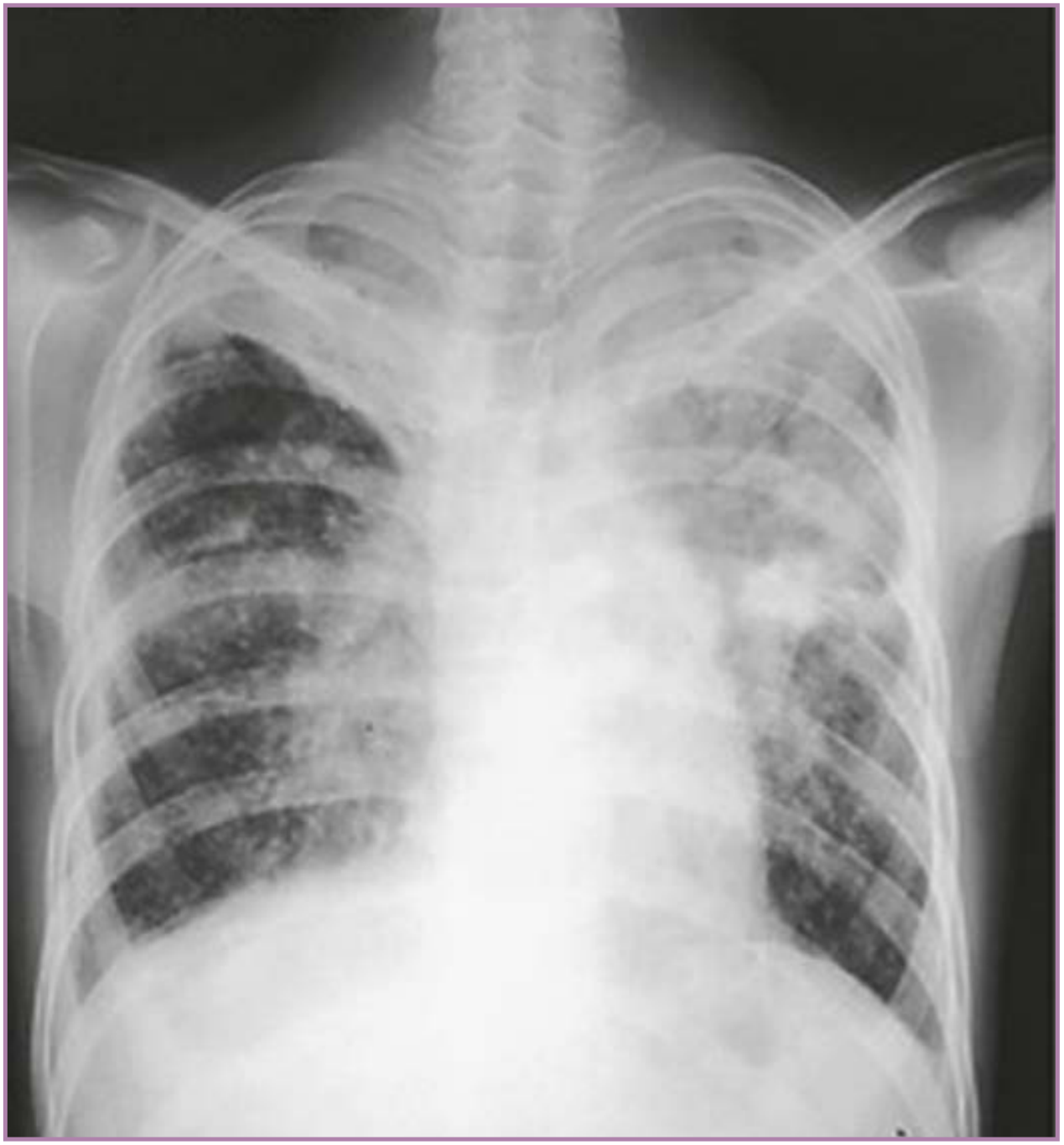
- AFB smear microscopy: At least 3 early-morning sputum samples; if sputum cannot be produced - sputum induction, gastric washings, or bronchoscopy
- Nucleic acid amplification tests (NAAT): MTD Test and Amplicor MTB Test - amplify M. tuberculosis 16S ribosomal DNA; sensitivity and specificity of 95% and 100% in smear-positive specimens
- Placental and endometrial biopsy at delivery if congenital TB is suspected (but workup must never be delayed until delivery)
Management
Latent TB Infection (LTBI) in Pregnancy
- Treatment may be deferred to postpartum in women at low risk for reactivation (unknown or prolonged PPD positivity >2 years)
- Immediate treatment is indicated in:
- HIV co-infected women (risk of reactivation is significant)
- Women with recent TB contact/conversion
- High-risk patients
| Regimen | Duration | Notes |
|---|---|---|
| Rifampicin (4R) - Preferred | 4 months daily | Recommended regimen |
| INH + Rifampicin (3HR) | 3 months daily | Alternative |
| INH alone (6H or 9H) | 6 or 9 months | With pyridoxine supplementation |
Active TB in Pregnancy - Treatment
Drug Safety Profile in Pregnancy
| Drug | Safety | Notes |
|---|---|---|
| Isoniazid (INH) | Safe | Risk of acute hepatitis (especially with rifampicin); add pyridoxine 50 mg/day |
| Rifampicin (RIF) | Safe | Give Vitamin K to neonate at birth (anticoagulant effect) |
| Ethambutol (EMB) | Safe | Teratogenic in animals but NOT in humans; monitor for optic neuritis |
| Pyrazinamide (PZA) | Controversial | WHO/IUATLD say justifiable; CDC does NOT recommend routinely - add if drug resistance suspected |
| Streptomycin | CONTRAINDICATED | Cranial nerve VIII (auditory/vestibular) damage in neonate |
| Ethionamide | Avoid | Teratogenic |
| Capreomycin, Kanamycin, Amikacin | Avoid | Fetal nephrotoxicity/ototoxicity |
| Cycloserine | Avoid | CNS side effects |
| Para-aminosalicylic acid (PAS) | Acceptable | No teratogenicity shown |
MDR-TB in Pregnancy
- MDR-TB (resistant to INH + RIF) complicates management significantly
- Many second-line drugs are contraindicated
- Directly observed therapy (DOT) is strongly recommended
- Patient must be counselled about small risk of teratogenicity
- Management must be individualized by a specialist team
- Second-line injectable agents (streptomycin, capreomycin, kanamycin) are all best avoided
Fetal Surveillance
- Serial ultrasound for fetal growth assessment
- Antepartum surveillance for fetal well-being in severe/active disease
- Neonatal evaluation at birth if maternal active TB is present
Labor and Delivery
- Uncomplicated TB is not an indication for cesarean section
- Mother with active (infectious) TB should wear a mask during labor
- Neonates born to mothers with active TB:
- Evaluate for congenital TB
- Should not be routinely separated from mother if mother on treatment for >2 weeks
- Give isoniazid prophylaxis to the neonate; BCG vaccination after completion of INH prophylaxis
- Monitor for neonatal TB symptoms in the first 90 days
Breastfeeding
- Breastfeeding should not be discouraged in women being treated for TB, unless the mother is still infectious (sputum AFB positive)
- Anti-TB drugs are secreted in breast milk in small concentrations (not therapeutic for the infant but potentially additive if the neonate is also on treatment)
- Transmission of mycobacteria via breast milk is very unlikely, except in rare cases of TB mastitis
Prevention
- BCG vaccination (all neonates in endemic areas)
- Active case finding and contact tracing
- IGRA/PPD screening of all high-risk pregnant women
- Directly observed therapy (DOT) for adherence
- HIV testing in all TB cases (and vice versa)
- Adequate nutrition and social support
Summary
| Aspect | Key Point |
|---|---|
| Prevalence | 5-13 per 100,000 pregnancies; higher in developing countries |
| Most common form | Pulmonary TB (85%) |
| Effect on pregnancy | Untreated: preeclampsia, PTB, IUGR, PPH, perinatal death |
| Diagnosis | TST/IGRA screening; AFB smear + CXR for active disease |
| Treatment (active) | INH + RIF + EMB x2 months, then INH + RIF x7 months |
| Contraindicated drug | Streptomycin (8th nerve damage) |
| Breastfeeding | Allowed unless mother is infectious |
| Key principle | Untreated TB is more dangerous than treatment |
Thanku
Prolapse uterus complicating pregnancy
prolapse uterus complicating pregnancy management obstetrics
https://rscope.org/collective-journal-of-gynecology-and-obst…
uterine prolapse pregnancy management complications
PMID: 37976302
Prolapse Uterus Complicating Pregnancy
Definition
Incidence
- Developing countries (India, Africa, Middle East)
- Grand multiparae
- Rural populations with poor obstetric care
- Women with connective tissue disorders
Classification / Degrees of Prolapse
| Degree | Description |
|---|---|
| 1st degree | Cervix descends into the vagina but does not reach the introitus |
| 2nd degree | Cervix reaches or protrudes just beyond the introitus with straining |
| 3rd degree (Procidentia) | Complete prolapse - entire uterus lies outside the vagina |
Aetiology and Predisposing Factors
Pre-existing (before pregnancy):
- Grand multiparity with inadequate perineal repair
- Previous difficult/prolonged labour and delivery
- Pelvic floor injury from prior obstetric trauma
- Inherent connective tissue weakness (Ehlers-Danlos syndrome, Marfan syndrome)
- Congenital weakness of supporting ligaments
Pregnancy-related factors (aggravating):
- Cervical elongation, hypertrophy due to increased vascularity
- Ligamentous relaxation from progesterone and relaxin - these hormones soften and relax the cardinal and uterosacral ligaments
- Increased intra-abdominal pressure (especially with twin pregnancy, polyhydramnios)
- Aggravation of pre-existing prolapse by the gravid uterus
Natural course during pregnancy:
- As the uterus enlarges beyond the 1st trimester, it rises out of the pelvis and is supported by the pelvic brim - this naturally reduces the degree of prolapse in many cases
- However, the elongated, oedematous cervix may continue to protrude even after the body of the uterus has risen
Effects of Prolapse on Pregnancy (Complications)
Early Pregnancy (1st Trimester):
- Spontaneous abortion (miscarriage) - increased risk due to poor vascularity, infection, and physical factors
- Difficulty conceiving (though conception can occur even with significant prolapse)
During Pregnancy (2nd & 3rd Trimester):
- Cervical desiccation and ulceration - exposed cervix undergoes ulceration, secondary infection; increases risk of ascending infection
- Urinary tract infections (UTI) and urinary retention - common; bladder neck distortion causes voiding dysfunction
- Acute urinary retention - may require catheterization; this is a serious and recurrent complication
- Preterm labour - due to infection (chorioamnionitis), cervical incompetence, or physical stimulation of the prolapsed cervix
- Maternal and fetal sepsis - from ascending infection through the ulcerated, exposed cervix
- Incarceration of the gravid uterus - retroversion with entrapment behind the sacral promontory; a related but distinct complication
- Maternal discomfort, pressure sensation, difficulty walking
- Preterm premature rupture of membranes (PPROM)
During Labour:
- Obstruction of labour - prolapsed, oedematous, rigid cervix may fail to dilate normally
- Cervical dystocia - oedematous prolapsed cervix may not efface and dilate
- Cord prolapse - if membranes rupture with presenting part not engaged
- Postpartum haemorrhage - due to uterine atony from prolonged labour
Maternal Outcome:
- Increased maternal morbidity
- Rare cases of maternal death (from sepsis, obstructed labour)
Fetal Outcome:
- Increased perinatal mortality
- Prematurity
- Fetal growth restriction
- Neonatal sepsis
Clinical Features
- Symptoms: Feeling of something coming down the vagina, dragging/pressure sensation in the pelvis, difficulty walking, dysuria, urinary retention, vaginal discharge (from cervical ulceration/infection), recurrent UTI
- Signs: On examination - cervix/uterus visible or palpable at or beyond the introitus; cervical oedema, hypertrophy, elongation; ulceration/inflammation of exposed cervix; positive gravid uterus on bimanual examination (if 1st/2nd trimester)
Management
- Gestational age at presentation
- Degree of prolapse
- Presence of complications (ulceration, urinary retention, infection)
- Patient's desire to continue the pregnancy
- Available resources
Conservative Management (First-line)
- Knee-chest position or Trendelenburg position to reduce the prolapse by gravity
- Rest reduces prolapse aggravated by upright posture and straining
- Gentle digital reduction of the prolapsed uterus with the patient in Trendelenburg position
- Regular reduction and maintenance
- Vaginal ring pessary (with or without support, Hodge pessary) is the most practical conservative measure
- Provides mechanical support to the uterus and prevents re-prolapse
- First reported in pregnancy in 1949; remains the most valuable non-invasive option
- Silicone-coated ring pessary is preferred - easy insertion, patient self-care possible
- Changed or cleaned every 2-4 weeks; monitor for pressure ulcers
- Effective until about 14-16 weeks, after which the enlarging uterus rises out of the pelvis naturally
- Local antiseptic/oestrogen cream for cervical ulceration
- Antibiotics for superadded infection or UTI
- Urinary catheterization for urinary retention (indwelling catheter if needed)
- Maintain good perineal hygiene
- In cases with associated cervical incompetence
- Helps retain pregnancy to viable gestational age
- Performed in 2nd trimester (14-16 weeks)
Surgical Management
- Failed conservative management
- Severe prolapse not reducible
- Recurrent urinary retention unresponsive to conservative measures
- Severe ulceration and cervical necrosis
- Laparoscopic uterine suspension (sacrohysteropexy): Most evidence from case reports; performed typically in early 2nd trimester (13-16 weeks) when the risk of pregnancy loss is lowest; preserves uterus and pregnancy
- Pelvic floor repair procedures: Generally deferred postpartum
- Avoid 1st trimester (high miscarriage risk)
- Early 2nd trimester is the safest window if surgery is necessary
Management During Labour and Delivery
Mode of Delivery:
- Vaginal delivery is possible and preferred if:
- Cervix dilates adequately
- No obstruction to labour
- Fetal presentation is cephalic
- Caesarean section (LSCS) is indicated for:
- Failed progress / cervical dystocia (oedematous, non-dilating cervix)
- Cord prolapse
- Fetal distress
- Severe cervical ulceration, infection, or oedema preventing vaginal delivery
- Obstetric indications (malpresentation, CPD)
- Most modern authorities favour LSCS to avoid further pelvic floor trauma
Intraoperative precautions:
- Be alert for urinary tract involvement (bladder, ureter injury) due to distorted anatomy
- Careful haemostasis
After Delivery:
- Uterine prolapse often reduces spontaneously after delivery as the uterus involutes
- However, the pelvic floor damage may worsen with each subsequent vaginal delivery
- Surgical repair (pelvic floor reconstruction, colporrhaphy, pelvic floor repair) is best planned 3-6 months postpartum after full involution
- Advise against future pregnancies until surgical repair is done
Summary Table
| Aspect | Key Points |
|---|---|
| Incidence | 1 in 10,000-15,000 deliveries; rare but serious |
| Common setting | Grand multipara, developing countries |
| Natural course | May reduce as uterus rises after 12-16 weeks |
| Key complications | Miscarriage, PTL, UTI, urinary retention, sepsis, cervical ulceration, obstructed labour |
| First-line management | Bed rest, manual reduction, ring pessary |
| Surgical option | Laparoscopic uterine suspension (early 2nd trimester) |
| Mode of delivery | Vaginal if cervix dilates; LSCS if obstruction or complications |
| Postpartum | Surgical repair at 3-6 months |
Key Points to Remember (Exam Focus)
- Uterine prolapse in pregnancy is RARE (1 in 10,000-15,000) but potentially life-threatening
- The degree appears to reduce spontaneously as pregnancy advances beyond 16 weeks (uterus rises above pelvis)
- The cervix undergoes hypertrophy and elongation making it protrude even when the uterine body has risen
- Urinary retention is the most troublesome acute complication
- Ring pessary is the cornerstone of conservative management
- Streptomycin-like 2nd line drugs are - never forget progesterone/relaxin are the hormones responsible for ligamentous laxity
- Surgical repair should be deferred to 3-6 months postpartum
- Procidentia (3rd degree/complete prolapse) persisting into 3rd trimester is extremely rare
Embryo or fetal reduction
Embryo / Fetal Reduction (Multifetal Pregnancy Reduction - MFPR)
Definition
- Reduce the risks of preterm delivery and associated morbidity/mortality for the surviving fetuses
- Improve the overall chances of a healthy live birth
- Reduce maternal health risks from carrying a high-order multiple pregnancy
Background and Rationale
| Pregnancy Type | Preterm Birth Rate | Rate before 34 weeks | Very Low Birthweight |
|---|---|---|---|
| Singleton | 8.4% | 2% | Low |
| Twins | 61% | 20% | Moderate |
| Triplets | 99% | 63% | 34% |
| Quadruplets | ~95% | ~85% | 61% |
| Quintuplets+ | 100% | 100% | 100% |
- 75% of triplets are expected to require NICU admission
- Natural history of triplet gestation: 7-8% risk of delivery between 24-28 weeks, and 11% risk of spontaneous loss before 24 weeks
- Quadruplets: 14% risk of delivery before 28 weeks
Maternal risks of higher order multiple pregnancy:
- Gestational diabetes
- Hypertensive disorders of pregnancy (gestational hypertension, preeclampsia)
- Anemia
- Increased need for cesarean section
- Postpartum haemorrhage
- Maternal mortality
Indications
- Higher-order multiple pregnancy (triplets and above) - most common indication; all patients with quadruplet+ gestation should be counselled about MFPR
- Triplet pregnancy - offered with counselling; debate exists but evidence supports it
- One fetus with a major chromosomal or structural abnormality in a multiple pregnancy (selective feticide)
- Monochorionic pair in a higher-order multiple (they share circulation, carry greater risk)
- Maternal medical conditions where multiple pregnancy poses excessive risk
- Patient choice after informed counselling (for twin pregnancy reduced to singleton - controversial)
Contraindications / Special Considerations
- Monochorionic twins within a higher-order multiple: potassium chloride (KCl) cannot be used because fetocide via KCl in one twin of a monochorionic pair risks death or neurological injury in the co-twin through the shared placental circulation. Alternative methods (radiofrequency ablation, cord coagulation) must be used.
- Ethical concerns - requires thorough pre-procedure counselling
Timing of the Procedure
- Optimal timing: 10-13 weeks of gestation (first trimester)
- At this stage:
- Crown-rump length (CRL) can be accurately measured
- Nuchal translucency can be assessed (to select abnormal fetuses for reduction)
- Spontaneous loss of one or more fetuses (which occurs in 5-10% of higher-order multiples in 1st trimester) has already been accounted for
- The procedure carries a lower loss rate than in the second trimester
- Performing earlier (before 10 weeks) carries higher loss rates
- Second-trimester MFPR is occasionally performed but carries higher complication rates
Pre-procedure Evaluation
- Detailed ultrasound to:
- Count all gestations
- Determine chorionicity and amnionicity
- Map placental location
- Measure CRL and nuchal translucency (NT) of all fetuses
- Chorionic villus sampling (CVS) - may be performed before MFPR to confirm karyotype
- CVS of fetuses destined to be retained (to confirm normality)
- Fetuses with chromosomal abnormalities are preferentially targeted for reduction
- CVS before MFPR has been shown safe with no increase in loss rate
- Genetic counselling
- Informed consent after full discussion of risks, alternatives, and ethical considerations
Technique
Transabdominal Approach (Preferred)
- Patient is placed supine; no prophylactic antibiotics are routinely given (analogous to CVS/amniocentesis)
- Detailed ultrasonography maps all gestational sacs, CRL, NT, and abnormalities
- Target fetus selection:
- Fetuses with abnormal NT or identified abnormality are selected first
- Otherwise, the fetus/fetuses that are technically most accessible are chosen
- The fetus overlying the internal os is avoided (rarely selected)
- If a monochorionic pair exists, that pair is usually selected for reduction together (KCl cannot be used for one twin of a monochorionic pair)
- Under continuous ultrasound guidance and strict sterile technique:
- A 22-gauge needle is inserted transabdominally into the thorax of the targeted fetus
- 2-3 mL of potassium chloride (KCl) is injected intracardiac/intrathoracic
- Asystole (cardiac standstill) is observed for at least 3 minutes to confirm fetal demise
- Procedure is repeated for additional fetuses using a new needle (or occasionally the same needle)
- Post-procedure ultrasound at 1 hour and 1 week to confirm:
- Demise of reduced fetuses
- Viability and cardiac activity of retained fetuses
Transvaginal Approach
- Reserved for rare situations where target fetuses cannot be safely accessed transabdominally
- Associated with significantly higher loss rates (12% vs 5% transabdominal)
- Hence, the transabdominal route is strongly preferred
Target Number (Finishing Number)
- Most higher-order multiples are reduced to twins - this is the most common finishing number
- Reduction to singleton is increasingly performed (patient preference, especially after years of infertility); loss rates 2-4% vs 4-6% for reduction to twins
- Optimal finishing number appears to be twins, balancing loss rates and perinatal outcomes
Outcomes and Loss Rates
International Collaborative Data (>3500 pregnancies):
| Starting Number | Pregnancy Loss Rate before 24 weeks |
|---|---|
| 6 fetuses | 22% |
| 5 fetuses | 15% |
| 4 fetuses | 12% |
| 3 fetuses | 6% |
| 2 fetuses (finishing) | 6% |
- Loss rates have fallen over time from 8% to 5%, reflecting a learning curve effect
- Risk of very preterm delivery (25-28 weeks) decreased from 6% to 1% as finishing number decreased
- For fetuses that reach viability after MFPR: 85-90% are born at ≥32 weeks, with only 3-5% born between 25-28 weeks
MFPR in Triplets (Meta-analysis, 24 studies):
| Outcome | Reduction to Twins | Expectant (Triplets) |
|---|---|---|
| Pregnancy loss <24 weeks | 7% | 8% (comparable) |
| Preterm birth <32 weeks | 11% | 29% |
| Preterm birth <28 weeks | 4% | 8% |
- MFPR of triplets to twins significantly reduces preterm birth at <32 weeks (11% vs 29%) with no significant increase in miscarriage rates
Post-Procedure Care
- Close observation for complications of multiple gestation
- Serial ultrasound for fetal growth - increased risk of IUGR after MFPR
- Monitor for chorioamnionitis, preterm labour, PPROM
- Amniocentesis may be offered 4 weeks after MFPR for karyotyping of surviving fetuses (does not appear to increase loss risk)
- Psychological support - strongly recommended:
- 70% of women demonstrate grief reactions after MFPR
- 84% describe the procedure as very stressful
- Psychological counselling should be offered routinely
Complications
| Complication | Details |
|---|---|
| Procedure-related pregnancy loss | 5% transabdominal; 12% transvaginal |
| Inadvertent demise of retained fetus | If KCl tracks to retained fetus |
| Infection/chorioamnionitis | Rare |
| Preterm labour/PPROM | From the procedure itself |
| IUGR of surviving fetus(es) | Observed post-procedure |
| Psychological morbidity | 70% grief reaction; 84% describe as very stressful |
| DIC (disseminated intravascular coagulation) | Rare; from retained dead fetal tissue |
Ethical and Counselling Considerations
- FIGO position: "For multiple pregnancy higher than twins, it may be considered ethically preferable to reduce the number of fetuses rather than to do nothing"
- ACOG/SMFM (2017): Support MFPR; recognize that reducing pregnancy decreases spontaneous loss rates and complications for all multifetal pregnancies; offer an ethical framework for obstetricians
- ASRM: More neutral - "MFPR appears associated with reduced prematurity risk, though true benefit is difficult to enumerate"
Key counselling points:
- MFPR is not the same as abortion - the goal is to improve survival and health of remaining fetuses
- The procedure involves the deliberate death of one or more viable fetuses - couples must understand and accept this
- Options of expectant management vs. MFPR with their respective risks must be discussed in detail
- Religious, cultural, and personal values must be respected
- The decision must be individualized - a woman must weigh the obligate death of one fetus + procedure-related loss risk against a 2-3 fold reduction in very preterm birth risk
- Psychological support throughout the process is essential
Summary
| Feature | Key Point |
|---|---|
| Definition | Deliberate termination of one or more fetuses in a higher-order multiple pregnancy |
| Goal | Reduce preterm birth risk; improve healthy live birth rates |
| Timing | 10-13 weeks (first trimester) |
| Route | Transabdominal preferred (loss rate 5%); transvaginal 12% |
| Agent | Potassium chloride (KCl) 2-3 mL intrathoracic/intracardiac |
| Confirm | Asystole ≥3 minutes; U/S at 1 hour and 1 week |
| KCl contraindicated | Monochorionic co-twin (use RFA/cord coagulation instead) |
| Target number | Usually twins; increasingly singleton |
| Psychological impact | 70% grief reactions; 84% find it very stressful |
| Supported by | FIGO, ACOG, SMFM |
Stem cell banking
stem cell banking cord blood types public private banking indications clinical applications 2024
Stem Cell Banking
Definition
What Are Stem Cells?
- Self-renewal - ability to divide and replicate themselves indefinitely
- Differentiation - ability to develop into specialised cell types
Classification by Potency:
| Type | Potency | Example |
|---|---|---|
| Totipotent | Can form any cell including extraembryonic tissue | Fertilized egg (zygote), first 2-3 divisions |
| Pluripotent | Can form any cell of all 3 germ layers (but not placenta) | Embryonic stem cells (ESCs), iPSCs |
| Multipotent | Can differentiate into multiple but limited cell types | Hematopoietic stem cells, MSCs |
| Oligopotent | Can differentiate into a few cell types | Lymphoid/myeloid progenitors |
| Unipotent | Can differentiate into only one cell type | Muscle satellite cells |
Types of Stem Cells Relevant to Banking:
- Hematopoietic Stem Cells (HSCs) - found in cord blood; give rise to all blood cells (RBCs, WBCs, platelets) and immune cells
- Mesenchymal Stem Cells (MSCs) - found in cord tissue (Wharton's jelly); can differentiate into bone, cartilage, fat, muscle; have immunomodulatory properties
- Embryonic Stem Cells (ESCs) - derived from inner cell mass of blastocyst; pluripotent; ethically controversial
- Induced Pluripotent Stem Cells (iPSCs) - adult somatic cells reprogrammed to a pluripotent state using transcription factors (Oct4, Klf4, Sox2, cMyc - Yamanaka factors, 2006); bypasses ethical concerns of ESCs
Sources of Stem Cells for Banking
| Source | Stem Cell Type | Notes |
|---|---|---|
| Umbilical cord blood | HSCs, MSCs | Most common; collected at birth; non-invasive |
| Cord tissue (Wharton's jelly) | MSCs (rich source) | Stored by private banks; promising for regenerative medicine |
| Bone marrow | HSCs, MSCs | Painful harvest; requires HLA matching |
| Peripheral blood | HSCs (mobilized by G-CSF) | Non-invasive collection via apheresis |
| Adipose tissue | MSCs (stromal vascular fraction) | Easily accessible |
| Dental pulp | MSCs | Emerging source |
| Placenta/amnion | MSCs | Being explored |
| Embryo (blastocyst) | ESCs | Ethically controversial |
Umbilical Cord Blood - Why It Is Important
Advantages of UCB over Bone Marrow:
- Non-invasive collection - no risk to mother or baby
- Collected at birth - readily available; no donor recruitment needed
- Immediately available - reduces time to transplantation (banked in advance)
- Greater HLA tolerance - allows some degree of HLA mismatch (up to 2-antigen mismatch) without severe graft-versus-host disease (GvHD)
- Lower risk of GvHD - cord blood lymphocytes are immunologically naive
- Lower risk of transmissible infection - young donor
- Less stringent HLA matching required compared to bone marrow
Disadvantages of UCB:
- Limited cell dose - small volume (40-150 mL); insufficient for adults (weight >50 kg)
- Slower engraftment - delayed neutrophil and platelet recovery
- Cannot collect additional cells from the same donor if needed
- Failed or delayed engraftment - low HPC dose leads to increased morbidity/mortality
- One-time collection opportunity
Collection of Cord Blood
Timing:
- Collected immediately after delivery (before or after placental delivery)
- Can be collected in utero (placenta still in place) or ex utero (after placental delivery)
- In utero collection typically yields a larger volume
Technique:
- After delivery of the baby, the umbilical cord is clamped and cut
- The cord vein is cannulated using a sterile needle and blood collection bag
- Gravity drains blood from the cord and placenta into the collection bag
- Volume typically: 40-150 mL (experienced centers)
- Collections <40 mL are usually discarded (inadequate)
- Approximately 4-11 × 10⁸ nucleated cells are harvested
- Dedicated trained staff in the delivery room improves volume collected and reduces contamination risk
Processing:
- The collected blood is not usually further volume-processed (unlike bone marrow)
- Frozen directly with DMSO (dimethyl sulfoxide) as cryoprotectant
- Stored in liquid nitrogen at -196°C
- Shelf life: potentially indefinite when properly stored (some banks claim 25+ years)
Maternal screening before storage:
- Serological tests for HIV, Hepatitis B, Hepatitis C, HTLV, CMV, syphilis
- NAT (Nucleic Acid Testing) on maternal sample
- Bacteriologic testing on cord blood unit
- Screening for genetic diseases in mother/family
- HLA typing of the unit
Types of Cord Blood Banks
1. Public Cord Blood Banks
- Cord blood is donated by the family after birth
- The family relinquishes ownership; the unit is listed in national/international registries
- Available to any matched recipient worldwide
- Free of cost to the family
- Units are HLA-typed and made searchable for unrelated donor transplants
- Regulated by government bodies (FDA in USA; ICMR in India; HTA in UK; TGA in Australia)
- Certified by FACT-NetCord standards
- Approximately 9% of allogeneic transplants (2012-2016) utilized cord blood HPCs
- Increases the donor pool for unrelated transplants
- Helps patients without HLA-matched bone marrow donors
- No cost to family
- Contributes to public health
- Family cannot retrieve the donated unit
- No guarantee of availability or match for the donor family
2. Private (Family) Cord Blood Banks
- Cord blood stored exclusively for the donor family (child or compatible family member)
- Family retains 100% ownership
- Acts as "biological insurance"
- Annual storage fees apply (typically USD 100-300/year + initial processing fee USD 1500-2500)
- Regulated like public banks but operated as for-profit entities
- Guaranteed availability for the child
- Available for HLA-compatible sibling or family member
- No risk of unavailability when needed
- Expensive - not accessible to all families
- Very low probability of personal use (estimated 1 in 2500 to 1 in 200,000 chance the stored unit will be used by the donor child)
- Risk of the bank closing or going out of business
- The child most likely to need it (for inherited diseases) may be affected in the stored cells themselves
3. Hybrid / Directed Banks (Emerging)
- Family-directed storage while the unit is also HLA-typed and listed in the national registry
- If the child does not need it, the unit can be released to a matched unrelated recipient
- Increases diversity of cord blood units in public registries
Diseases Treatable with Cord Blood Stem Cell Transplantation
Established (Well-proven) Indications:
- Acute lymphoblastic leukaemia (ALL)
- Acute myeloid leukaemia (AML)
- Chronic myeloid leukaemia (CML)
- Myelodysplastic syndromes (MDS)
- Hodgkin and Non-Hodgkin lymphoma
- Neuroblastoma
- Retinoblastoma
- Aplastic anaemia
- Fanconi anaemia (first disease treated with UCB, 1989)
- Thalassaemia major - curative via allogeneic UCB transplant
- Sickle cell disease
- Severe combined immunodeficiency (SCID)
- Hurler's syndrome (MPS I) and other metabolic storage diseases
- Diamond-Blackfan anaemia
- Wiskott-Aldrich syndrome
- Chronic granulomatous disease
Investigational / Emerging Applications (clinical trials):
- Cerebral palsy (autologous UCB infusion - promising early results)
- Type 1 diabetes mellitus
- Autism spectrum disorder
- Acquired hearing loss (autologous UCB)
- Hypoxic-ischaemic encephalopathy (neonatal)
- Multiple sclerosis
- Cardiovascular disease (MSCs)
- Spinal cord injury
- Parkinson's disease
- Liver cirrhosis
Regulatory Framework
| Country | Regulatory Body |
|---|---|
| USA | FDA (Food and Drug Administration) |
| India | ICMR (Indian Council of Medical Research) |
| UK | Human Tissue Authority (HTA) under Human Tissue Act 2004 |
| Australia | Therapeutic Goods Administration (TGA) |
| International standard | FACT-NetCord Cord Blood Standards |
Induced Pluripotent Stem Cells (iPSCs) - A Special Mention
- Oct4, Klf4, Sox2, c-Myc (Yamanaka factors)
- iPSCs can potentially be generated from banked cord blood cells in the future
- Creates the prospect of patient-specific, autologous stem cells for any disease
- Eliminates rejection and GvHD concerns
Ethical Considerations
- Public vs. private banking: Medical societies (ACOG, AAP, ICMR) generally favour public donation over private banking for most families, as it benefits the public and the probability of personal use is very low
- Misleading marketing by private banks - overpromising future uses
- Embryonic stem cell research - ethically controversial (requires destruction of embryos)
- Equity and access - private banking is expensive and accessible mainly to affluent families
- Consent - informed consent from the mother is mandatory for cord blood collection
- Conflict of interest - healthcare providers must not financially benefit from recommending private banks
Position of Medical Bodies
- AAP (American Academy of Pediatrics): Supports public donation; does not recommend routine private banking unless a first-degree family member has a condition known to benefit from stem cell transplantation
- ACOG: Supports public cord blood banking; private banking may be considered when there is a family history of disease treatable by UCB transplantation
- ICMR (India): Regulates both public and private banks; supports public banking; calls for honest counselling about limitations of private banking
- FIGO: Supports public cord blood banking
Summary Table
| Feature | Public Bank | Private Bank |
|---|---|---|
| Ownership | Public (donated) | Family (exclusive) |
| Cost | Free | Expensive (USD 2000-5000 + storage) |
| Availability | Open registry | Reserved for donor family |
| HLA matching | For unrelated recipients | For child/family |
| Probability of use | High (public benefit) | Very low (1 in 2500) |
| Recommended by AAP/ACOG | Yes (preferred) | Only if family history |
| Regulated by | FDA/ICMR/HTA/TGA | Same |
Key Exam Points
- First clinical use: Gluckman et al. 1989 - Fanconi anaemia patient cured with sibling cord blood
- Collection volume: 40-150 mL; <40 mL discarded
- Cryopreservation: DMSO + liquid nitrogen at -196°C
- Advantage over bone marrow: 2-antigen HLA mismatch tolerated; less GvHD; immediately available
- Major limitation: Low cell dose - insufficient for adults; slower engraftment
- Yamanaka factors: Oct4, Klf4, Sox2, c-Myc (Nobel Prize 2012)
- AAP/ACOG position: Favour public over private banking for routine cases
- COBLT study: 55% event-free survival (malignant) and 78% (non-malignant) at 2 years
Stem cell therapy in obstetrics and gynaecology
stem cell therapy obstetrics gynaecology applications Asherman endometriosis premature ovarian failure preeclampsia 2024
https://pmc.ncbi.nlm.nih.gov/articles/PMC12288315
stem cell therapy female reproductive disorders obstetrics
Stem Cell Therapy in Obstetrics and Gynaecology
Introduction
Types of Stem Cells Used in Reproductive Medicine
| Type | Source | Key Properties |
|---|---|---|
| Mesenchymal Stem Cells (MSCs) | Bone marrow, adipose tissue, cord tissue (Wharton's jelly), endometrium, menstrual blood | Anti-inflammatory, pro-angiogenic, immunomodulatory, paracrine effects; most widely used |
| Hematopoietic Stem Cells (HSCs) | Cord blood, bone marrow, peripheral blood (G-CSF mobilized) | Differentiate into blood/immune cells; used in haematological malignancies |
| Endometrial Stem Cells (eMSCs) | Endometrial basalis layer | Contribute to cyclic endometrial regeneration; autologous use |
| Menstrual Blood-Derived Stem Cells (MenSCs) | Menstrual blood | Non-invasive source; similar to eMSCs; promote endometrial repair |
| Embryonic Stem Cells (ESCs) | Blastocyst inner cell mass | Pluripotent; ethically controversial; limited clinical use |
| Induced Pluripotent Stem Cells (iPSCs) | Reprogrammed somatic cells (skin/blood) | Patient-specific; avoids HLA mismatch; emerging |
| Amniotic Epithelial Stem Cells | Amniotic membrane | Low immunogenicity; anti-inflammatory; emerging for POI |
Mechanisms of Action of MSCs
- Secretion of growth factors - VEGF (angiogenesis), IGF-1, HGF, EGF - promote tissue repair and regeneration
- Anti-inflammatory effects - suppress pro-inflammatory cytokines (TNF-α, IL-1β, IL-6); increase anti-inflammatory IL-10, TGF-β
- Anti-fibrotic effects - inhibit TGF-β1/Smad3 signalling; reduce matrix metalloproteinase (MMP) dysregulation; reduce fibrosis and scar formation
- Pro-angiogenic effects - stimulate new blood vessel formation; improve endometrial vascularity
- Immunomodulation - suppress T-cell, NK cell, and dendritic cell activity; relevant in implantation failure and autoimmune POI
- Exosome-mediated repair - MSC-derived exosomes carry microRNAs (miRNAs) that regulate gene expression in target cells
- Direct differentiation - into endometrial cells, granulosa cells, oocyte-like cells (less well established)
GYNAECOLOGICAL APPLICATIONS
1. Asherman's Syndrome (Intrauterine Adhesions / Thin Endometrium)
- Bone Marrow-Derived HSCs (CD133+ cells): Mobilised using G-CSF (5 mcg/kg/day x 4-5 days); CD133+ cells harvested; infused into uterine arteries via intrauterine artery catheterisation
- Results: Significant increase in endometrial thickness; improved menstrual flow; documented pregnancies
- Bone Marrow MSCs (BMSCs): Mobilised with G-CSF; delivered intra-arterially or hysteroscopically
- Menstrual Blood MSCs (MenSCs): Promote endometrial cell proliferation via ERK1/2 and JNK signalling; upregulate cyclin D1 and cyclin E; reduce fibrosis via STAT3/Smad pathway modulation
- Endometrial MSCs (eMSCs): Derived from the basalis layer; directly contribute to endometrial regeneration
- Increased endometrial thickness (≥7 mm achieved in many treated patients)
- Improved menstrual pattern
- Successful pregnancies reported in previously infertile women
- Rodriguez-Eguren et al. systematic review (Hum Reprod Update 2024) confirms regenerative therapies show promise for endometrial disorders
2. Premature Ovarian Insufficiency (POI) / Premature Ovarian Failure (POF)
- Bone Marrow MSCs / BMSCs: Injected into ovarian tissue (transvaginal ultrasound-guided); dosing: 2, 5, or 10 million cells in 100 μL per ovary
- Goal: stimulate follicular recruitment; restore hormonal function
- UC-MSC exosomes: Human umbilical cord MSC-derived exosomes have been shown to facilitate ovarian renovation in chemotherapy-induced POI in animal models (PMID: 37564988)
- Amniotic Epithelial Stem Cells: Alleviate autoimmune POI by targeting granulosa cells via AKT/ERK signalling pathways
- Adipose-Derived MSCs: Improve ovarian reserve markers (AMH, AFC)
- iPSC-derived oocytes: Experimental - iPSCs have been successfully differentiated into oocyte-like cells in vitro
- MSCs home to the damaged ovary
- Secrete paracrine factors that stimulate follicular granulosa cell survival and proliferation
- Suppress autoimmune inflammation (relevant in autoimmune POI)
- Reduce oxidative stress; improve follicular microenvironment
- Resumption of menstruation in some patients
- Improvement in FSH, AMH, and antral follicle count
- World's first baby born via ovarian stem cell therapy in a perimenopausal woman aged 45 (Gupta et al., 2018)
3. Endometriosis
- MSCs used not to stimulate endometriosis but to correct the underlying immune dysregulation and inflammation that drives it
- MSCs reduce pro-inflammatory cytokines; regulate NK cell activity; suppress endometriotic lesion growth via immunosuppression
- Anti-fibrotic effects: Reduce peritoneal adhesion formation
- Endometrial stem cell dysregulation hypothesis: Abnormal migration of endometrial stem cells to ectopic sites may be a cause of endometriosis; understanding this is key
- MenSCs and eMSCs in preclinical models show paracrine anti-inflammatory effects on ectopic endometrial lesions
4. Polycystic Ovarian Syndrome (PCOS)
- MSCs reduce androgen levels; improve insulin sensitivity; restore ovulation
- Mechanisms: Anti-inflammatory and anti-oxidant effects; modulation of ovarian steroidogenesis; improvement of granulosa cell function
- Adipose-derived MSCs: Show promising results in PCOS animal models
- MSC therapy can provide a holistic treatment approach addressing both metabolic and reproductive aspects of PCOS
5. Recurrent Implantation Failure (RIF) and Recurrent Miscarriage
- Endometrial MSC instillation before embryo transfer in IVF
- PRP (Platelet-Rich Plasma): Contains growth factors that stimulate endometrial MSC activity; intrauterine PRP infusion improves endometrial receptivity (analogous to stem cell activation)
- Autologous peripheral blood mononuclear cells (PBMC) instillation - stimulates endometrial immune tolerance (Treg cells)
- MSC-derived exosomes improve endometrial receptivity markers (integrin αvβ3, LIF, HOXA10)
- Improved implantation rates in thin endometrium patients
- Ongoing clinical trials show promising live birth rate improvement
OBSTETRIC APPLICATIONS
6. Preeclampsia
- MSCs have potent anti-inflammatory and pro-angiogenic properties
- In animal models: MSC infusion reduces hypertension, proteinuria, and IUGR in preeclampsia models
- Mechanisms:
- Improve placental trophoblast invasion
- Restore spiral artery remodelling
- Reduce anti-angiogenic factors (sFlt-1, sEng)
- Increase VEGF and PlGF (pro-angiogenic)
- Suppress systemic maternal inflammatory response
- Adipose-derived and bone marrow MSCs have shown the most evidence in preclinical models
7. Fetal Stem Cell Therapy (In Utero Stem Cell Transplantation)
- Fetal HSC transplantation in utero for severe inherited haematological/immunological conditions
- Donor cells engraft in fetal liver/bone marrow; establish mixed chimerism
- Fetal tolerance may persist postnatally (beneficial for postnatal booster transplants)
- Alpha-thalassaemia major (hydrops fetalis) - HSC transplantation
- Severe combined immunodeficiency (SCID)
- Sickle cell disease
- Fanconi anaemia
- Lysosomal storage disorders (Hurler's, Gaucher's)
- Osteogenesis imperfecta - in utero MSC transplantation; MSCs differentiate into osteoblasts and partially correct bone fragility
- Clinical trials (NCT programmes) have shown safety
- MSC engraftment in fetal bone tissue demonstrated
- Partial improvement in fracture rate and linear growth
8. Intrauterine Growth Restriction (IUGR) and Placental Insufficiency
- MSC-derived exosomes and growth factor secretion may improve placental function in IUGR
- Improve uteroplacental blood flow; stimulate trophoblast invasion
- Primarily preclinical; human studies pending
9. Post-partum Cardiomyopathy (Peripartum Cardiomyopathy)
- Cardiac stem cell therapy / MSC infusion for peripartum cardiomyopathy (PPCM) - a severe, life-threatening condition
- MSCs improve cardiac function, reduce fibrosis, and promote angiogenesis
- Case reports and early trials show benefit
- Not strictly OBG, but directly relevant to obstetric care
10. Gynaecological Oncology
- Stem cell-targeted therapy: Cancer stem cells (CSCs) in cervical cancer maintain tumour growth and mediate resistance; targeting CSCs (CD44+/CD24- population) is a therapeutic strategy
- MSC as drug delivery vehicles: MSCs naturally home to tumour sites; engineered MSCs can deliver anticancer payloads (TRAIL, oncolytic viruses) directly to cervical/ovarian cancer
- Ovarian cancer stem cells drive chemoresistance (to platinum/taxol)
- Targeting CSC markers (CD133, CD44, EpCAM) is investigational
- MSCs used as delivery vehicles for anticancer agents to ovarian tumours
- Endometrial cancer stem cells (ECSCs) contribute to recurrence; targeting via differentiation therapy or immune modulation
Sources of MSCs Used in Reproductive Medicine
| Source | Advantages | Application |
|---|---|---|
| Bone marrow | Well-studied; established protocols | POI, Asherman's, fetal transplant |
| Adipose tissue | Easily accessible; abundant | PCOS, POI, endometriosis |
| Umbilical cord (Wharton's jelly) | High yield; non-invasive; young cells | POI, Asherman's |
| Endometrium / menstrual blood | Autologous; non-invasive; directly relevant | Asherman's, thin endometrium |
| Amniotic membrane / fluid | Low immunogenicity; abundant | POI, inflammation |
| Placenta | Rich MSC source; discarded material | Research; PPCM |
Routes of Stem Cell Delivery
| Route | Application |
|---|---|
| Intrauterine artery infusion (catheter-based) | Asherman's, thin endometrium |
| Hysteroscopic direct injection | Asherman's |
| Intrauterine instillation (via catheter) | RIF, thin endometrium |
| Transvaginal ultrasound-guided ovarian injection | POI |
| Intravenous infusion | Systemic conditions (preeclampsia, PPCM) |
| In utero (intrahepatic/intravascular) | Fetal stem cell therapy |
| Intraperitoneal | Endometriosis (experimental) |
Current Status and Evidence
| Condition | Evidence Level | Stage |
|---|---|---|
| Asherman's syndrome | Phase I/II clinical trials | Most advanced; clinical use emerging |
| Premature ovarian insufficiency | Phase I/II trials; case reports | Active clinical trials |
| PCOS | Preclinical + early trials | Investigational |
| Endometriosis | Mostly preclinical | Early investigational |
| Preeclampsia | Animal studies | Preclinical |
| In utero HSC transplantation (thalassaemia) | Phase I trials | Early clinical |
| In utero MSC (osteogenesis imperfecta) | Phase I trial (NCT) | Early clinical |
| Recurrent implantation failure | Phase I/II trials | Emerging clinical |
Limitations and Challenges
- Safety concerns: Risk of malignant transformation of transplanted stem cells; tumorigenicity of ESCs/iPSCs
- Immune rejection: Allogeneic MSCs may be rejected; autologous cells preferred but limited
- Standardisation: No standard protocols for cell isolation, culture, dose, or delivery route
- Limited cell dose and survival: Transplanted cells often fail to engraft long-term
- Ethical concerns: ESC use requires embryo destruction; iPSC reprogramming still imperfect
- Regulatory hurdles: Stem cell therapies require rigorous clinical trial data before approval
- Cost: Expensive; not currently covered by insurance/health systems
- Lack of long-term data: Long-term safety and efficacy data from human trials are limited
- Commercial exploitation: Unregulated clinics offering unproven stem cell treatments (cautioned against by ICMR, FDA)
Future Prospects
- Exosome-based therapy - cell-free approach using MSC-derived exosomes; avoids cell viability issues
- Gene-edited iPSCs - patient-specific cells corrected of genetic defects (e.g., for haemoglobinopathies)
- 3D bioprinting with stem cells - bioengineered uterus/ovarian tissue
- Ovarian tissue banking + stem cell-assisted reconstitution - fertility preservation in cancer patients
- Artificial ovary - decellularised ovarian scaffold + patient's own stem cells
- Endometrial organoids from stem cells for modelling implantation failure
- Targeted CSC therapy in gynaecological cancers
Summary Table
| Domain | Application | Primary Cell Type |
|---|---|---|
| Endometrium | Asherman's, thin endometrium, RIF | BMSCs, eMSCs, MenSCs, CD133+ HSCs |
| Ovary | POI, PCOS, chemotherapy-induced infertility | BMSCs, UC-MSCs, adipose MSCs |
| Uterus (fetal) | In utero transplant for thalassaemia, SCID, OI | HSCs, MSCs |
| Obstetric | Preeclampsia, IUGR, PPCM | MSCs |
| Oncology | Cervical/ovarian/endometrial cancer | MSC (drug delivery), anti-CSC therapy |
| Endometriosis | Anti-inflammatory repair | MSCs |
Ethics and medico legal issues in obstetrics
ethics medico legal issues obstetrics India PCPNDT MTP Act informed consent maternal fetal conflict
https://www.acog.org/clinical/clinical-guidance/committee-op…
Ethics and Medico-Legal Issues in Obstetrics
Introduction
I. PRINCIPLES OF MEDICAL ETHICS (BEAUCHAMP AND CHILDRESS)
| Principle | Definition | Application in Obstetrics |
|---|---|---|
| Autonomy | Respect for the patient's right to make her own decisions | Informed consent; respecting refusal of caesarean section |
| Beneficence | Doing good; acting in the patient's best interest | Recommending caesarean for fetal distress; recommending termination when indicated |
| Non-maleficence | "Primum non nocere" - first, do no harm | Avoiding unnecessary interventions; careful drug use in pregnancy |
| Justice | Fair distribution of healthcare resources | Equal access to obstetric care regardless of socioeconomic status |
- Confidentiality - patient information must not be disclosed without consent
- Veracity (honesty) - truthful communication with patients
- Fidelity - keeping promises made to the patient
II. ETHICAL ISSUES SPECIFIC TO OBSTETRICS
1. Informed Consent in Obstetrics
- Voluntary - free from coercion or undue influence
- Informed - patient has received adequate information about:
- Nature of the procedure
- Risks and benefits
- Alternatives available
- Consequences of refusing treatment
- Competent - patient has decision-making capacity
- Documented - in writing, with signature
- Emergency situations: When a patient cannot consent (unconscious, in shock) and delay would cause serious harm, the doctor may proceed under the doctrine of implied consent (emergency exception)
- Minors: In India, consent of parent/guardian is required for minors (<18 years); however, in case of MTP, a minor's own consent (without guardian) is respected (MTP Act 2021 amendment)
- Caesarean section consent: Must explain that caesarean is a major surgery with real risks; cannot be performed on a competent patient without consent, even to save the fetus
- Routine obstetric procedures: Vaginal examinations, episiotomy, and instrumental deliveries all require specific consent
2. Maternal-Fetal Conflict
- Patient refuses recommended caesarean section (e.g., for placenta praevia, fetal distress)
- Patient refuses blood transfusion (Jehovah's Witness) despite life-threatening haemorrhage; refusal may jeopardise fetal welfare
- Patient refuses treatment for drug/alcohol use that is harming the fetus
- Patient insists on vaginal delivery after previous uterine rupture
- The pregnant woman's autonomy is paramount
- Obstetricians should refrain from performing procedures that are unwanted by a pregnant woman
- Court orders to force treatment on competent pregnant women raise serious ethical concerns:
- Forced treatment violates autonomy in ways not required of any other competent adult
- Creates precedent that pregnancy forfeits a woman's right to bodily autonomy
- Adversely affects the physician-patient relationship
- Inconsistency: competent adults can refuse therapy in all other situations
- The obstetrician should seek a negotiated solution - identifying duties to both patients and seeking balance - rather than taking sides
3. Abortion / Medical Termination of Pregnancy - Ethical Issues
- Respect for fetal life vs. woman's reproductive autonomy
- Religious and cultural values vs. medical need
- Societal interest vs. individual rights
- Pro-life: The fetus has moral status from conception; abortion is morally impermissible
- Pro-choice: The woman's right to bodily autonomy overrides fetal moral status at least until viability
- Moderate positions: Fetal moral status increases progressively with gestational age; early abortion may be permissible but late abortion requires stronger justification
- Not required to perform abortions against their moral/religious convictions (conscientious objection)
- However, must refer the patient to a willing provider promptly and without delay
- Must not impose personal values on patients
- Must provide factually accurate, non-directive information
4. Genetic Testing and Prenatal Diagnosis - Ethical Issues
- Non-directive counselling: Genetic counsellors must present information without directing the patient's choice
- Right not to know: Patient has the right to decline prenatal genetic testing
- Selective termination for genetic abnormality: Acceptable for lethal conditions (anencephaly, trisomy 18); ethically more complex for conditions like Down syndrome
- Sex-selective termination: Ethically and legally unacceptable (see PCPNDT Act)
- Incidental findings: Discovery of unexpected genetic information (e.g., non-paternity, carrier status) raises confidentiality issues
- Disability discrimination: Termination for minor disabilities is ethically controversial
5. Confidentiality in Obstetrics
- All obstetric information is strictly confidential
- Cannot be disclosed without patient consent except:
- When required by law (notifiable diseases, birth/death registration)
- To prevent serious harm to a third party (rare)
- Statutory reporting (stillbirths, maternal deaths - MDSR)
- Partner disclosure: If patient is HIV positive, the obstetrician faces a conflict between confidentiality (patient does not want partner told) and duty to warn (partner at risk)
- Medical records: Patient has the right to access her own records
6. Surrogate Motherhood - Ethical Issues
- Altruistic surrogacy (permitted in India under Surrogacy (Regulation) Act 2021): only close relative can be surrogate
- Commercial surrogacy now banned in India (previously legal)
- Ethical concerns:
- Exploitation of economically vulnerable women
- Commodification of reproduction
- Legal parentage disputes
- What happens if surrogate changes her mind?
- What if intended parents refuse to accept the child (e.g., disabled)?
7. Assisted Reproductive Technology (ART) - Ethical Issues
- Multiple embryo transfer: Creates higher-order multiple pregnancies; ethical to limit to 1-2 embryos?
- Embryo storage and disposal: Frozen embryos - who decides their fate on divorce/death of partner?
- Pre-implantation genetic diagnosis (PGD): Selection of embryos based on genetics - where does selection end?
- Donor gametes: Right of donor-conceived children to know their genetic origin
- Post-menopausal pregnancies: Ethically controversial - child may be orphaned early
- Regulated in India by ART (Regulation) Act, 2021
III. MEDICO-LEGAL ISSUES IN OBSTETRICS
1. Medical Negligence in Obstetrics
- Duty of care existed (doctor-patient relationship)
- Breach of duty - standard of care was not met
- Causation - the breach caused the harm (damage)
| Area | Common Claims |
|---|---|
| Caesarean section | Delayed or inappropriate timing; failure to perform when indicated |
| Fetal monitoring | Failure to recognise or act on CTG abnormalities; inappropriate CTG interpretation |
| Shoulder dystocia | Failure to follow HELPERR/STOMP protocol; brachial plexus injury to baby |
| Postpartum haemorrhage | Delayed recognition and management; uterine rupture not diagnosed |
| Anaesthesia | Spinal complications; failed intubation; awareness under GA |
| Instrumental delivery | Inappropriate use; failure to recognise failure to progress |
| Neonatal outcomes | Birth asphyxia, HIE; cerebral palsy claims |
| Ectopic pregnancy | Delayed diagnosis |
| Consent | Inadequate pre-operative counselling |
| Maternal death | Failure to follow guidelines; delayed escalation |
- Accurate, contemporaneous medical records are the first line of defence in litigation
- "If it is not documented, it was not done"
- Partogram, CTG strips, operation notes, consent forms must be properly maintained
2. PCPNDT Act (Pre-Conception and Pre-Natal Diagnostic Techniques Act, 1994)
- Use of any technique (ultrasound, amniocentesis, CVS) to determine the sex of a fetus before or after conception
- Communication of sex of the fetus in any form
- Sex-selective abortion
- All ultrasound machines/clinics must be registered with the appropriate authority
- Written consent of the woman is mandatory before any prenatal diagnostic procedure
- Form F must be filled for every ultrasound scan (name, address, indication)
- Prohibition: No person shall conduct sex-selective abortion
- Communication of sex is banned by all persons including clinician, radiologist, and technician
- Sting operations permitted by the competent authority
- First offence: 3 years imprisonment + Rs. 10,000 fine
- Second and subsequent offences: 5 years imprisonment + Rs. 50,000 fine
- Name removed from medical register
- Equipment may be seized and sealed
3. MTP Act (Medical Termination of Pregnancy Act, 1971) - Amended 2021
| Gestational Age | Conditions | Provider |
|---|---|---|
| Up to 20 weeks | Opinion of 1 RMP; any woman (broadly defined categories) | Any approved centre |
| 20-24 weeks | Opinion of 2 RMPs; special categories only (rape survivors, minors, women with disabilities, those with fetal anomalies, contraceptive failure - now includes unmarried women) | Approved centre |
| Beyond 24 weeks | Substantial fetal anomalies - requires Medical Board approval | Government hospital or approved place |
- Continuance of pregnancy involves risk to life of pregnant woman
- Risk of grave injury to physical or mental health
- Pregnancy from rape or incest (presumed to cause grave mental injury)
- Contraceptive failure - now applies to ALL women (not just married women - 2021 amendment)
- Risk to health of existing children
- Fetal abnormality - substantial abnormality incompatible with life or likely serious handicap
- Woman ≥18 years: only her own consent is required (no husband/partner consent needed)
- Woman <18 years or mentally ill: guardian consent required
- 2021 amendment: identity of woman must not be disclosed; strict confidentiality
- Registered Medical Practitioner (RMP) with experience/training in gynaecology and obstetrics
- At a government hospital or a place approved under the MTP Act
4. Maternal Death Review and Reporting
- All maternal deaths must be reported to the state health authority
- Maternal Death Surveillance and Response (MDSR) is mandatory in India
- Audits of maternal deaths identify avoidable factors
- Withholding a maternal death from notification is a legal and ethical violation
5. Birth Registration and Certification
- Every live birth must be registered within 21 days (Registration of Births and Deaths Act 1969)
- Every stillbirth must be registered
- The birth certificate is a legal document; falsification is a criminal offence
- The doctor/midwife who attends the birth is responsible for registration
6. Stillbirth - Medico-Legal Aspects
- Definition in India (for registration): Baby born after 28 weeks with no sign of life
- Stillbirths must be registered separately from live births
- A stillbirth is not a legal death (no death certificate needed)
- Medical certificate of stillbirth (Form 4/5) must be issued
- Unexplained or suspicious stillbirth may require medico-legal investigation
7. The Consumer Protection Act and Obstetrics
- The Consumer Protection Act 1986 (amended 2019) classifies medical services as "services"
- Patients are "consumers" who can file complaints for deficiency in service (including negligence)
- Cases are heard before Consumer Disputes Redressal Commissions (District/State/National)
- Simpler, cheaper, and faster than civil courts for the patient
- Does not require proof of criminal intent - only negligence/deficiency in service
8. Rights of the Pregnant Woman - A Summary
| Right | Legal/Ethical Basis |
|---|---|
| Right to refuse treatment | Autonomy (ACOG); common law |
| Right to safe abortion | MTP Act 1971 (amended 2021) |
| Right to confidentiality | Ethics + MTP Act |
| Right to not have sex of fetus disclosed | PCPNDT Act 1994 |
| Right to free obstetric care in government hospitals | Constitutional right to health (Article 21) |
| Right to emergency care | Cannot be denied even without payment |
| Right to information | Informed consent doctrine |
IV. SPECIFIC MEDICO-LEGAL SCENARIOS
Scenario 1: Patient Refuses Emergency Caesarean
- Assess competency of the patient
- Ensure she is fully informed of consequences
- Involve family (with her permission) in counselling
- Document all discussions
- Seek ethics committee input if time permits
- Do not perform the procedure without consent - forced surgery = assault in law
- Document refusal with witnesses; patient must sign a refusal form
Scenario 2: Jehovah's Witness with PPH
- Respect refusal of blood transfusion (autonomous adult decision)
- Discuss all alternatives: cell salvage, iron infusion, coagulation factor concentrates, tranexamic acid, uterotonics
- Document detailed consent and refusal
- Involve haematology and anaesthetics early
- If patient loses consciousness and becomes incompetent, legal position changes
- For minor children of JW parents, courts may override parental refusal
Scenario 3: Discovery of Female Feticide Request
- The doctor is legally obligated to refuse
- Must not reveal sex of fetus
- Must complete Form F accurately
- If pressure is applied, the doctor should report to the appropriate authority
- The doctor who complies is equally liable under PCPNDT Act
V. ETHICAL FRAMEWORK FOR DECISION-MAKING
- Gather facts: Medical facts, patient's values, family situation
- Identify stakeholders: Mother, fetus, family, healthcare team
- Apply ethical principles: Which principles are at stake? Which takes priority?
- Consider legal framework: Is there a relevant statute (MTP Act, PCPNDT Act)?
- Explore all options: Are there alternatives that avoid the conflict?
- Consult: Ethics committee; senior colleagues; legal advice if needed
- Document: Every discussion, decision, and its rationale
- Act: Implement the decision with ongoing reassessment
Summary
| Topic | Key Point |
|---|---|
| Core ethics | Autonomy, beneficence, non-maleficence, justice |
| Informed consent | Mandatory; competent adult can refuse any treatment |
| Maternal-fetal conflict | Woman's autonomy generally prevails; ACOG opposes court-ordered procedures |
| PCPNDT Act 1994 | Bans sex determination/disclosure; 3-5 years imprisonment + fine |
| MTP Act 1971 (amended 2021) | Legal abortion up to 20 weeks (1 doctor); 20-24 weeks special categories (2 doctors); >24 weeks Medical Board for fetal anomaly |
| Negligence | Duty + breach + causation; Bolam test |
| Consumer Protection Act | Patients are consumers; medical services are "services" |
| Documentation | First line of defence against litigation |
Measures by Telangana government to reduce ceserean section
Telangana government measures reduce caesarean section rate policy initiative
https://www.hmda.gov.in/wp-content/uploads/2020/07/Telangana…
Telangana caesarean section rate 60% measures reduce Robson classification LaQshya public private hospitals 2024 2025
https://www.thehindu.com/news/national/telangana/telangana-r…
Measures by Telangana Government to Reduce Caesarean Section Rate
Background: The Problem
Key Statistics (NFHS-6, 2023-24):
| Indicator | Telangana | National Average |
|---|---|---|
| Overall C-section rate | 62.2% | 27.2% |
| C-section rate in private hospitals | 83.9% | 54.1% |
| C-section rate in government hospitals | 48.1% | 16.9% |
| Institutional delivery rate | 98.8% | 90.6% |
- Telangana's C-section rate has risen from 60.7% (NFHS-5, 2019-21) to 62.2% (NFHS-6) - moving in the wrong direction
- Neighbouring Andhra Pradesh has the second-highest rate at 52.2%
- WHO recommended rate: 10-15%
- Telangana's target (Telangana Rising 2047): Reduce from 55-62% to <15%
Why Is the Rate So High in Telangana? (Root Causes)
Provider-side factors:
- Financial incentives in private hospitals - C-sections are more profitable than vaginal deliveries
- Fear of litigation - doctors prefer C-section to avoid blame for a bad vaginal delivery outcome
- Convenience scheduling - elective C-sections during daytime/weekdays
- Insufficient skilled manpower for managing prolonged labour (no patience for watchful waiting)
- Inadequate training in operative vaginal delivery (vacuum/forceps)
- Lack of proper labour room protocols
Patient-side factors:
- Patient/family demand for C-section ("too posh to push" phenomenon)
- Fear of pain and labour
- Misconceptions about C-section being "safer"
- High literacy rates in Telangana correlate paradoxically with higher C-section demand
System-side factors:
- Inadequate regulation of private hospitals
- Lack of transparency in C-section data
- 98.8% institutional delivery rate - nearly all births occur in hospitals, increasing exposure to medicalised birth
- Absence of robust midwifery model in private sector
Measures Taken / Proposed by Telangana Government
1. Inquiry Committee for Unwarranted C-sections
- The Telangana government constituted a four-member inquiry committee to investigate unwarranted/unnecessary C-section surgeries, particularly in government hospitals following reports of complications (five women developing complications post-C-section at a government hospital)
- Mandate: investigate causes, identify systemic failures, and recommend corrective action
2. Mandatory Public Display of C-section Data
- Both public and private hospitals directed to publicly display their C-section delivery rates
- Rationale: Transparency and public accountability - allows patients to make informed choices about where to deliver
- A hospital with an 80%+ C-section rate displayed on its board creates reputational pressure to reduce unnecessary procedures
3. Robson Ten-Group Classification System - Mandatory Audits
- The Telangana Rising 2047 Vision Document mandates implementation of systematic Robson classification audits in both public and private facilities
- Robson Classification categorises all deliveries into 10 mutually exclusive, totally inclusive groups based on:
- Parity (nulliparous/multiparous)
- Onset of labour (spontaneous/induced/pre-labour caesarean)
- Fetal presentation (cephalic/breech/transverse)
- Gestational age (preterm/term)
- Number of fetuses (singleton/multiple)
- Allows hospitals to identify which specific group is driving the high C-section rate
- The groups contributing most to Telangana's high rate are likely Group 1 (nulliparous, singleton, cephalic, term, spontaneous labour) and Group 5 (previous caesarean, singleton, cephalic, term)
- Target: Bring overall C-section rate closer to WHO-recommended levels over time
4. LaQshya (Labour Room Quality Improvement Initiative)
- National programme actively implemented in Telangana
- LaQshya focuses on:
- Improving quality of care in labour rooms and maternity operation theatres
- Respectful maternity care
- Reducing avoidable interventions (including unnecessary C-sections)
- Skilled birth attendant availability round-the-clock in all maternity OTs
- Standard protocols for labour management
- Evidence-based decision making during labour
5. Strengthening Midwifery and Skilled Birth Attendance
- Telangana Rising 2047 commits to:
- Scaling nurse, midwife practitioner training
- Expanding midwife-led continuity of care models
- A strong midwifery cadre reduces unnecessary C-sections by providing skilled, confident management of normal labour
- Midwife-led care models are associated with significantly lower C-section rates internationally
6. Maternity Operation Theatre Quality Standards
- Improving maternity OT quality standards with skilled birth attendants available 24×7
- Ensures that when a C-section IS genuinely needed, it can be done safely and promptly
- Reduces defensive C-sections done "just in case" due to lack of emergency backup
7. Regulation of Private Hospitals
- Private hospitals in Telangana account for the bulk of C-sections (83.9% of private hospital deliveries)
- Government measures include:
- Mandatory registration and reporting of mode of delivery data
- Clinical audit requirements for hospitals with high C-section rates
- Penal action against hospitals performing unwarranted C-sections
- Tying empanelment under government insurance schemes (Aarogyasri) to appropriate C-section rates
8. Janani Shishu Suraksha Karyakram (JSSK) Integration
- JSSK provides free deliveries (including C-sections when needed) in public hospitals
- Encouraging women to deliver in public institutions where C-section rates are monitored and controlled
- Removes financial incentive for unnecessary C-sections in public facilities
- Reduces out-of-pocket expenditure and patient vulnerability to persuasion for C-section
9. Pradhan Mantri Surakshit Matritva Abhiyan (PMSMA)
- Monthly antenatal camps for high-risk identification
- Identifies true high-risk pregnancies early, allowing proper planning
- Reduces late, panic-driven C-sections in the absence of proper antenatal planning
10. Community Awareness and Demand Generation
- Counselling of pregnant women during antenatal visits about the safety, normalcy, and benefits of vaginal delivery
- ASHA and ANM-led awareness campaigns to dispel myths that C-section is safer or preferable
- Addressing the social norm in Telangana where C-section has become a status symbol in some communities
11. IMI 5.0 (Intensified Mission Indradhanush)
- Linked to maternal health improvement
- Convergent programme addressing the broader determinants of maternal health
Summary of Targets (Telangana Rising 2047 Vision)
| Indicator | Current Status | Target by 2047 |
|---|---|---|
| Overall C-section rate | 62.2% (NFHS-6) | <15% |
| Institutional delivery rate | 98.8% | 100% |
| MMR (per 100,000 live births) | 45 | <6 |
National Framework Supporting These Measures
| Programme | Relevance |
|---|---|
| LaQshya | Labour room quality; reduce unnecessary interventions |
| JSSK | Free delivery in public hospitals |
| PMSMA | Antenatal care; high-risk identification |
| RMNCAH+N | Reproductive, maternal, newborn, child, adolescent health and nutrition |
| IMI 5.0 | Intensified Mission Indradhanush 5.0 |
Challenges in Implementation
- Private sector regulation is difficult - private hospitals resist government oversight
- Deep-rooted financial incentives - C-sections are significantly more profitable
- Cultural demand from patients and families for C-sections
- Medico-legal fear among doctors driving defensive C-sections
- Lack of skilled birth attendants for managing complex labour
- Shortage of qualified midwives in India (unlike UK/Netherlands where midwifery models reduce C-sections)
- Even public hospital C-section rate (48.1%) is nearly 3 times the national government hospital average (16.9%) - suggesting systemic rather than purely private-sector problem
Key Points to Remember
- Telangana has the highest C-section rate in India at 62.2% (NFHS-6, 2026) - more than double the national average
- Private hospitals account for 83.9% C-sections vs. 48.1% in government hospitals (both alarming)
- WHO recommended rate: 10-15%; Telangana's target: <15%
- Key measures: Inquiry committee, mandatory data display, Robson classification audits, LaQshya, midwifery strengthening, private hospital regulation
- The Robson Ten-Group Classification is the cornerstone of identifying and reducing unnecessary C-sections
- Transparency (public display of data) and accountability (audits) are the most potent levers