
Different between small and large intestine
The small intestine is a long, narrow tube (about 6-7 meters) responsible for most digestion and nutrient absorption, while the large intestine is shorter and wider (about 1.5 meters) and mainly absorbs water and electrolytes, forming and expelling feces.
Searching sources...
Reading File
Finding Sources
Finding Sources
There's a dedicated comparison table in Medical Physiology. Let me read it along with the histology content.
Reading File
Reading File
Excellent - there's a detailed anatomy diagram. Let me fetch it and read a bit more physiology content.
Loading Image
Reading File
I now have all the information needed for a thorough, well-sourced answer. Here is the complete comparison:
Differences Between the Small and Large Intestine
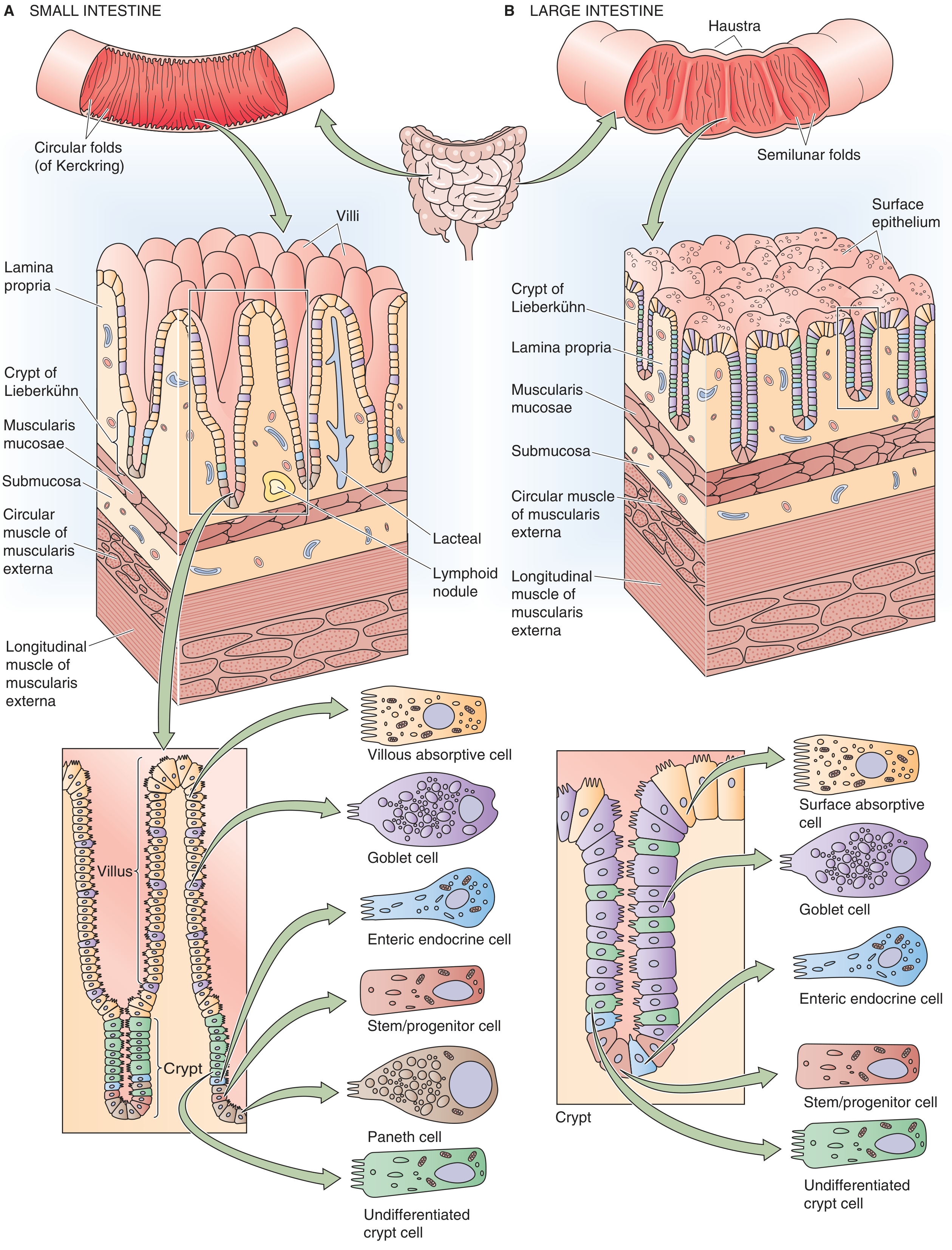
Figure 44-1 from Medical Physiology - structural comparison of the small (A) and large (B) intestine.
1. Gross Anatomy
| Feature | Small Intestine | Large Intestine |
|---|---|---|
| Length | ~6 m | ~2.4 m |
| Diameter | Narrow (~2.5-3 cm) | Wide (~6.5 cm) |
| Parts | Duodenum, Jejunum, Ileum | Cecum, Colon (ascending, transverse, descending, sigmoid), Rectum, Anal canal |
| Macroscopic folds | Circular folds (Folds of Kerckring) | Semilunar folds (haustra) |
| Surface markings | Smooth outer wall | Haustra (pouches), taeniae coli, appendices epiploicae |
2. Microscopic (Histological) Differences
| Feature | Small Intestine | Large Intestine |
|---|---|---|
| Villi | Present (finger-like projections) | Absent |
| Crypts of Lieberkühn | Present | Present |
| Plicae circulares | Present | Absent |
| Microvilli (brush border) | Present | Present (less prominent) |
| Paneth cells | Present (in crypts) | Normally absent in humans |
| Goblet cells | Present (less abundant) | Present (more abundant, ratio approaches 1:1 near rectum) |
| Mucosal surface area | ~200 m² | ~25 m² |
| Digestive enzymes in glycocalyx | Present (brush-border enzymes) | Not present |
The large intestine has the same basic wall layers (mucosa, submucosa, muscularis, serosa) as the small intestine, but lacks villi and plicae circulares. As noted in Histology: A Text and Atlas, goblet cell numbers progressively increase toward the rectum, reflecting the mucus-secreting role of the large bowel.
3. Amplification of Surface Area
The small intestine achieves a 600-fold increase in surface area over a smooth cylinder through three levels:
- Macroscopic folds of Kerckring
- Microscopic villi and crypts
- Submicroscopic microvilli
The large intestine amplifies surface area through colonic folds, crypts, and microvilli only (no villi), resulting in a more limited total area (~25 m²). - Medical Physiology, p. 1339
4. Functions
| Function | Small Intestine | Large Intestine |
|---|---|---|
| Primary role | Digestion and nutrient absorption | Water/electrolyte reabsorption; feces formation |
| Nutrient absorption | Yes (carbohydrates, proteins, fats, vitamins) | No |
| Water absorption | Absorbs ~6.5 L/day (of ~8.5 L presented) | Absorbs ~1.9 L/day |
| Na⁺ absorption | Active (via Na⁺/H⁺ exchanger and co-transporters) | Active (electrogenic Na⁺ absorption in rectosigmoid) |
| K⁺ handling | Net absorption | Net secretion |
| HCO₃⁻ handling | Net secretion | Net secretion |
| Cl⁻ handling | Net absorption | Net absorption |
| Bacterial fermentation | Minimal | Major - produces short-chain fatty acids from undigested carbohydrate/protein |
| Mucus secretion | Moderate | Abundant (lubrication of feces) |
The small intestine absorbs net amounts of water, Na⁺, Cl⁻, and K⁺ and secretes HCO₃⁻. The colon absorbs net amounts of water, Na⁺, and Cl⁻ and secretes both K⁺ and HCO₃⁻. - Medical Physiology, p. 1340
5. Cell Types
| Cell Type | Small Intestine | Large Intestine |
|---|---|---|
| Absorptive (enterocytes) | Villous absorptive cells | Surface absorptive cells |
| Goblet cells | Yes | Yes (more numerous) |
| Paneth cells | Yes (secrete defensins, lysozyme) | Absent |
| Enteroendocrine cells | Yes | Yes |
| Stem/progenitor cells | Yes (base of crypts) | Yes (base of crypts) |
| M cells | Yes (overlying Peyer's patches) | Absent |
6. Lymphoid Tissue
- Small intestine: Contains Peyer's patches (aggregated lymphoid nodules), especially prominent in the ileum - important for mucosal immunity and antigen sampling.
- Large intestine: Scattered solitary lymphoid nodules; no Peyer's patches.
Summary
The small intestine is the main site of digestion and nutrient absorption, with enormous surface area created by villi and folds. The large intestine is primarily a water-recovery and waste-storage organ, with a higher density of goblet cells for mucus production, no villi, and a distinct flora that ferments undigested residue. Both share crypts, microvilli, and active electrolyte transport, but differ markedly in cell composition, surface amplification, and physiological priorities.
Different between jejunum illaium
The jejunum and ileum are the two main parts of the small intestine after the duodenum. The jejunum (upper 2/5) is wider, thicker-walled, and more active in nutrient absorption, while the ileum (lower 3/5) is narrower, thinner-walled, and specialized for vitamin B12 and bile salt reabsorption.
Searching sources...
Finding Sources
Finding Sources
Reading File
Reading File
Reading File
I have rich content. Let me now fetch the arterial supply diagram and read a bit more on functional differences.
Loading Image
Finding Sources
Reading File
I now have comprehensive data from multiple authoritative textbooks. Here is the full comparison:
Differences Between the Jejunum and Ileum
The jejunum and ileum together form the majority of the small intestine, picking up after the duodenum. Though they are continuous with no sharp anatomical boundary, they differ considerably in structure and function.
1. Position and Length
| Feature | Jejunum | Ileum |
|---|---|---|
| Position | Upper 2/5 of small intestine | Lower 3/5 of small intestine |
| Location | Primarily left upper/central abdomen | Primarily right lower quadrant |
| Ending | Continuous with ileum | Opens into cecum via ileocecal valve |
The jejunum forms up to two-fifths and the ileum up to three-fifths of the total small intestinal length. - Color Atlas of Human Anatomy, Vol. 2, p. 328
2. Gross Anatomy
| Feature | Jejunum | Ileum |
|---|---|---|
| Diameter | Wider | Narrower |
| Wall thickness | Thicker | Thinner |
| Color | Deeper pink/red (more vascular) | Paler pink |
| Mesenteric fat | Less fat (translucent windows visible) | More mesenteric fat ("fatty") |
3. Mucosal Features
| Feature | Jejunum | Ileum |
|---|---|---|
| Plicae circulares (valvulae conniventes) | Tall, densely packed, prominent | Fewer, shorter, eventually absent in distal ileum |
| Villi shape | Tall, finger-like | More leaf-like and shorter |
| Goblet cells | Fewer | More numerous |
| Peyer's patches | Absent or sparse | Numerous, large aggregated lymphoid nodules on antimesenteric side |
As noted in Histology: A Text and Atlas, villi in the ileum are more frequently leaf-like, and lymphatic tissue in the lamina propria is organized into numerous nodules (Peyer's patches). In the jejunum, plicae circulares are tall and densely arranged; closer to the ileum they become shorter, spaced further apart, and in the second half of the ileum they are usually absent. - Color Atlas of Human Anatomy, p. 329
4. Arterial Supply

| Feature | Jejunum | Ileum |
|---|---|---|
| Arterial arcades | Fewer (1-2 tiers) | More (4-5 tiers) |
| Vasa recta (straight arteries) | Longer | Shorter |
| Blood supply | Jejunal branches of SMA | Ileal branches of SMA |
The fewer arcades and longer vasa recta of the jejunum are a key distinguishing feature visible at surgery or on imaging. - Gray's Anatomy for Students, p. 373
5. Function
| Function | Jejunum | Ileum |
|---|---|---|
| Primary role | Major site of digestion and absorption | Reabsorption, water recovery, specialized absorption |
| Carbohydrates & proteins | Final brush-border hydrolysis + absorption | Can compensate if jejunum is resected |
| Fats | Fatty acids and monoglycerides absorbed after bile salt separation | - |
| Iron | Absorbed (duodenum/proximal jejunum) | - |
| Folate | Absorbed | - |
| Vitamin B12 | Not absorbed | Only site - via specific intrinsic factor receptors (cubilin) |
| Bile salts | Separated from fat digestion products | Only site of active reabsorption (enterohepatic circulation) |
| Water & electrolytes | Absorbed | Absorbed (tight junctions support concentration gradient) |
| Fat-soluble vitamins (A, D, E, K) | Absorbed | Critical - reduced by ileal resection |
The jejunum is the principal site for digestion and absorption of fluid, electrolytes, iron, folate, fat, protein and carbohydrate, but the absorption of bile salts and vitamin B12 only occurs in the terminal ileum, where there are specific transporters. - Bailey and Love's Short Practice of Surgery, p. 1328
6. Clinical Consequences of Resection
| Resection | Consequence |
|---|---|
| Jejunal resection | Ileum can compensate and assume all absorptive functions |
| Ileal resection | Diminished bile salt pool, vitamin B12 deficiency, deficiency of fat-soluble vitamins A/D/E/K, troublesome diarrhoea |
This asymmetry is clinically important: losing the ileum is far more consequential than losing the jejunum, because the ileum's specialized functions (B12 and bile salt absorption) cannot be compensated by any other segment.
Summary Table
| Feature | Jejunum | Ileum |
|---|---|---|
| Length proportion | 2/5 | 3/5 |
| Wall | Thick | Thin |
| Plicae circulares | Prominent | Reduced/absent distally |
| Villi shape | Finger-like | Leaf-like |
| Peyer's patches | Absent | Present (antimesenteric side) |
| Goblet cells | Fewer | More |
| Arterial arcades | 1-2, long vasa recta | 4-5, short vasa recta |
| Mesenteric fat | Less | More |
| Main absorption | Nutrients, fat, iron, folate | B12, bile salts, water |
| Clinical importance | Compensated by ileum | Irreplaceable specialized functions |
This is a shared conversation. Sign in to Orris to start your own chat.