
Total Abdominal Hysterectomy with Bilateral Salpingo-Oophorectomy.
Reading File
The query is about a specific surgical procedure. Let me clarify what aspect the user needs before diving in.
Asking for Preferences
What would you like to know about Total Abdominal Hysterectomy with Bilateral Salpingo-Oophorectomy (TAH-BSO)?
Finding Sources
Finding Sources
Finding Sources
Good. Now let me read the key sections from Sabiston, Schwartz's, Berek & Novak's, and Gray's Anatomy in parallel.
Reading File
Reading File
Reading File
Reading File
Good content retrieved. Let me now read the detailed surgical steps from Schwartz's and Sabiston, plus complications and post-op care, and fetch key images in parallel.
Reading File
Reading File
Finding Sources
Finding Sources
Loading Image
Excellent content. Now let me fetch the remaining key images and read the complications/indications sections from Berek & Novak's, plus run a PubMed search in parallel.
Reading File
Reading File
Searching PubMed
"total abdominal hysterectomy" outcomes complications
Loading Image
Loading Image
Reading File
Searching PubMed
hysterectomy surgical approach outcomes
I now have comprehensive information to write a complete, well-organized overview of TAH-BSO. Let me update the todos and compile the response.# Total Abdominal Hysterectomy with Bilateral Salpingo-Oophorectomy (TAH-BSO)
Definition
TAH-BSO is the surgical removal of the entire uterus (corpus + cervix), both fallopian tubes, and both ovaries through an abdominal incision. It is distinguished from:
- Simple (total) hysterectomy — uterus removed, adnexa preserved
- Supracervical hysterectomy — corpus removed, cervix left in situ
- Radical hysterectomy — uterus + parametria + upper vaginal cuff removed (for malignancy)
"A hysterectomy is performed through a transverse suprapubic incision. During the procedure tremendous care is taken to identify the distal ureters and to ligate the nearby uterine arteries without damage to the ureters." — Gray's Anatomy for Students
Indications
Gynecologic Malignancy (Primary indication for BSO)
- Endometrial cancer — staging laparotomy requires TAH-BSO + omentectomy + lymph node dissection
- Ovarian cancer — BSO is part of cytoreductive surgery
- Cervical cancer — radical hysterectomy ± BSO
- Uterine leiomyosarcoma — TAH with bilateral oophorectomy indicated
Benign Conditions (Hysterectomy ± selective BSO)
| Condition | Notes |
|---|---|
| Symptomatic uterine fibroids | Menorrhagia, bulk symptoms, failed conservative management |
| Abnormal uterine bleeding | Refractory to medical therapy |
| Endometriosis | BSO for refractory pelvic pain; consider ovarian conservation if ovaries appear normal |
| Pelvic organ prolapse | Usually vaginal or laparoscopic approach preferred |
| Pelvic inflammatory disease (PID) | Bilateral salpingo-oophorectomy ± hysterectomy for refractory/severe cases |
| Pelvic mass / benign ovarian tumor | If perimenopausal/postmenopausal or fertility not desired |
| Excessive postpartum hemorrhage | Uterus removed when other measures fail |
Prophylactic BSO Indications
- BRCA1/BRCA2 mutation carriers — lifetime ovarian cancer risk 10–50%; risk-reducing BSO recommended (after age 35–40)
- Lynch II syndrome — prophylactic hysterectomy with BSO is effective primary prevention
Important: In premenopausal women at average risk for ovarian cancer, ovarian conservation is recommended — prophylactic BSO before age 55 is associated with an 8.58% excess mortality by age 80 (predominantly cardiovascular) and offers no overall survival benefit. — Berek & Novak's Gynecology
Preoperative Considerations
- Informed consent documenting options, risks, benefits, alternatives, and personnel
- Document completion of childbearing; sterilization consent forms required in most jurisdictions
- Health assessment: Correct underlying anemia preoperatively (iron supplementation or GnRH agonists reduce blood product use)
- VTE risk stratification: Age, BMI, thrombophilia, smoking, hormone use
- Imaging: Pelvic ultrasound; CT/MRI for malignancy staging
- Bowel prep: No longer routinely required
- Prophylactic antibiotics: Standard of care (cephalosporin, 1 hour preoperatively)
Route Selection
TAH (laparotomy) is now reserved for specific circumstances (Berek & Novak's):
- Cardiopulmonary disease precluding pneumoperitoneum or general anesthesia
- Known/suspected uterine malignancy where morcellation is required
- Severely distorted anatomy from disease or adhesions making minimally invasive surgery unsafe
- No surgeon access to laparoscopic/vaginal expertise
Minimally invasive hysterectomy (laparoscopic or vaginal) is preferred for benign disease when feasible, with lower complication rates, less pain, faster recovery, and similar or lower hospital readmission rates.
Surgical Anatomy Relevant to TAH-BSO
Key structures at risk:
- Ureters — cross beneath the uterine artery ("water under the bridge") at the lateral cervix; must be identified before ligation of uterine vessels
- Uterine arteries — arise from internal iliac arteries; ligated at the cervicouterine junction
- Infundibulopelvic (IP) ligament — contains ovarian vessels (from aorta/renal vein); ligated to remove the adnexa
- Cardinal ligament (Mackenrodt's ligament) — provides primary uterine support; serially clamped and ligated
- Uterosacral ligaments — support posterior uterus; incorporate into vaginal cuff closure for apical support
- Bladder — mobilized anteriorly off the lower uterine segment and cervix
Surgical Technique: Step-by-Step
Access and Exposure
- Abdominal incision: Low transverse (Pfannenstiel) for benign disease and smaller uteri; vertical midline for malignancy, large pathology, poor visualization, or anticipated extensive surgery
- Exploration: Upper abdomen examined for extrapelvic disease; suitable retractor (e.g., Bookwalter) placed
- Uterus grasped at both cornua with Kocher clamps and elevated into the incision
Key Steps
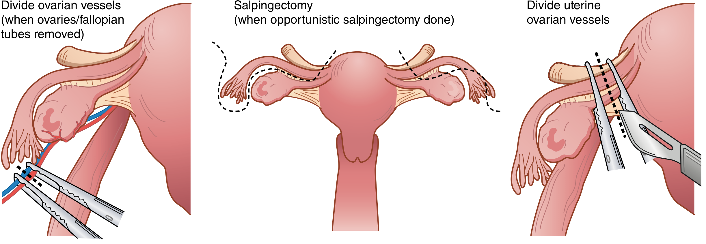
Step 1 — Round ligament transection
The round ligament is identified, clamped laterally, and divided with electrocautery or ligated with delayed absorbable suture and divided medial to the suture.
Step 2 — Broad ligament incision
The anterior leaf of the broad ligament is incised medially toward the level of the internal cervical os, creating the bladder flap. The posterior leaves are also incised.
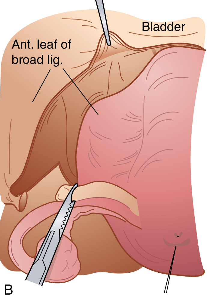
Step 3 — Ureter identification
The retroperitoneal space is bluntly opened and the ureter identified on the medial leaf of the broad ligament before any vascular ligation. This is the single most important step to prevent ureteral injury.
Step 4 — BSO: Infundibulopelvic ligament ligation
The IP ligament containing the ovarian vessels is doubly clamped with curved Heaney or Zeppelin clamps, incised with curved Mayo scissors, and doubly ligated. The specimen sides are also ligated to prevent back-bleeding. The posterior broad ligament leaves are incised inferiorly to skeletonize the uterine vessels.
Step 5 — Bladder mobilization
The bladder is sharply dissected off the anterior surface of the uterus and cervix (Metzenbaum scissors or electrocautery), retracted inferiorly below the level of the cervix.
Step 6 — Uterine vessel ligation
A curved Heaney or Zeppelin clamp is placed across the uterine vessels at the level of the internal cervical os, pedicle cut and suture-ligated.
Step 7 — Cardinal and uterosacral ligament division
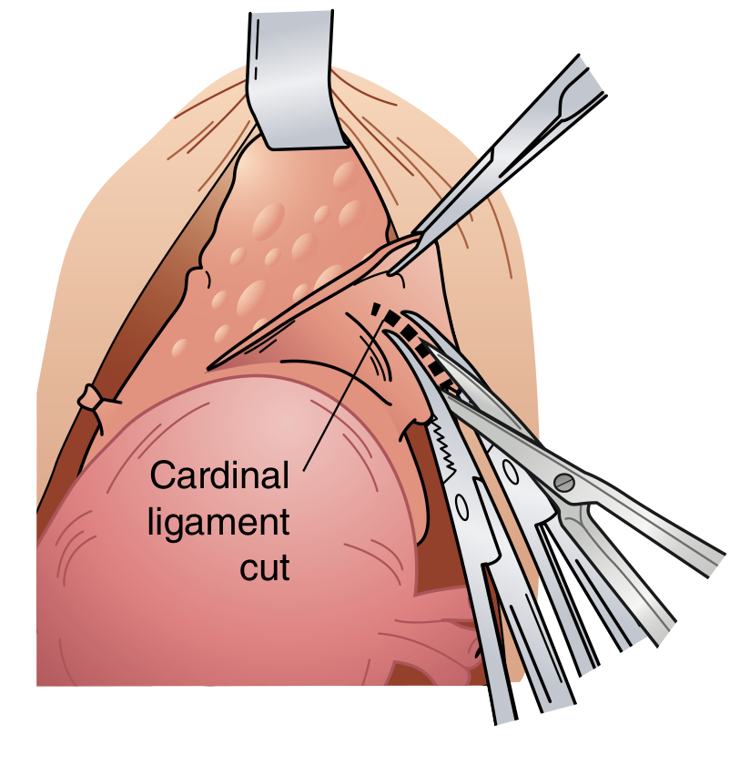
Cardinal ligaments are serially clamped, cut, and ligated bilaterally. Uterosacral ligaments are similarly divided.
Step 8 — Colpotomy and specimen removal
Once both sides are cleared at the external cervical os, curved clamps are placed across the vagina just below the cervix bilaterally. Curved scissors cut above the clamps to amputate the specimen. The cervix is thus separated from the upper vagina.
Step 9 — Vaginal cuff closure
- Heaney transfixion stitches at lateral vaginal corners (incorporating the ipsilateral uterosacral ligament for apical support)
- The colpotomy is closed with running, interrupted, or figure-of-eight absorbable sutures
- Pelvic reperitonization is not routinely necessary
Step 10 — Hemostasis and closure
Pelvic hemostasis confirmed, irrigation performed, and the abdomen closed in layers.
Complications
Intraoperative
| Complication | Details |
|---|---|
| Ureteral injury | Most common at two sites: (1) at the IP ligament, (2) where the ureter passes under the uterine artery. Risk ↑ with distorted anatomy, endometriosis, prior surgery. Most injuries occur during laparoscopic hysterectomy at the point of cervical detachment |
| Bladder injury | Most common urinary tract injury overall; usually at bladder base during dissection off the cervix. Recognized by visualization of Foley catheter or urine in field. Repaired in 2 layers |
| Hemorrhage | From IP ligament pedicle, uterine vessels, or vaginal cuff. Avoid blind clamping — identify the ureter first. Use Heaney sutures for bulky pedicles |
| Bowel injury | Rare in open approach; more common in patients with adhesions/endometriosis |
| Nerve injury | Genitofemoral, femoral, obturator, or lateral femoral cutaneous nerve from retractor pressure |
Postoperative
| Complication | Notes |
|---|---|
| Vault hematoma / infection | Fever POD 3–5; vaginal cuff tenderness; treated with antibiotics ± drainage |
| Vesicovaginal fistula (VVF) | Bladder injury most common cause; may present 7–14 days post-op as continuous urine leak per vagina |
| Ureterovaginal fistula | Unrecognized ureteral injury; presents as watery vaginal discharge |
| VTE (DVT/PE) | Prophylaxis with LMWH + compression stockings; early mobilization |
| Ileus | Common after laparotomy; managed conservatively |
| Wound complications | Seroma, dehiscence, hematoma; ↑ in obese patients |
| Vault dehiscence | Rare but serious; presents as evisceration |
Long-term Sequelae of BSO in Premenopausal Women
- Surgical menopause — abrupt estrogen withdrawal; hot flashes, sleep disturbance, vaginal atrophy
- Increased cardiovascular risk — especially if BSO before age 50 without HRT
- Osteoporosis — accelerated bone loss
- Sexual dysfunction — reduced libido, dyspareunia
- Cognitive effects — possible increased dementia risk (controversial)
- Hormone replacement therapy (HRT) should be strongly considered in women under 50 undergoing BSO for benign conditions
Perioperative Care
Preoperative
- NPO after midnight; IV access
- Antibiotic prophylaxis (cefazolin 2g IV, or 3g if >120 kg) within 60 min of incision
- DVT prophylaxis initiated preoperatively
- Foley catheter placed in OR
Intraoperative
- Regional (epidural T8–T11) or general anesthesia
- Patient supine; modified Trendelenburg to displace bowel
- Normothermia maintained
Postoperative
- Foley catheter: Remove within 24 hours when not anticipated to have spontaneous voiding difficulty
- Diet: Advance as tolerated; no evidence for dietary restriction
- Analgesia: NSAIDs (e.g., diclofenac reduces postoperative morphine consumption), acetaminophen, opioids as needed
- Early mobilization: Key for VTE prevention and GI recovery
- Hospital stay: 2–4 days typical for open TAH
- Return to activity: 4–6 weeks for full return; pelvic rest (no intercourse) for 6–8 weeks until vaginal cuff healed
Route Comparison (TAH vs. Minimally Invasive)
| Parameter | TAH (Open) | Laparoscopic / Vaginal |
|---|---|---|
| Hospital stay | 2–4 days | Same-day to 1 day |
| Blood loss | Higher | Lower |
| Complication rate | Higher | Lower or similar |
| Pain | More | Less |
| Recovery time | 4–6 weeks | 1–2 weeks |
| Cost | Lower procedural | Higher procedural (but lower institutional) |
| Appropriate when | Malignancy, large uterus, dense adhesions, cardiopulmonary disease | Most benign indications |
Key Points Summary
- TAH-BSO removes uterus, cervix, both fallopian tubes, and both ovaries via laparotomy
- Primary indications include gynecologic malignancy, and selected benign conditions refractory to conservative therapy
- Ureter identification at the IP ligament and at the level of the uterine artery is the most critical step to prevent the most feared intraoperative complication
- BSO in premenopausal women at average ovarian cancer risk carries excess long-term mortality; HRT should be offered post-operatively
- Open TAH is now reserved for cases where minimally invasive approaches are unsafe or unavailable
- Common postoperative complications include vault infection, VTE, VVF, and ileus
Sources: Sabiston Textbook of Surgery (21st ed.) | Schwartz's Principles of Surgery (11th ed.) | Berek & Novak's Gynecology | Gray's Anatomy for Students | Goldman-Cecil MedicineThat covers TAH-BSO comprehensively. Here's a quick orientation to each section:
- Indications — separated into malignant (where BSO is mandatory) vs. benign (where ovarian conservation decisions are nuanced), plus prophylactic BSO for high-risk genetics
- Surgical technique — step-by-step with intraoperative images from Sabiston and Schwartz's showing the key pedicle divisions
- Complications — organized intraoperatively vs. postoperatively, including the long-term hormonal sequelae unique to BSO in premenopausal women
- Perioperative care — covers the full continuum from NPO to discharge to activity restrictions
- Route comparison — contextualizes when open TAH remains appropriate vs. minimally invasive alternatives, supported by the 2023 Cochrane review
Let me know if you'd like to go deeper on any specific aspect — e.g., management of ureteral injury, hormonal considerations post-BSO, oncologic staging procedures, or the laparoscopic technique.
Bartholin cyst
Finding Sources
Finding Sources
Reading File
Reading File
Reading File
Reading File
Reading File
Reading File
Reading File
Reading File
Loading Image
Now fetch the marsupialization images and the Word catheter image, and read the Bartholin gland carcinoma section for completeness.
Loading Image
Loading Image
Reading File
Searching PubMed
"Bartholin" cyst treatment
Excellent — I have everything needed. Here is the comprehensive overview:
Bartholin Cyst (Bartholin Duct Cyst & Abscess)
Anatomy
The Bartholin glands (greater vestibular glands) are paired, pea-sized, mucus-secreting glands located bilaterally in the posterior labia minora. Their ducts drain into the vaginal vestibule at the 4 o'clock and 8 o'clock positions (some sources describe this as 5 and 7 o'clock). They begin functioning at puberty — providing vestibular moisture/lubrication — and involute after menopause. In the normal state they are not palpable.
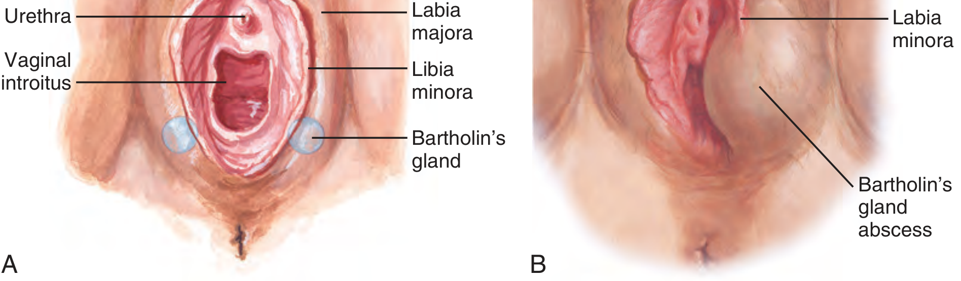
Pathophysiology
Obstruction of the Bartholin duct — most often by an inflammatory process — causes mucus accumulation, producing a cyst. If the stagnant secretions become infected, the gland fills with pus, forming a Bartholin abscess. Importantly, an abscess can develop without a preceding cyst.
"Obstruction of the duct may result in a cyst or abscess. A cyst does not need to be present before an abscess can develop." — Tintinalli's Emergency Medicine
Histology: Cysts are lined by transitional or squamous epithelium. They can grow up to 3–5 cm in diameter.
Epidemiology
- Occur at all ages, predominantly in reproductive-age women
- Bartholin abscesses affect approximately 2% of women
- Uncommon after menopause (gland involutes); any Bartholin gland mass in a woman >40 years must raise suspicion for malignancy
Microbiology (Abscess)
Bartholin abscesses are typically polymicrobial:
| Organism | Notes |
|---|---|
| Escherichia coli | Most common isolate |
| Staphylococcus aureus (incl. MRSA) | Culture purulent fluid |
| Streptococcus spp. | Polymicrobial mix |
| Neisseria gonorrhoeae | Less common; treat if suspected |
| Chlamydia trachomatis | Less common; treat if suspected |
Clinical Features
Bartholin Duct Cyst
- Unilateral, smooth, fluctuant mass at the posterior vestibule (4 or 8 o'clock)
- Often asymptomatic if small
- Larger cysts cause local pressure, discomfort, dyspareunia
- No erythema or warmth (unless infected)
Bartholin Abscess
- Rapidly enlarging, extremely painful mass at same location
- Induration, erythema, fluctuance
- Patient may have difficulty walking or sitting
- Systemic symptoms (fever, chills) are rarely present — their absence does not exclude an abscess
- Develops over several days (or longer if preceded by a cyst)
Diagnosis
Primarily clinical — based on history and examination:
- Characteristic location at posterior introitus (labia minora transecting the mass)
- Fluctuance confirms abscess
Ultrasound can confirm a discrete fluid collection when the diagnosis is uncertain or if the abscess is not clearly defined.
Caution: Distinguish a Bartholin abscess from an abscess of the labia majora, which requires gynecologic consultation. Any solid component or atypical appearance in a woman >40 warrants biopsy to exclude carcinoma.
Treatment
Management depends on whether it is a cyst or abscess, and on recurrence:
1. Asymptomatic Cyst
- No treatment required — observation is appropriate for small, asymptomatic cysts
2. Symptomatic Cyst or Abscess — First-line: Word Catheter
The Word catheter (a small catheter with an inflatable balloon tip) is the most widely used office/ED procedure.
Technique:
- Patient in dorsal lithotomy position; identify abscess at 5 or 7 o'clock with labia minora transecting it
- Analgesia: 2–4 mL 1% lidocaine infiltrated through the mucosal surface (not skin surface); IV opioids/procedural sedation for severe pain
- #11 scalpel — stab incision on the mucosal surface of the vestibule, just lateral to the hymenal ring, where fluctuance is maximal. Incision must be large enough for catheter, but not so large the balloon is displaced
- Alternatively: stabilize abscess with thumb/index finger and "skewer" onto a hemostat for entry
- Drain pus (expect a palpable pop and free flow of pus); insert Word catheter to the hilt
- Inflate balloon with 2–4 mL saline; tuck catheter end into vagina
- Catheter remains in place 4–6 weeks to allow epithelialization of a new permanent duct opening
- Send pus for culture (including MRSA, GC, chlamydia)
- Prescribe analgesics; antibiotics after drainage are of no proven value routinely (cover GC/CT if suspected)
3. Recurrent Cyst/Abscess — Marsupialization
The preferred definitive procedure for recurrent disease. Performed under local, spinal, or general anesthesia.
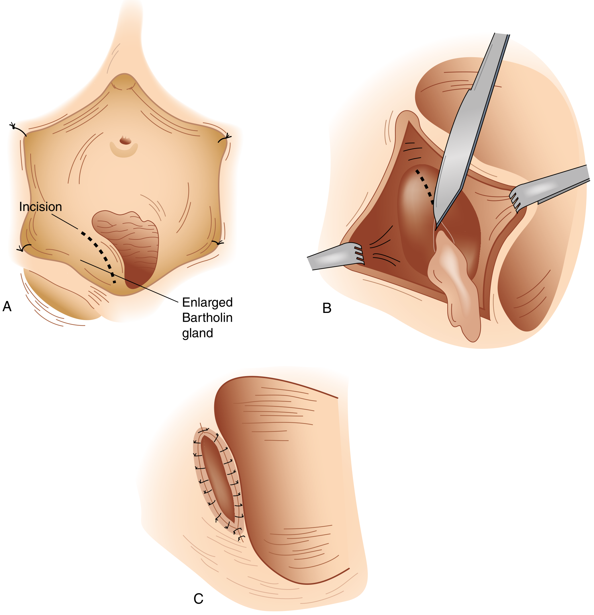
Technique:
- Patient in dorsal lithotomy; local anesthetic (2% lidocaine ± epinephrine) or pudendal block
- Retract labium laterally; identify incision site internal to the hymenal ring (mucosa, not skin)
- Longitudinal fusiform incision 1–2 cm wide over the cyst wall; excise an ellipse of mucosa
- Grasp cyst wall with two hemostats before segment is removed; cyst will decompress
- Break up any loculations with small hemostats; irrigate with normal saline
- In patients >40 years: inspect interior for neoplastic-appearing epithelium; send for biopsy
- Suture cyst wall edge to adjacent vaginal mucosa with interrupted 4-0 Vicryl — this is the marsupialization step, suturing the cavity open permanently
- Minimum gap of ≥1 cm across the open marsupialization
- No dressing required; sitz baths daily for 3–4 days
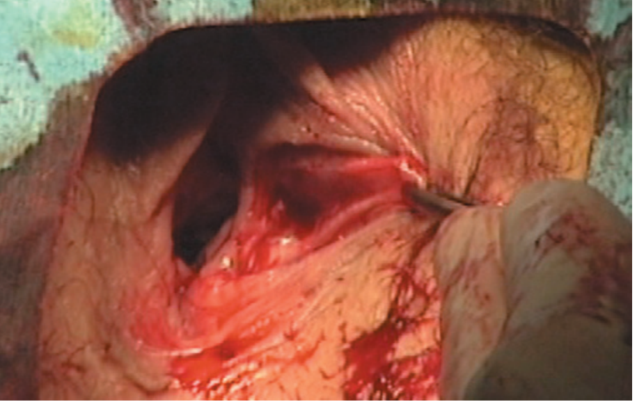
4. Failed/Persistent Cases — Complete Gland Excision
Reserved for cysts or abscesses that fail to resolve after drainage, recurrent disease not amenable to marsupialization, or suspicion of malignancy.
- Technically more demanding; risks include hemorrhage and hematoma
- A common surgical error is making the incision on the lateral vulvar skin rather than the vestibular mucosa
CPT / Procedure Codes
| Procedure | CPT |
|---|---|
| I&D of vulvar/perineal abscess | 56405 |
| I&D of Bartholin's gland cyst/abscess | 56420 |
| Marsupialization of Bartholin's gland cyst | 56440 |
| Excision of Bartholin's gland | 56740 |
Bartholin Gland Carcinoma
Although rare, carcinoma can arise from the Bartholin gland. Types include:
- Squamous cell carcinoma (most common)
- Adenocarcinoma
- Adenoid cystic carcinoma (characteristically slow-growing, perineural invasion)
- Transitional cell carcinoma
Key rule: A biopsy with histologic evaluation is indicated for all cases with a palpable or visible solid mass within a Bartholin cyst or abscess, and in all instances where a presumed Bartholin gland lesion occurs in women over 40 years of age. — Berek & Novak's Gynecology
Special Considerations
| Situation | Action |
|---|---|
| Large abscess, uncertain diagnosis, significant cellulitis | Gynecology consultation |
| Unstable vitals, bleeding dyscrasia, immunocompromised | Gynecology to manage (possibly OR) |
| Abscess not yet walled-off | Broad-spectrum antibiotics + warm sitz baths + close follow-up; I&D when fluctuant |
| N. gonorrhoeae / C. trachomatis suspected | Treat per STI guidelines (ceftriaxone + doxycycline) |
| MRSA on culture | Treat with appropriate antibiotics (TMP-SMX, clindamycin) |
| Woman >40 years | Biopsy to exclude malignancy |
| Postmenopausal new Bartholin mass | Malignancy until proven otherwise |
Summary Algorithm
Bartholin lesion
│
├── Asymptomatic cyst → Observe
│
├── Symptomatic cyst or abscess (1st episode)
│ → Word catheter (4–6 weeks)
│
├── Recurrent cyst/abscess
│ → Marsupialization
│
├── Failed marsupialization / persistent
│ → Complete gland excision
│
└── Solid mass / age >40 / postmenopausal
→ Biopsy to exclude carcinoma
Sources: Tintinalli's Emergency Medicine | Robbins & Cotran Pathologic Basis of Disease | Berek & Novak's Gynecology | Sabiston Textbook of Surgery | Schwartz's Principles of Surgery | Roberts & Hedges' Clinical Procedures in Emergency Medicine | Pfenninger & Fowler's Procedures for Primary Care
This is a shared conversation. Sign in to Orris to start your own chat.