
Adrenaline short notes simplify
Reading File
Now let me search the medical library for adrenaline content in parallel:
Finding Sources
Finding Sources
Good. Let me now read from the best pharmacology sources - Goodman & Gilman and Medical Physiology - in parallel:
Finding Sources
Reading File
Let me now read detailed content on adrenaline's pharmacology, synthesis, and clinical uses from two good sources simultaneously:
Reading File
Finding Sources
Let me read the key pharmacology content from Goldman-Cecil and Katzung simultaneously:
Reading File
Reading File
Now let me get the clinical uses and effects of adrenaline from the textbooks:
Finding Sources
Finding Sources
Let me get the full Lippincott section on epinephrine which should have the most organized pharmacology content:
Reading File
Reading File
I now have rich content from multiple authoritative textbooks. Let me also read the adverse effects / contraindications section from Lippincott:
Reading File
I also need to read the Lippincott adverse effects section. Let me also fetch the catecholamine synthesis diagram:
Reading File
Loading Image
I now have comprehensive information from multiple authoritative textbooks. Here is the complete simplified short note on Adrenaline:
Adrenaline (Epinephrine) - Short Notes
1. Basic Identity
| Feature | Detail |
|---|---|
| Other name | Epinephrine |
| Class | Catecholamine hormone + drug |
| Source | Adrenal medulla (chromaffin cells) - ~80% of release |
| Precursor | Norepinephrine (methylated to yield epinephrine) |
2. Synthesis (Catecholamine Pathway)
Tyrosine → (Tyrosine hydroxylase - rate-limiting step) → DOPA → (DOPA decarboxylase) → Dopamine → (Dopamine β-hydroxylase) → Norepinephrine → (PNMT*) → Epinephrine
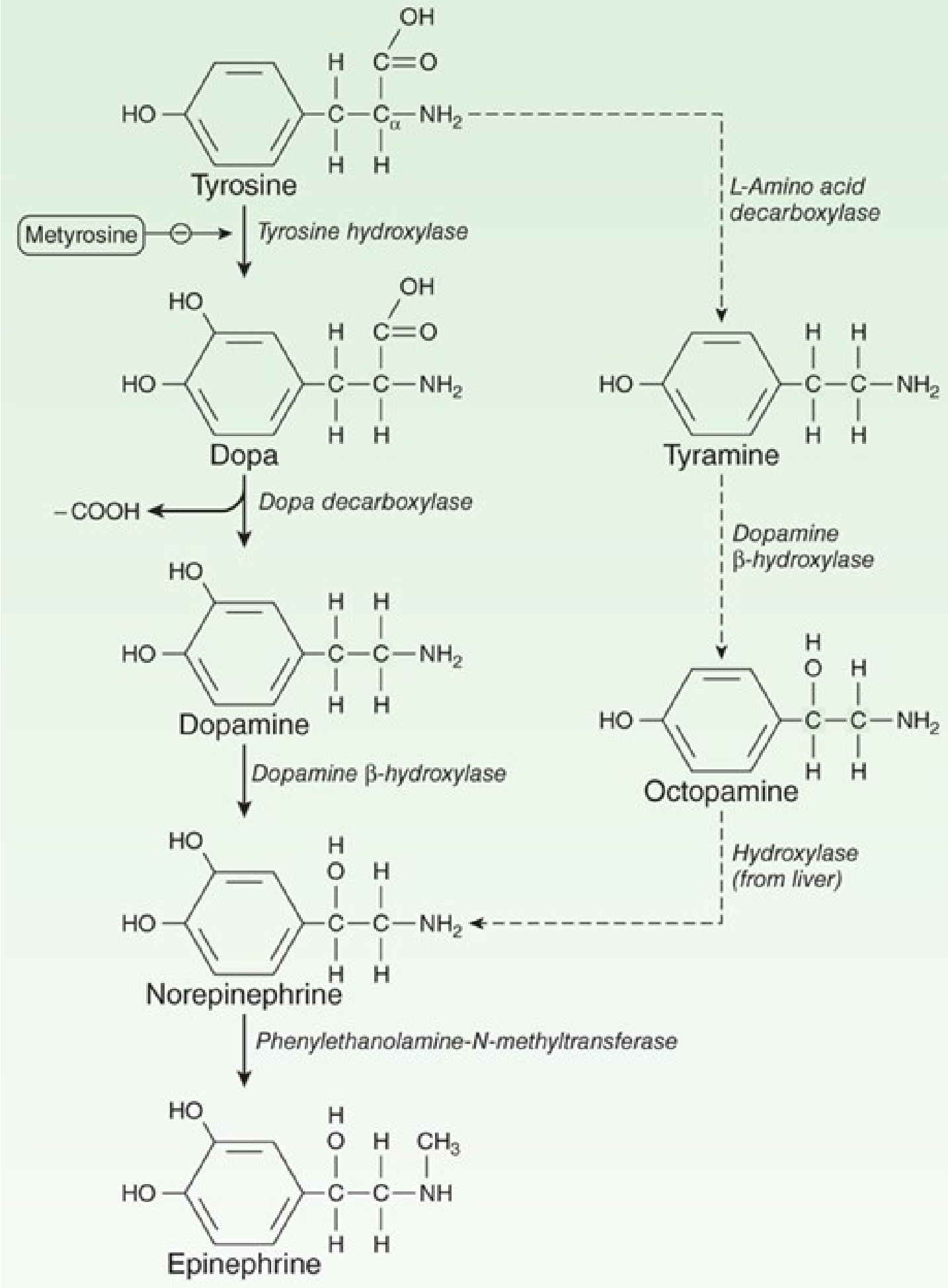
PNMT = Phenylethanolamine-N-methyltransferase (found only in adrenal medulla; its expression is upregulated by cortisol from the adjacent cortex)
Key fact: Metyrosine inhibits tyrosine hydroxylase (rate-limiting step) - used in pheochromocytoma.
3. Receptors Activated
| Receptor | Location | Effect |
|---|---|---|
| α₁ | Skin, mucosa, viscera vessels | Vasoconstriction |
| α₂ | Pancreatic β-cells | ↓ Insulin secretion |
| β₁ | Heart, kidney | ↑ HR, ↑ contractility, renin release |
| β₂ | Bronchi, skeletal muscle vessels, liver | Bronchodilation, vasodilation, glycogenolysis |
Dose-dependent effect:
- Low dose → β effects dominate (vasodilation)
- High dose → α effects dominate (vasoconstriction)
4. Pharmacological Actions
Cardiovascular
- ↑ Heart rate (positive chronotrope - β₁)
- ↑ Myocardial contractility (positive inotrope - β₁)
- ↑ Cardiac output + ↑ O₂ demand
- ↑ Systolic BP (β₁)
- Slight ↓ diastolic BP (β₂ vasodilation in skeletal muscle)
- Vasoconstriction in skin, mucosa, viscera (α₁)
- Vasodilation in skeletal muscle and liver (β₂)
- ↑ Conduction velocity in AV node, His-Purkinje system
Respiratory
- Powerful bronchodilation (β₂)
- Inhibits histamine/leukotrienes from mast cells
Metabolic
- Hyperglycemia: glycogenolysis in liver (β₂) + ↑ glucagon (β₂) + ↓ insulin (α₂)
- Lipolysis (β₁/β₃)
5. Clinical Uses
| Indication | Route | Notes |
|---|---|---|
| Anaphylaxis (drug of choice) | IM (anterolateral thigh) | Counteracts hypotension, bronchospasm, angioedema |
| Cardiac arrest | IV | Restores cardiac rhythm in ACLS |
| Bronchospasm / acute asthma | SC / inhalation | Emergency relief |
| Adjunct in local anesthesia | Local injection (1:100,000) | Prolongs duration, reduces systemic absorption, hemostasis |
| Intraocular surgery | Topical/local | Induces and maintains mydriasis |
| Septic shock | IV infusion | Increases cardiac output + oxygen delivery |
6. Pharmacokinetics
- Onset: Rapid
- Duration: Brief (rapidly degraded)
- Routes: IM (anaphylaxis) > IV (emergencies) > SC > inhalation > endotracheal tube
- Metabolism: MAO (monoamine oxidase) + COMT (catechol-O-methyltransferase)
- Metabolites: Metanephrine → Vanillylmandelic acid (VMA) - excreted in urine
- Half-life: 10-100 seconds in circulation
- NOT oral (destroyed by gut enzymes)
7. Adverse Effects
- CNS: Anxiety, fear, tremor, headache
- Cardiac: Arrhythmias (VT, VF), angina, tachycardia (especially with halothane anesthesia)
- Pulmonary edema (due to increased afterload from vasoconstriction)
- Hyperglycemia (caution in diabetics - may need more insulin)
8. Precautions / Drug Interactions
| Situation | Problem |
|---|---|
| Non-selective β-blockers (e.g. propranolol) | Block β₂ vasodilation → unopposed α → severe hypertension |
| Hyperthyroidism | Enhanced response - reduce dose |
| Inhalation anesthetics (e.g. halothane) | Sensitize heart → arrhythmias |
| Coronary artery disease | Increases O₂ demand → risk of ischemia |
9. Breakdown / Termination of Action
- Uptake-1 (neuronal reuptake) - accounts for ~90% of removal; blocked by cocaine, tricyclic antidepressants
- Uptake-2 (extraneuronal) - metabolized by COMT
- Enzymatic degradation by MAO and COMT
Sources: Lippincott Illustrated Reviews: Pharmacology, Goldman-Cecil Medicine, Barash Clinical Anesthesia, Medical Physiology (Boron & Boulpaep), Katzung's Basic and Clinical Pharmacology
Insulin in diabetes malaitus short notes renal physiology
Finding Sources
Finding Sources
Finding Sources
Reading File
Reading File
Finding Sources
Reading File
Reading File
Reading File
Reading File
Reading File
Finding Sources
Reading File
Reading File
Now let me get the renal physiology section - glucose handling, glucosuria, and renal threshold:
Finding Sources
Reading File
Now let me fetch a key diagram - the beta cell insulin secretion mechanism and the islet anatomy:
Loading Image
Loading Image
I now have rich, comprehensive content from Guyton & Hall plus Goldman-Cecil. Here are the complete simplified short notes:
Insulin in Diabetes Mellitus - Short Notes (with Renal Physiology)
Source: Guyton & Hall Textbook of Medical Physiology, Goldman-Cecil Medicine
1. Source and Structure
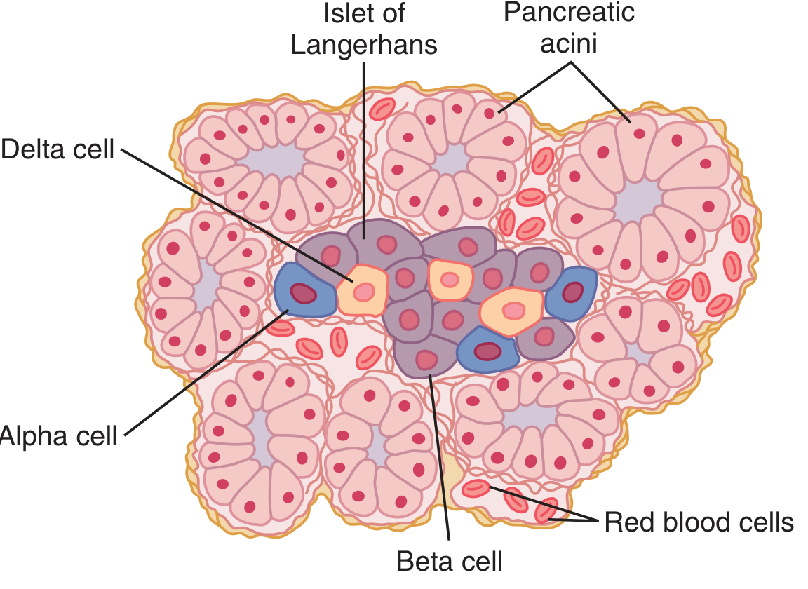
| Cell | % of Islet | Secretes |
|---|---|---|
| Beta (β) | 60% | Insulin + Amylin (IAPP) |
| Alpha (α) | 25% | Glucagon |
| Delta (δ) | 10% | Somatostatin |
- Insulin = MW 5808; two chains (A + B) connected by disulfide bonds
- Synthesized as: Preproinsulin → Proinsulin → Insulin + C peptide (cleaved in Golgi)
- C peptide is secreted in equimolar amounts with insulin - useful as a marker of endogenous insulin secretion
- Half-life in plasma: ~6 minutes; metabolized by liver and kidney
2. Mechanism of Insulin Secretion (Beta Cell)
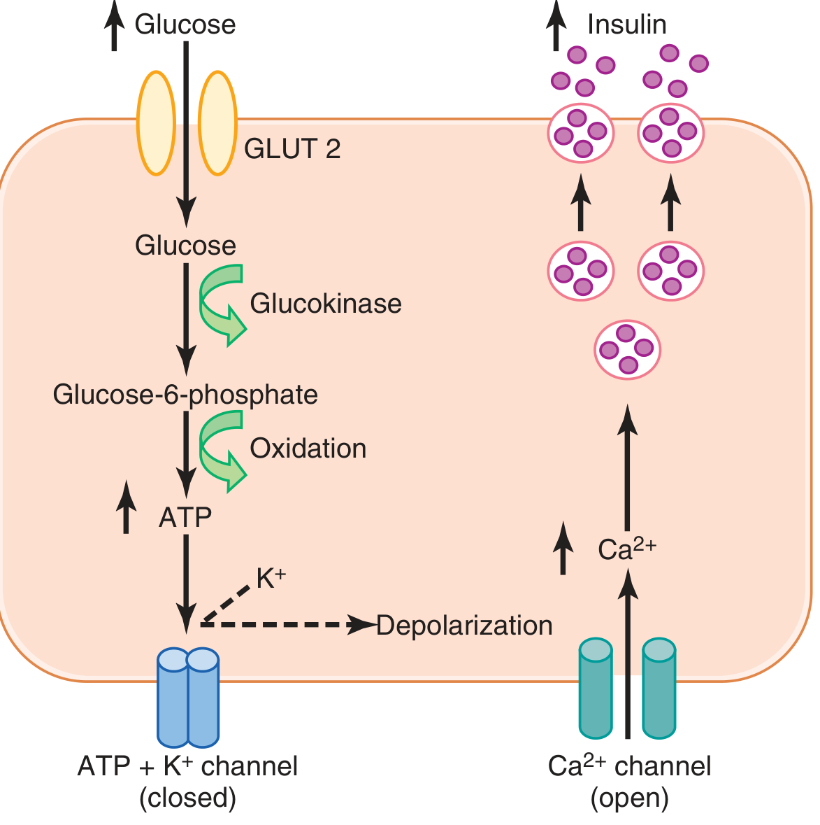
Step by step:
- Glucose enters via GLUT-2 transporter (proportional to blood glucose)
- Glucokinase phosphorylates glucose → Glucose-6-phosphate (rate-limiting/glucose-sensing step)
- Oxidation → ↑ ATP
- ATP closes ATP-sensitive K⁺ channels → cell depolarizes
- Depolarization opens voltage-gated Ca²⁺ channels → Ca²⁺ influx
- Ca²⁺ triggers exocytosis of insulin granules
Drug link: Sulfonylureas (e.g. glibenclamide) block ATP-sensitive K⁺ channels directly → depolarize beta cell → ↑ insulin secretion
3. Stimulators vs. Inhibitors of Insulin Secretion
| Stimulators | Inhibitors |
|---|---|
| ↑ Blood glucose | Somatostatin |
| Amino acids | Norepinephrine (α₂ receptors) |
| GLP-1, GIP (incretins) | Fasting / hypoglycemia |
| Acetylcholine (vagus) | Insulin itself (feedback) |
| Glucagon | |
| Sulfonylurea drugs |
4. Metabolic Effects of Insulin
A. Carbohydrate Metabolism
- ↑ Glucose uptake (GLUT-4 in muscle, fat)
- ↑ Glycogen synthesis (liver and muscle) - activates glycogen synthase
- ↓ Glycogenolysis (inactivates liver phosphorylase)
- ↓ Gluconeogenesis (decreases liver enzymes + decreases amino acid precursors)
- Net: Lowers blood glucose
B. Fat Metabolism
- ↑ Fatty acid synthesis (liver)
- ↑ Lipoprotein lipase activity in adipose capillaries → fat storage
- ↓ Hormone-sensitive lipase → ↓ lipolysis
- Net: Fat sparer + fat storage promoter
- Insulin deficiency → ↑ lipolysis → ↑ free fatty acids → ↑ ketone bodies → ketoacidosis
C. Protein Metabolism
- ↑ Amino acid uptake into cells
- ↑ Protein synthesis
- ↓ Protein breakdown
- Insulin deficiency → tissue wasting
5. Blood Glucose Regulation
- Normal fasting glucose: 80-90 mg/100 mL
- Post-meal peak: 120-140 mg/100 mL (returns to normal within 2 hours)
- The liver is the primary "glucose buffer" (stores ~100g glycogen = ~5-6% of liver mass)
Four defense mechanisms against hypoglycemia:
- Insulin falls → liver releases glucose
- Glucagon rises → glycogenolysis
- Epinephrine (sympathetic) → emergency glucose release
- Prolonged: Cortisol + Growth hormone → ↓ glucose utilization, ↑ fat use
6. Diabetes Mellitus
| Feature | Type 1 | Type 2 |
|---|---|---|
| Mechanism | Beta cell destruction (autoimmune/viral) | Insulin resistance |
| Age at onset | Usually <20 yr | Usually >30 yr |
| Body mass | Low/wasted to normal | Obese (visceral) |
| Plasma insulin | Low/absent | Normal to high initially |
| Plasma glucagon | High (suppressible) | High (resistant to suppression) |
| Insulin sensitivity | Normal | Reduced |
| Treatment | Insulin (mandatory) | Lifestyle → metformin → sulfonylureas → SGLT2i → GLP-1 → insulin |
7. Renal Physiology in Diabetes (Key Points)
A. Glucosuria (Glycosuria)
- Glucose is freely filtered at glomerulus
- Normally 100% reabsorbed in proximal tubule via SGLT2 (90%) and SGLT1 (10%)
- Renal threshold for glucose = ~180-200 mg/100 mL (tubular maximum reached)
- When blood glucose >200 mg/100 mL → glucose appears in urine (glucosuria)
- In severe DM: 100+ grams glucose lost per day in urine
B. Osmotic Diuresis
- Unreabsorbed glucose retains water in tubular lumen → osmotic diuresis
- Result: Polyuria (large urine output)
- Leads to: Dehydration → polydipsia (thirst)
C. Classic Triad of DM (from renal mechanism)
| Symptom | Mechanism |
|---|---|
| Polyuria | Osmotic diuresis from glucosuria |
| Polydipsia | Dehydration + cellular dehydration from hyperosmolarity |
| Polyphagia | Cells starved of glucose despite high blood glucose |
D. Cellular Dehydration
- Glucose doesn't cross cell membranes easily
- Hyperglycemia → ↑ extracellular osmolarity → water drawn OUT of cells → cellular dehydration
E. SGLT2 Inhibitors (Pharmacological Link to Renal Physiology)
- Drugs: empagliflozin, dapagliflozin, canagliflozin
- Block SGLT2 in proximal tubule → prevent glucose reabsorption → ↑ urinary glucose excretion → ↓ blood glucose
- Insulin-independent mechanism
- Side effects: diuresis (↓ BP, risk of dehydration/hypotension), UTI risk
- Extra benefit: cardioprotective + renoprotective effects in clinical trials
F. Renal Insulin Handling
- Kidney removes ~30-40% of circulating insulin
- In chronic kidney disease (CKD): ↓ renal insulin clearance → plasma insulin levels rise slightly → increased risk of hypoglycemia in patients on insulin therapy (dose reduction needed in CKD)
8. Complications of Chronic Hyperglycemia
| System | Complication |
|---|---|
| Kidney | Diabetic nephropathy → end-stage renal disease |
| Eyes | Retinopathy → blindness |
| Heart | Atherosclerosis → MI, stroke |
| Nerves | Peripheral neuropathy, autonomic dysfunction |
| Limbs | Ischemia, gangrene |
Mechanism: Chronic high glucose → vascular endothelial damage → microvascular + macrovascular disease
9. Diabetic Ketoacidosis (DKA) - Type 1
- Insulin deficiency → ↑ lipolysis → ↑ keto acids (acetoacetic acid, β-hydroxybutyric acid)
- Released faster than tissues can use → metabolic acidosis
- Dehydration + acidosis → diabetic coma (if pH <7.0, death possible within hours)
- Compensation: Kussmaul breathing (deep, rapid) to blow off CO₂
- Treatment: IV insulin + IV fluids + electrolytes
Source: Guyton & Hall Textbook of Medical Physiology, Goldman-Cecil Medicine
Hypothalamus short notes easy words
Finding Sources
Finding Sources
Reading File
Reading File
Reading File
Reading File
Reading File
Loading Image
Reading File
Loading Image
I now have everything needed for a complete, simplified note. Here it is:
Hypothalamus - Short Notes (Simple Words)
Sources: Guyton & Hall Textbook of Medical Physiology, Neuroanatomy through Clinical Cases
1. What Is It? Where Is It?
Think of the hypothalamus as the "master controller" of the body. It is a tiny pea-sized structure at the base of the brain, sitting just below the thalamus (hence the name: "hypo" = below).
- Part of the diencephalon
- Forms the walls and floor of the 3rd ventricle
- Located just behind the optic chiasm
- Connected to the pituitary gland via the pituitary stalk (infundibulum)
Easy memory trick: HEAL
H - Homeostasis (hunger, thirst, sleep, sex) E - Endocrine control (pituitary hormones) A - Autonomic control (heart, BP, gut) L - Limbic functions (emotions, behavior)
2. Hypothalamic Nuclei - The Control Centers
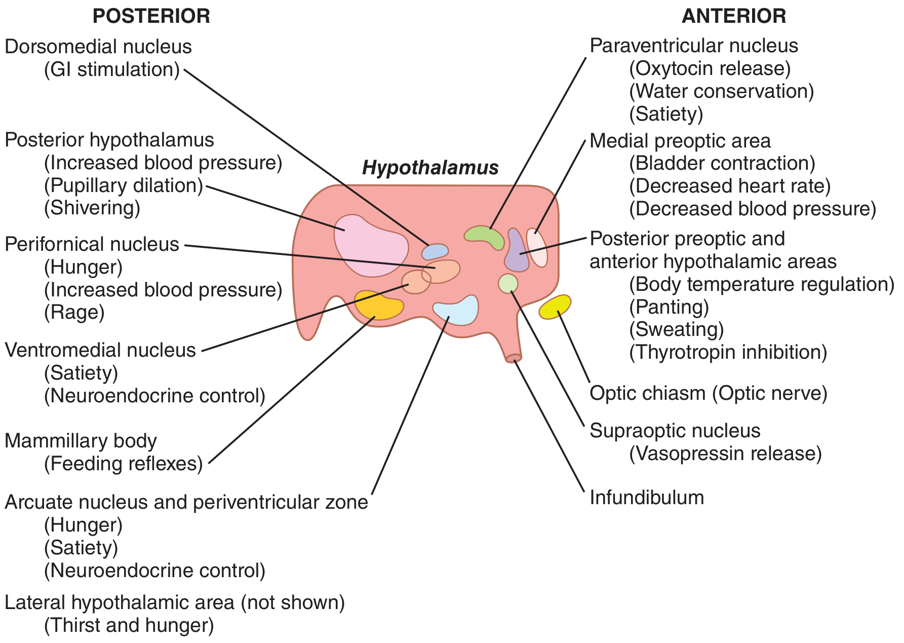
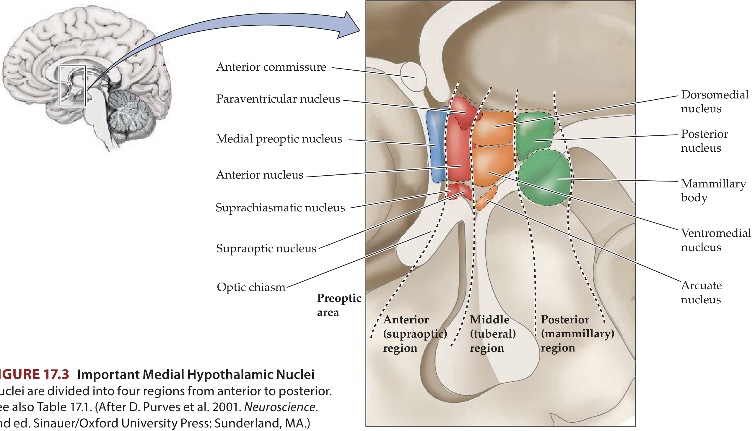
The hypothalamus is divided into anterior → middle → posterior regions, each with specific nuclei:
| Region | Key Nuclei | Main Job |
|---|---|---|
| Anterior (Preoptic/Supraoptic) | Supraoptic nucleus | Makes ADH (vasopressin) → water balance |
| Paraventricular nucleus | Makes Oxytocin + ADH | |
| Suprachiasmatic nucleus | Body clock (circadian rhythm) | |
| Anterior/Preoptic area | Cooling the body (heat loss) | |
| Middle (Tuberal) | Ventromedial nucleus | Satiety center (stop eating) |
| Arcuate nucleus | Controls anterior pituitary hormones | |
| Dorsomedial nucleus | GI stimulation | |
| Posterior (Mammillary) | Posterior nucleus | Heating the body (heat conservation), ↑ BP, shivering |
| Mammillary body | Feeding reflexes, memory (connected to hippocampus) | |
| Lateral area (all regions) | Lateral hypothalamic area | Hunger + Thirst center |
3. The 7 Big Jobs of the Hypothalamus
🌡️ Job 1: Temperature Regulation
- Anterior hypothalamus = detects overheating → triggers sweating, panting, vasodilation to cool you down
- Posterior hypothalamus = detects cold → triggers shivering, vasoconstriction to warm you up
- Lesion of anterior = hyperthermia (can't cool down)
- Lesion of posterior = poikilothermia (body temperature just follows the environment like a lizard)
💧 Job 2: Water Balance (Thirst + ADH)
- Osmoreceptors in anterior hypothalamus detect when blood is too concentrated (dehydration)
- Response: feel thirsty (drink water) + release ADH from supraoptic nucleus (kidneys retain water)
- Lesion of supraoptic nucleus → Diabetes Insipidus (no ADH → massive water loss in urine)
🍔 Job 3: Hunger and Appetite
| Center | Location | Effect |
|---|---|---|
| Hunger center | Lateral hypothalamus | "I'm hungry, eat!" |
| Satiety center | Ventromedial nucleus | "I'm full, stop eating!" |
- Leptin (from fat cells) acts on hypothalamus → ↓ appetite (tells brain: "we have enough fat stored")
- Ghrelin (from stomach) acts on hypothalamus → ↑ appetite ("feed me!")
- Lesion of lateral hypothalamus → stops eating → weight loss/starvation
- Lesion of ventromedial nucleus → can't feel full → obesity + rage
😴 Job 4: Sleep-Wake Cycle (Circadian Rhythm)
- Suprachiasmatic nucleus = the body's master clock
- Gets light signals from eyes (via retinohypothalamic tract) → adjusts day/night cycles
- Anterior hypothalamus (VLPO area) = promotes sleep (releases GABA to inhibit arousal centers)
- Posterior hypothalamus = promotes wakefulness (histamine, orexin neurons)
- Lesion of anterior hypothalamus → insomnia
- Lesion of posterior hypothalamus → hypersomnia (sleeps too much)
🫀 Job 5: Cardiovascular Control
- Posterior/lateral hypothalamus stimulation → ↑ BP, ↑ heart rate
- Anterior/preoptic area stimulation → ↓ BP, ↓ heart rate, bladder contraction
- Works via relays in the brainstem (pons and medulla)
🧬 Job 6: Endocrine Control (via Pituitary Gland)
The hypothalamus controls the anterior pituitary through releasing and inhibiting hormones carried in the portal blood system:
| Hypothalamic Hormone | Effect on Pituitary | Final Result |
|---|---|---|
| CRH (Corticotropin-releasing hormone) | → ACTH release | → Cortisol from adrenal cortex |
| TRH (Thyrotropin-releasing hormone) | → TSH release | → Thyroid hormones |
| GHRH (Growth hormone-releasing hormone) | → GH release | → Growth |
| Somatostatin (GIH) | Inhibits GH + TSH | Slows growth hormone |
| GnRH (Gonadotropin-releasing hormone) | → LH + FSH release | → Sex hormones |
| Dopamine (PIF) | Inhibits Prolactin | ↓ Milk production |
| TRH / PRF | → Prolactin release | Milk production |
The posterior pituitary is different - it just stores and releases hormones made directly in the hypothalamic nuclei:
- Supraoptic nucleus → ADH (vasopressin) - water retention
- Paraventricular nucleus → Oxytocin - uterine contractions, milk letdown, bonding
😡 Job 7: Emotions and Behavior
- Lateral hypothalamus stimulation → rage, fighting, aggression
- Ventromedial nucleus stimulation → calm, peaceful, satiety
- Periventricular nuclei stimulation → fear, punishment reaction
- Sexual drive controlled by anterior and posterior hypothalamus
- Oxytocin (made in hypothalamus) → increases nurturing behaviors
4. Hypothalamus - Lesion Summary Table
| Lesion Site | What Goes Wrong |
|---|---|
| Anterior hypothalamus | Hyperthermia (can't cool) + Insomnia |
| Posterior hypothalamus | Poikilothermia + Hypersomnia |
| Lateral hypothalamus | Stops eating + stops drinking → wasting |
| Ventromedial nucleus | Obesity + rage (can't feel full) |
| Supraoptic nucleus | Diabetes Insipidus (no ADH, massive urination) |
| Suprachiasmatic nucleus | Loss of circadian rhythm |
5. Quick Memory Tricks
"HEAT" for Anterior Hypothalamus:
Heat dissipation (cooling), Emotions calm, ADH + oxytocin production, Thirst sensing
"SHIP" for Posterior Hypothalamus:
Shivering, Heat conservation, Increase BP, Pupil dilation
Lateral = Hunger (L for Lust for food)
Ventromedial = satiety (V = Very full)
Sources: Guyton & Hall Textbook of Medical Physiology, Neuroanatomy through Clinical Cases 3rd Edition
This is a shared conversation. Sign in to Orris to start your own chat.