
Spleen
Reading File
The query "Spleen" is broad - let me clarify the angle before diving in.
Asking for Preferences
What aspect of the spleen are you interested in?
Searching sources...
Finding Sources
Finding Sources
Gray's Anatomy for Students and Cellular/Molecular Immunology look best. Let me pull both simultaneously, along with the Bailey & Love surgical section for good measure.
Reading File
Reading File
Excellent content. Now let me fetch the key diagrams while also reading a bit more of the immunology section.
Loading Image
Loading Image
Loading Image
Reading File
I now have rich anatomy and histology content. Let me fetch the splenic histology micrograph and the arterial supply diagram.
Loading Image
Loading Image
I have all the material needed for a thorough anatomy and histology overview. Here is the complete response:
The Spleen - Anatomy & Histology
Overview
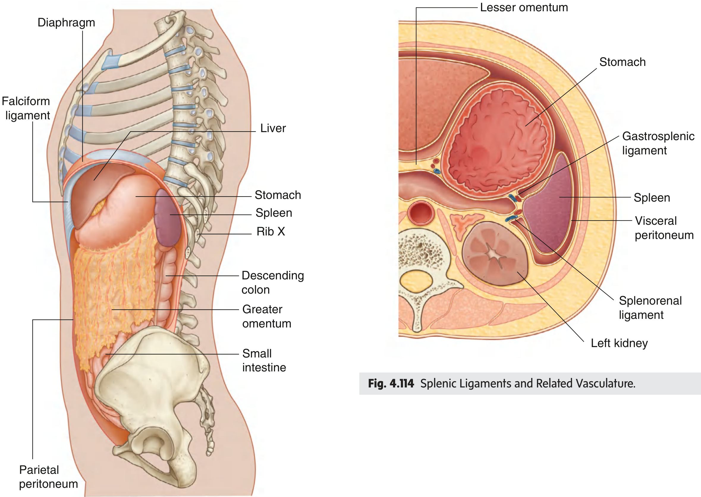
The spleen is the largest secondary lymphoid organ in the body, weighing approximately 150 g in adults. It lies in the left upper quadrant (left hypochondrium) of the abdomen, against the diaphragm at the level of ribs IX-X. It develops from the dorsal mesentery of the stomach and retains peritoneal connections to nearby viscera.
Gross Anatomy
Surfaces
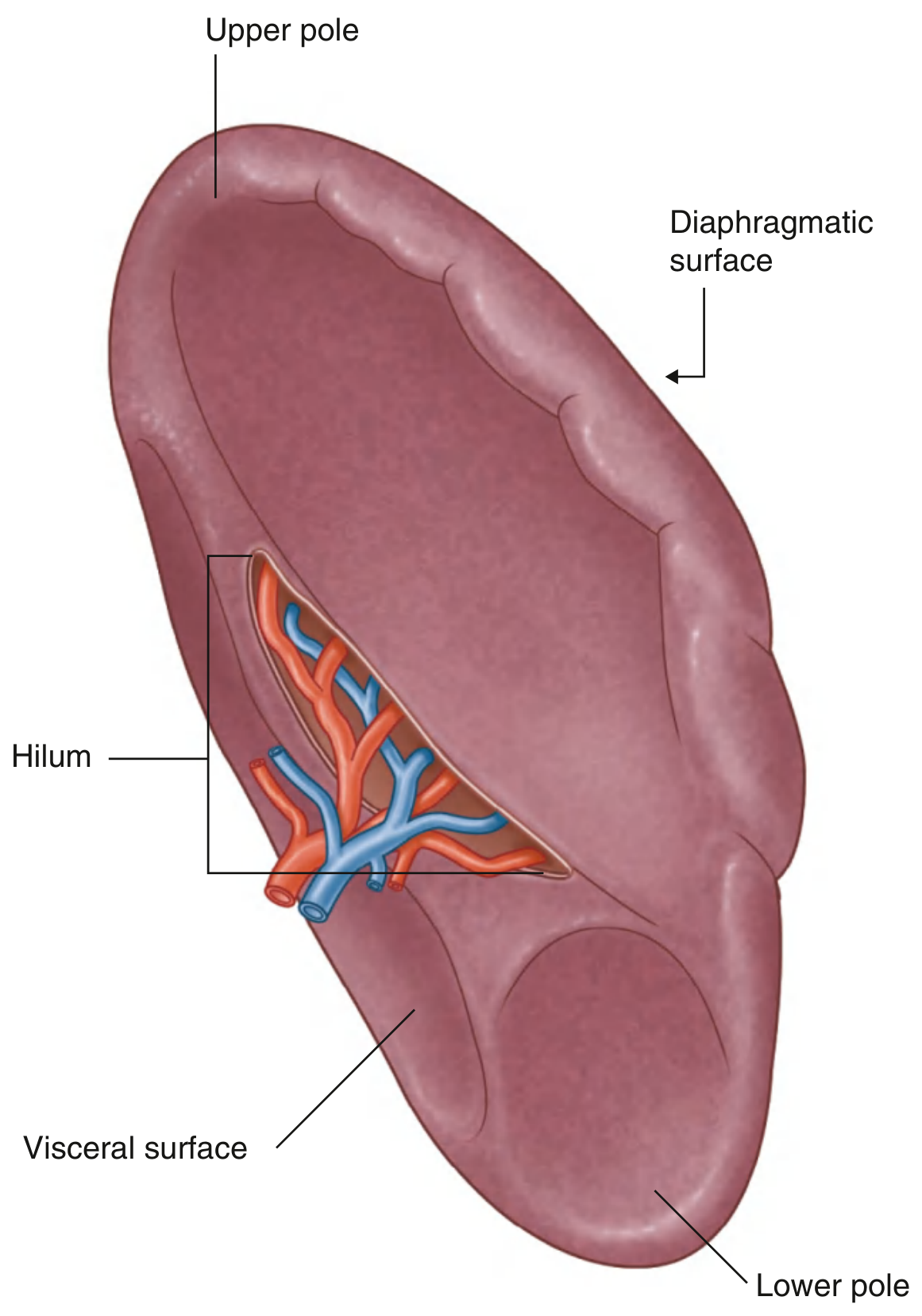
The spleen has two surfaces:
- Diaphragmatic surface - smooth and convex, facing upward and laterally against the diaphragm
- Visceral surface - concave medial face bearing impressions from adjacent organs (stomach, left kidney, tail of pancreas, splenic flexure of colon)
Peritoneal Ligaments
The spleen is covered by visceral peritoneum except at the hilum, and is held in place by two peritoneal folds (both parts of the greater omentum):
| Ligament | Connects To | Contents |
|---|---|---|
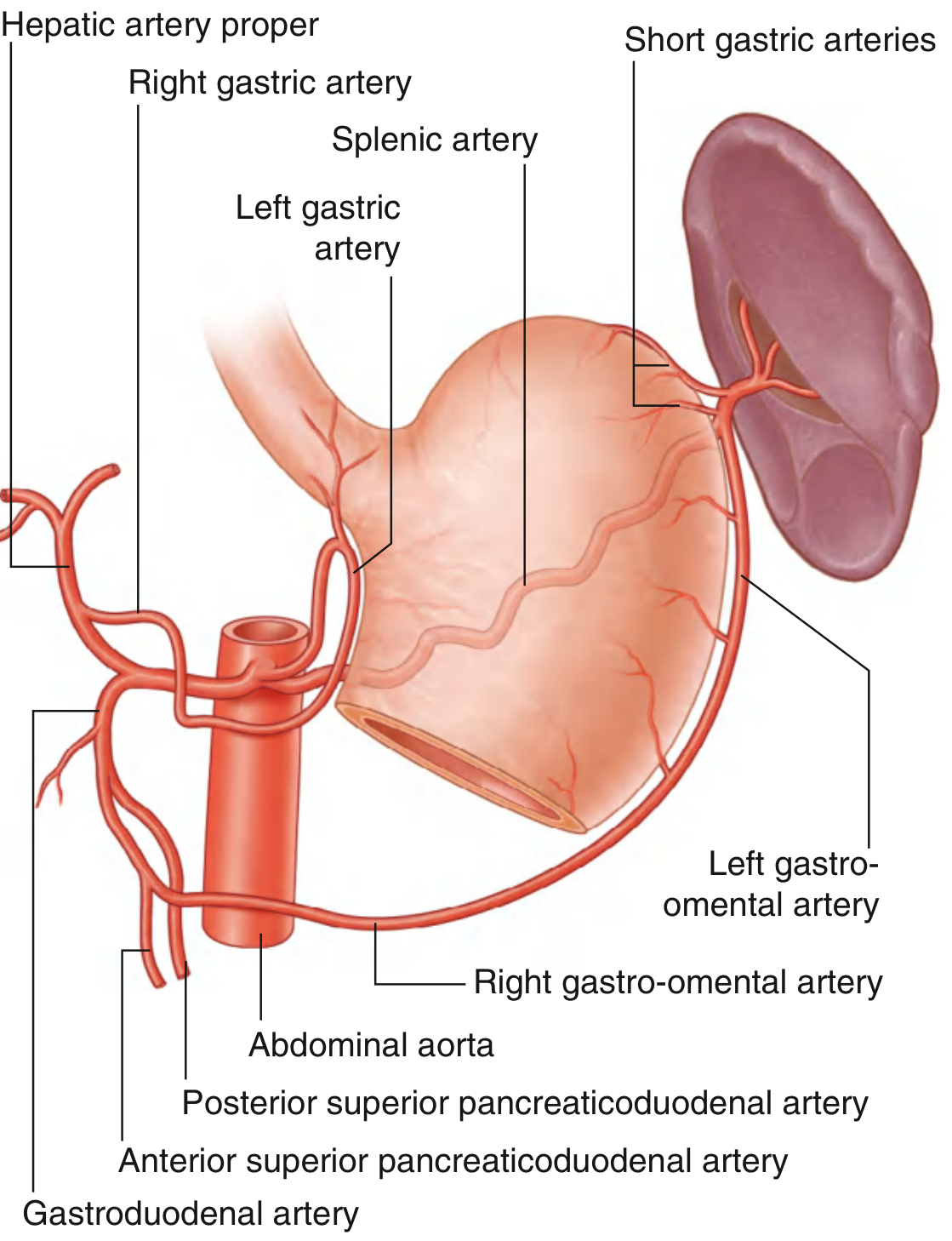
| Gastrosplenic ligament | Greater curvature of stomach | Short gastric & left gastro-omental vessels |
| Splenorenal ligament | Left kidney | Splenic artery, splenic vein, tail of pancreas |
Vasculature
- Arterial supply: Splenic artery (largest branch of the celiac trunk) - it runs a tortuous course along the superior border of the pancreas before entering the hilum
- Venous drainage: Splenic vein drains into the portal vein (after joining the superior mesenteric vein behind the neck of the pancreas)
Internal Architecture / Histology
The splenic parenchyma ("pulp") is divided into two functionally distinct compartments:
Red Pulp (~75% of splenic volume)
- Composed of blood-filled vascular sinusoids lined by macrophages and littoral cells
- Sinusoids are filled with large numbers of erythrocytes
- Drain into venules - splenic vein - portal circulation
- The red pulp macrophages are the key filter: they phagocytose senescent red cells, damaged cells, opsonized microbes, and immune complexes
- This is where splenic "slow circulation" occurs: red cells must squeeze through gaps between sinusoidal lining cells - rigid/damaged cells cannot pass and are destroyed
White Pulp (lymphoid tissue)
Organized around central arteries (branches of the splenic artery distinct from those supplying sinusoids):
| Zone | Cell Population | Function |
|---|---|---|
| Periarteriolar lymphoid sheath (PALS) | Mainly T cells | T cell zone; chemokines CCL19/CCL21 attract naive T cells via CCR7 |
| B cell follicles | B cells | Primary follicles (naive B cells) and secondary follicles with germinal centers after antigen stimulation |
| Marginal zone | Marginal zone B cells + specialized macrophages | Boundary between red and white pulp; samples blood-borne antigens; contains functionally distinct MZ B cells with limited antigen repertoire |
The white pulp architecture is analogous to a lymph node - T and B zones are segregated by chemokine gradients produced by fibroblastic reticular cells (FRCs). CXCL13/CXCR5 drives B cells into follicles; CCL19/CCL21/CCR7 drives T cells into the PALS.
Marginal Sinus
A vascular channel that forms the border between white and red pulp. Antigens from the blood are delivered into the marginal sinus by circulating dendritic cells, or sampled directly by marginal zone macrophages.
Blood Flow Through the Spleen
Blood enters via the splenic artery → divides into trabecular arteries (surrounded by fibrous trabeculae) → central arteries → two routes:
- "Open" / slow circulation - arterioles open into the red pulp cords; blood percolates through the reticular meshwork, squeezes through sinusoidal walls back into venous sinusoids (allows macrophage screening of red cells)
- "Closed" / fast circulation - arterioles connect directly into sinusoids, bypassing the cords
Both routes ultimately drain into trabecular veins → splenic vein.
Key Histological Cell Types
| Cell | Location | Role |
|---|---|---|
| Red pulp macrophages | Sinusoidal lining | Phagocytose senescent RBCs, opsonized bacteria |
| Marginal zone macrophages | Marginal zone | Sample blood antigens, present to B cells |
| Follicular dendritic cells | B cell follicles | Trap antigen for B cell activation |
| Fibroblastic reticular cells | White pulp stroma | Form conduit network; produce CCL19/CCL21/CXCL13 |
| Marginal zone B cells | Marginal zone | T-independent antibody responses to polysaccharides |
| Plasma cells | Red pulp | Terminal antibody secretion |
Clinical Note: Asplenia
Individuals without a functional spleen (congenital asplenia, splenectomy, or functional asplenia e.g. sickle cell) are at high risk for overwhelming post-splenectomy infection (OPSI), particularly from encapsulated bacteria - Streptococcus pneumoniae, Neisseria meningitidis, Haemophilus influenzae. This is because:
- The spleen is a major site of opsonin (IgM) production
- Splenic macrophages clear encapsulated bacteria that have been opsonized - this function is lost
- The marginal zone B cells, which respond to polysaccharide antigens in a T-independent manner, are absent
Sources: Gray's Anatomy for Students; Cellular and Molecular Immunology (Abbas & Lichtman)
This is a shared conversation. Sign in to Orris to start your own chat.