
WALLERIAN DEGENERATION & REGENERATION. 2.SALTATORY Conduction+Refractory Period.
Reading File
Now I'll search the medical library for both topics in parallel.
Finding Sources
Finding Sources
Finding Sources
Reading File
Reading File
Reading File
Reading File
Now let me get the saltatory conduction description section and the Wallerian degeneration nerve regeneration continuation, plus fetch the key image.
Reading File
Reading File
Loading Image
Now let me fetch the saltatory conduction diagram:
Loading Image
Loading Image
Excellent - I now have rich content from authoritative textbooks and diagrams. Here is the complete, well-organized answer:
1. Wallerian Degeneration & Nerve Regeneration
What Triggers It?
Wallerian degeneration occurs after grade II-V nerve injuries (axonotmesis and neurotmesis). It involves changes both distal and proximal to the injury site.
- Bradley and Daroff's Neurology in Clinical Practice, p. 1334
A. Distal Segment Changes
After axonal injury, the earliest and most dramatic changes occur distal to the injury:
- Disruption of axoplasmic flow (both retrograde and anterograde) occurs immediately.
- Calcium and sodium influx through the disrupted axonal plasma membrane activates a cascade resembling programmed cell death (apoptosis).
- Leukocyte recruitment begins, with cytokine-mediated signaling in non-neuronal cells.
- This triggers synthesis of: neurotrophins, chemokines, extracellular matrix molecules, proteolytic enzymes, and interleukins.
- By Day 3: Schwann cells retract from the node of Ranvier. Activated Schwann cells and macrophages begin digesting myelin (phagocytosis of myelin ovoids).
- Complete axonal degeneration takes approximately 1 week.
B. Proximal Segment Changes
- Axon breakdown extends proximally up to the first node of Ranvier from the injury site.
- Very proximal injuries (e.g., proximal arm amputation) may cause apoptosis of the cell body itself.
- More commonly: the cell body undergoes chromatolysis:
- Breakup and dispersion of rough endoplasmic reticulum (Nissl substance dissolves)
- Eccentric displacement of the cell nucleus
- Upregulation of transcription factors that shift gene expression from axon maintenance to protein synthesis (preparing for regeneration)
C. Nerve Regeneration
The method of regeneration depends on injury grade:
1. Grade I (Neuropraxia) - Remyelination
- Schwann cell divides and remyelinates the axon.
- Recovery within weeks to months.
- New myelin is thinner with more internodes per original internode.
2. Collateral Sprouting (partial injury)
- Intact motor axons produce nodal sprouts (from nodes of Ranvier) or terminal sprouts within 4 days of injury.
- These sprouts reinnervate denervated muscle fibers - increasing motor unit size and contractile force.
- Clinical recovery takes 3-6 months.
3. Axonal Regeneration (complete/severe injury)
- Starts only after Wallerian degeneration is completed.
- Schwann cells dedifferentiate and upregulate: cadherins, immunoglobulin superfamily factors, and laminin - promoting axon sprout migration.
- Sprouts form along bands of Büngner (basal lamina tubes formed by proliferating Schwann cells after myelin clearance).
- The growth cone (tip of the sprout) uses:
- Filopodia (finger-like projections) and lamellipodia (sheet-like projections) to navigate
- Guidance molecules: semaphorins, ephrins, netrins, slits
- Secretes plasminogen activators to dissolve cell debris blocking its path
- Growth rate: 1-2 mm/day (~1 inch/month); proximal injuries grow faster (2-3 mm/day), distal injuries slower (~1 mm/day).
- Axon regeneration is the main recovery mechanism from 6-24 months after injury.
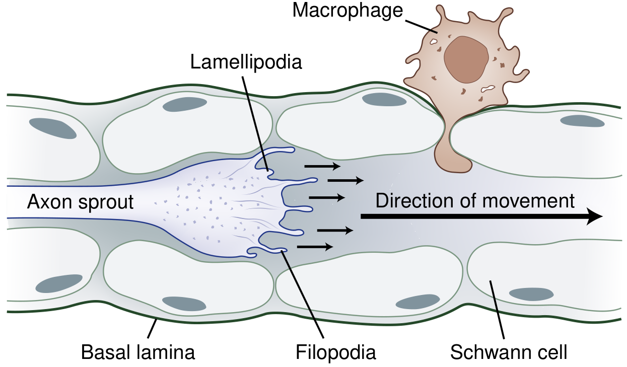
Fig. Schematic of axonal regeneration. The growth cone (with lamellipodia and filopodia) advances through bands of Büngner. Macrophages clear myelin debris. - Bradley and Daroff's Neurology
2. Saltatory Conduction + Refractory Period
Saltatory Conduction
The Role of Myelin
The myelin sheath is laid down by:
- Schwann cells - in the peripheral nervous system
- Oligodendroglia - in the CNS
Myelin acts like insulation on a leaky hose - it reduces transmembrane current leakage and forces current to flow longitudinally down the axon interior, dramatically increasing conduction velocity.
Nodes of Ranvier
- Gaps in the myelin sheath, only 1-2 μm long
- Myelinated internodal segments are 0.2-2.0 mm (up to 1,000x longer than nodes)
- Voltage-gated Na+ channels are highly concentrated at nodes and essentially absent under myelin
- K+ channels (both voltage-dependent and non-voltage-dependent) are also present at or near nodes
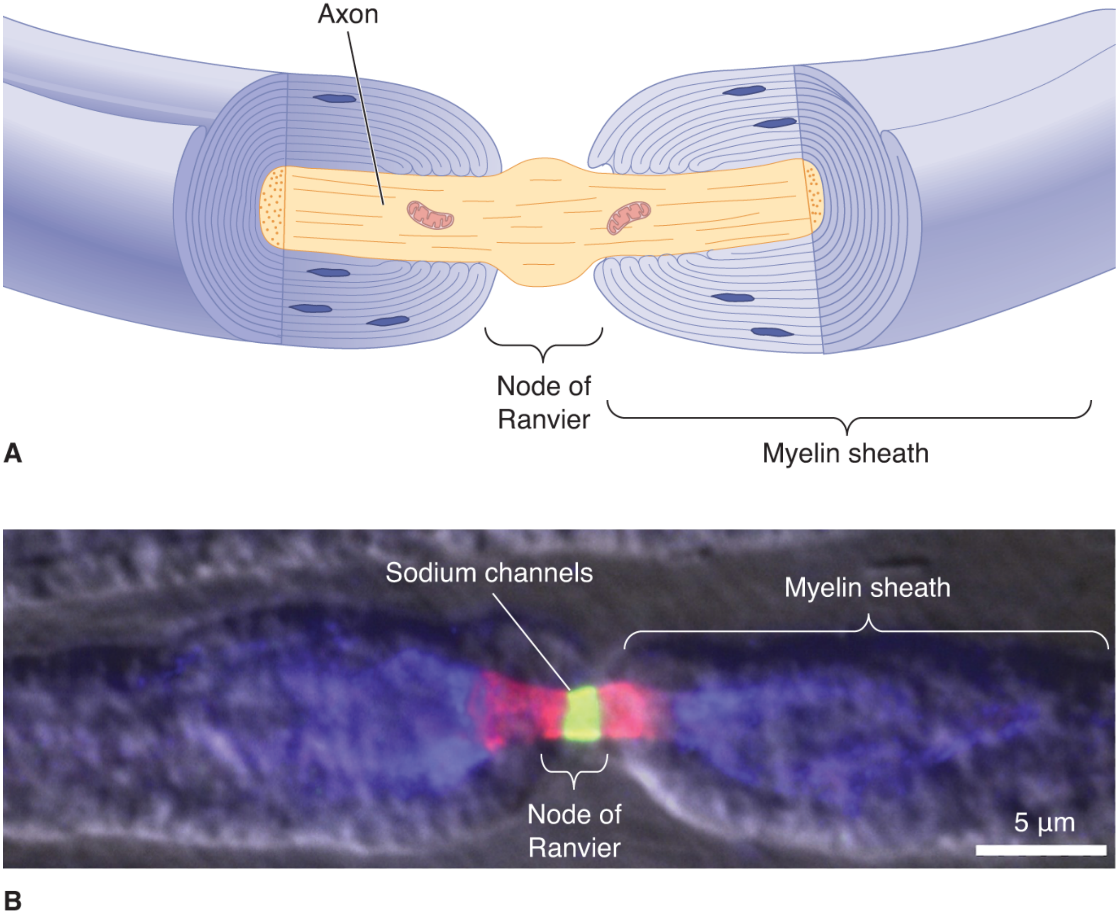
Fig. 4.16 - Myelin sheath and node of Ranvier. (B) Voltage-gated Na+ channels (green) concentrated at the node; K+ channels (red) flanking them. - Neuroscience: Exploring the Brain, 5th Ed.
The "Leap" - How It Works
In unmyelinated fibers, action potentials crawl along the membrane heel-to-toe, activating every square micron of membrane. In myelinated axons:
- Depolarization at node 1 drives a large inward Na+ current
- This current spreads passively and rapidly down the inside of the axon (without being lost through the insulated internode)
- The current arrives at node 2 still strong enough to trigger a new action potential
- The AP appears to "jump" from node to node - this is saltatory conduction (from Latin saltare = to leap)
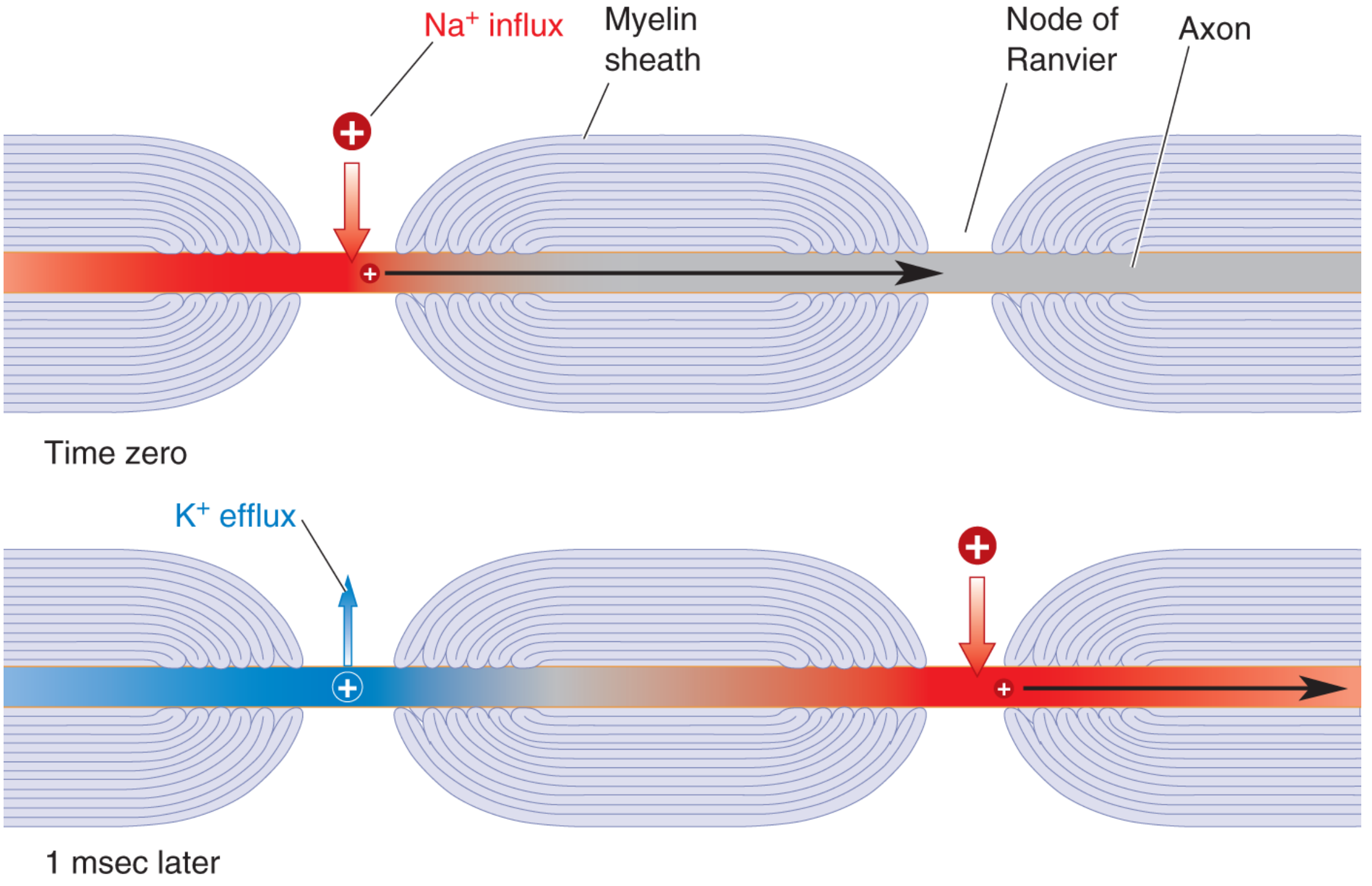
Fig. 4.17 - Saltatory conduction: current leaps from node to node. The first node depolarizes (Na+ influx, red), while 1 msec later the action potential has jumped to the next node, and the first is repolarizing (K+ efflux, blue). - Neuroscience: Exploring the Brain, 5th Ed.
Conduction Velocity
| Fiber type | Diameter | Velocity |
|---|---|---|
| Myelinated (peripheral) | 1-20 μm | 5-120 m/sec |
| Squid giant axon (unmyelinated) | 1,000 μm | ~25 m/sec |
Key point: In myelinated axons, conduction velocity is linearly proportional to axon diameter. In unmyelinated axons, it is proportional to the square root of diameter.
Saltatory conduction is far more energy-efficient - Na+/K+ ATPase only needs to restore ion gradients at the nodes, not along the entire membrane.
Clinical link: Multiple sclerosis demyelinates CNS axons, slowing or blocking saltatory conduction, producing weakness, incoordination, and visual disturbances.
Refractory Period
After an action potential fires, there is a window during which the neuron cannot (or cannot easily) fire again.
Absolute Refractory Period (ARP)
-
Overlaps with almost the entire duration of the action potential (depolarization + repolarization phases)
-
During the ARP: no stimulus of any strength can elicit a new action potential
-
Mechanism: Voltage-gated Na+ channels enter the inactivated state upon depolarization. The inactivation gates (h-gates) close and CANNOT reopen until the membrane repolarizes back to resting potential, returning channels to the "closed but available" state.
-
Duration in large myelinated nerve fibers: ~1 millisecond
-
This limits maximum firing rate to ~1,000 impulses/second
-
Costanzo Physiology 7th Ed., p. 29; Guyton & Hall Medical Physiology, p. 88
Relative Refractory Period (RRP)
-
Begins at the end of the ARP
-
Overlaps with the hyperpolarizing afterpotential (after-hyperpolarization phase)
-
During the RRP: an action potential can be elicited, but only with a greater-than-normal depolarizing stimulus
-
Mechanism: Voltage-gated K+ channels are still partially open, making membrane conductance to K+ higher than at rest. The membrane potential is closer to the K+ equilibrium potential (more negative), so more inward current is needed to reach threshold.
-
Duration: 2-4 milliseconds after the ARP
-
Guyton & Hall, p. 88
Summary Table
| Period | Timing | Can fire? | Ionic Basis |
|---|---|---|---|
| Absolute Refractory | During action potential | No | Na+ channels inactivated (h-gates closed) |
| Relative Refractory | During after-hyperpolarization | Yes, with stronger stimulus | K+ channels still open, membrane hyperpolarized |
Clinical Relevance - Accommodation
- If a cell is slowly depolarized, Na+ inactivation gates close progressively without an AP firing. This is accommodation (e.g., the reason why chronic depolarization from hyperkalemia causes muscle weakness - channels inactivate before threshold is reached).
Sources:
- Bradley and Daroff's Neurology in Clinical Practice (ISBN 9780323642613), Block 14
- Neuroscience: Exploring the Brain, 5th Edition (ISBN 9781284286878), Block 1
- Costanzo Physiology 7th Edition (ISBN 9780323793339), Block 1
- Guyton and Hall Textbook of Medical Physiology (ISBN 9780443111013), Block 1
This is a shared conversation. Sign in to Orris to start your own chat.