
Leukodystrophyies it’s presentation clinical features imaging and differential diagnosis
leukodystrophy MRI brain white matter changes
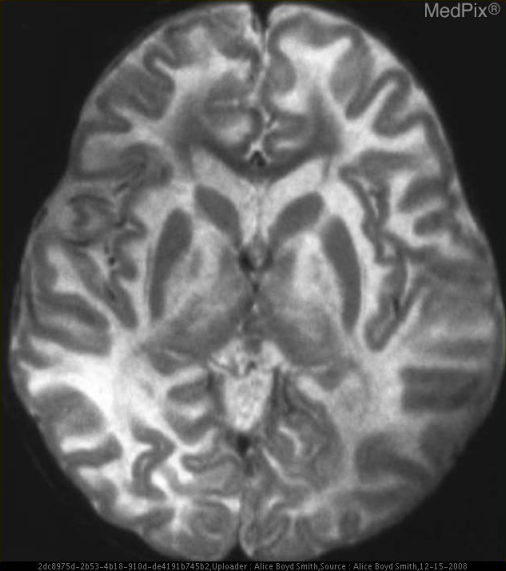
**Imaging Modality:** Magnetic Resonance Imaging (MRI), T2-weighted axial section.
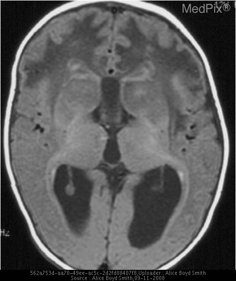
**Imaging Modality:** Axial Magnetic Resonance Imaging (MRI), T1-weighted sequence.
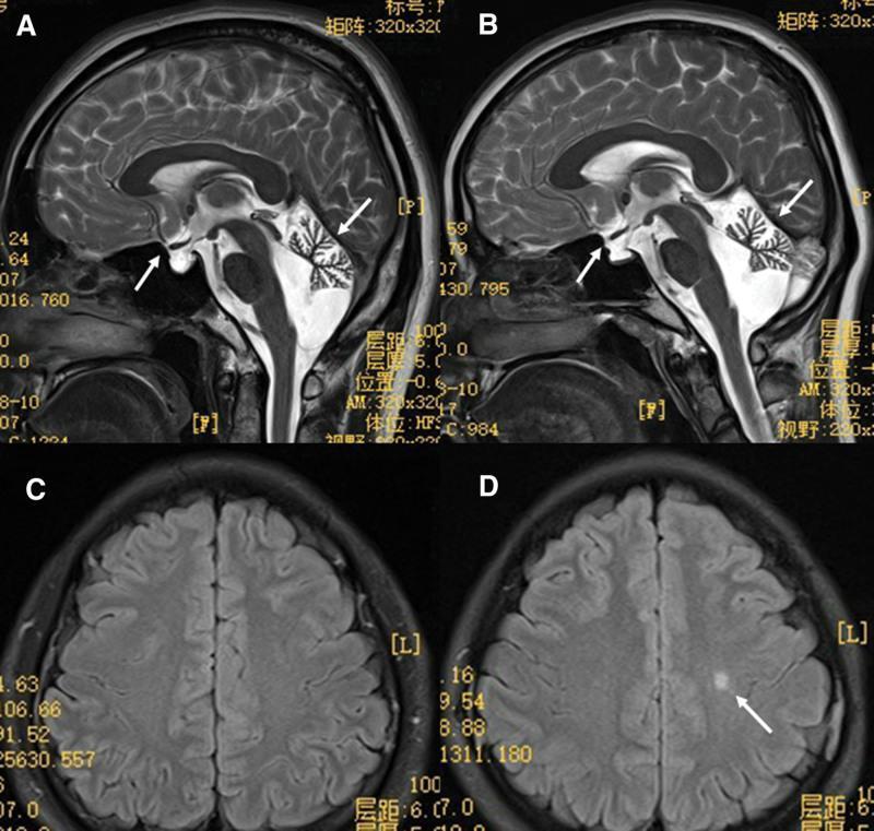
This composite brain MRI displays diagnostic images from two siblings with 4H leukodystrophy (Hypomyelination, Hypodontia, and Hypogonadotropic Hypogonadism). Panels A and B are sagittal T2-weighted images showing prominent cerebellar atrophy, characterized by enlarged sulci and reduced folial volume (white arrows, right). Both siblings also exhibit hypophysis (pituitary) dysplasia, with an abnormally small or flattened pituitary gland visible in the sella turcica (white arrows, left). Panels C and D are axial FLAIR MRI sequences evaluating white matter integrity. Panel C (brother) shows relatively normal signal intensity. Panel D (sister) reveals a focal hyperintense lesion (white arrow) indicating local demyelination or hypomyelination within the subcortical white matter. These imaging findings are classic neuroradiological hallmarks of POLR3B-related leukodystrophy, illustrating the characteristic combination of neurodegenerative changes (cerebellar atrophy) and developmental defects (pituitary dysplasia and myelin deficiency).
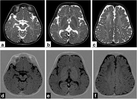
Diagnostic axial MRI sections of the brain in a 1.5-year-old child demonstrating classic features of hypomyelination and cerebral atrophy. Panels (a-c) show T2-weighted turbo spin-echo (TSE) images, while panels (d-f) show corresponding T1-weighted inversion recovery (IR) images. There is diffuse, symmetric signal alteration throughout the cerebral white matter, manifesting as hyperintensity on T2WI and hypointensity on T1WI. These changes specifically involve the internal and external capsules, the corpus callosum, frontal deep white matter, and the pre-aqueductal region. In addition to the white matter signal changes, there is notable diffuse prominence of the cerebral sulci and enlargement of the subarachnoid spaces, indicative of generalized cerebral atrophy. Clinically, this pattern of hypomyelination combined with hypodontia and hypogonadotropic hypogonadism (suggested by the '4H syndrome' context) is characteristic of RNA polymerase III-related leukodystrophy (POLR3A mutation). The images serve as an educational example of leukodystrophy classification based on signal intensity on standard MRI sequences.
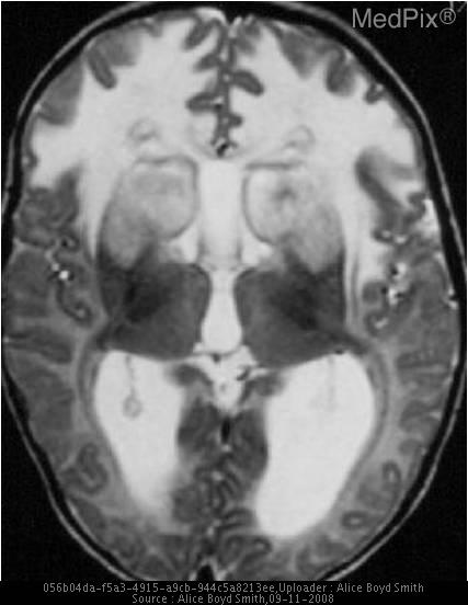
**Imaging Modality:** Magnetic Resonance Imaging (MRI), Axial T2-weighted sequence.
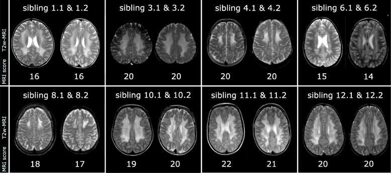
A comparison chart of T2-weighted axial MRI brain slices from eight pairs of siblings diagnosed with Metachromatic Leukodystrophy (MLD). The image demonstrates the distribution and intensity of white matter signal changes during the early stages of the disease. In each sibling pair, there is a striking visual similarity in the pattern of leukodystrophy. The affected white matter exhibits symmetric T2-hyperintensity (bright signal) compared to the relatively darker gray matter, indicating demyelination and increased water content. Spatial patterns vary between pairs, ranging from diffuse white matter involvement (e.g., pairs 3.1/3.2 and 11.1/11.2) to more regional predominance in the frontal and parietal-occipital areas (e.g., pairs 4.1/4.2 and 12.1/12.2). Below each scan, numerical MRI scores are provided, highlighting the closely matched disease severity within each sibling pair. This clinical imaging set serves as an educational tool for recognizing the characteristic 'butterfly' or confluent hyperintense patterns of pediatric leukodystrophies and the high phenotypic concordance in familial cases.
Topic Breakdown
- Metachromatic Leukodystrophy (MLD) - arylsulfatase A deficiency; late-infantile, juvenile, adult forms
- Krabbe Disease (Globoid Cell Leukodystrophy) - galactocerebrosidase deficiency
- X-linked Adrenoleukodystrophy / Adrenomyeloneuropathy (X-ALD/AMN) - peroxisomal, VLCFA accumulation
- Pelizaeus-Merzbacher Disease - PLP1 mutation, hypomyelinating
- Vanishing White Matter Disease (VWM) - EIF2B mutations, stress-triggered deterioration
- Alexander Disease - GFAP mutation, frontal-predominant
- Canavan Disease - aspartoacylase deficiency, macrocephaly
- 4H Leukodystrophy / POLR3-related - hypomyelination, hypodontia, hypogonadotropic hypogonadism
- Approach to imaging patterns (which white matter distribution points to which disease) and overall differential diagnosis (leukodystrophy vs leukoencephalopathy vs acquired demyelination like MS/ADEM vs toxic-metabolic)
Brief Overview
- Distribution (periventricular vs subcortical, frontal vs parieto-occipital, symmetric vs asymmetric) is often disease-specific - e.g., MLD and X-ALD classically start in parieto-occipital/periventricular white matter with a "butterfly" pattern, while Alexander disease is frontal-predominant.
- Signal characteristics: confluent T2/FLAIR hyperintensity with T1 hypointensity indicates demyelination; diffuse mild T2 hyperintensity without significant atrophy suggests hypomyelination (Pelizaeus-Merzbacher, 4H).
- Contrast enhancement at the leading edge (X-ALD) suggests active inflammatory demyelination.
- Associated structural findings - cerebellar atrophy (POLR3-related), macrocephaly (Canavan, Alexander), calcifications, basal ganglia/thalamic involvement (Krabbe) - help narrow the differential.
Which leukodystrophy (or aspect) would you like me to go into detail on?
Topic Breakdown
- Metachromatic Leukodystrophy (MLD) - arylsulfatase A deficiency; late-infantile, juvenile, adult forms
- Krabbe Disease (Globoid Cell Leukodystrophy) - galactocerebrosidase deficiency
- X-linked Adrenoleukodystrophy / Adrenomyeloneuropathy (X-ALD/AMN) - peroxisomal, VLCFA accumulation
- Pelizaeus-Merzbacher Disease - PLP1 mutation, hypomyelinating
- Vanishing White Matter Disease (VWM) - EIF2B mutations, stress-triggered deterioration
- Alexander Disease - GFAP mutation, frontal-predominant
- Canavan Disease - aspartoacylase deficiency, macrocephaly
- 4H Leukodystrophy / POLR3-related - hypomyelination, hypodontia, hypogonadotropic hypogonadism
- Approach to imaging patterns (which white matter distribution points to which disease) and overall differential diagnosis (leukodystrophy vs leukoencephalopathy vs acquired demyelination like MS/ADEM vs toxic-metabolic)
Brief Overview
- Distribution (periventricular vs subcortical, frontal vs parieto-occipital, symmetric vs asymmetric) is often disease-specific - e.g., MLD and X-ALD classically start in parieto-occipital/periventricular white matter with a "butterfly" pattern, while Alexander disease is frontal-predominant.
- Signal characteristics: confluent T2/FLAIR hyperintensity with T1 hypointensity indicates demyelination; diffuse mild T2 hyperintensity without significant atrophy suggests hypomyelination (Pelizaeus-Merzbacher, 4H).
- Contrast enhancement at the leading edge (X-ALD) suggests active inflammatory demyelination.
- Associated structural findings - cerebellar atrophy (POLR3-related), macrocephaly (Canavan, Alexander), calcifications, basal ganglia/thalamic involvement (Krabbe) - help narrow the differential.
leukodystrophy diagnosis MRI pattern
Leukodystrophies: Presentation, Clinical Features, Imaging, and Differential Diagnosis
1. Metachromatic Leukodystrophy (MLD)
- Late-infantile (most common, onset 1-2 yrs): gait regression, hypotonia progressing to spasticity, loss of speech, optic atrophy, peripheral neuropathy (hyporeflexia despite pyramidal signs is a classic clue)
- Juvenile (4-16 yrs): behavioral/cognitive decline preceding motor signs, school failure, clumsiness
- Adult: psychiatric presentation (psychosis, personality change) mimicking primary psychiatric illness, followed by dementia and spasticity
2. Krabbe Disease (Globoid Cell Leukodystrophy)
- Infantile (most severe): irritability, hyperesthesia, feeding difficulty, hypertonia progressing to opisthotonic posturing, blindness, seizures, rapid deterioration
- Late-onset/adult forms: spastic paraparesis, peripheral neuropathy, vision loss, slower progression
3. X-linked Adrenoleukodystrophy / Adrenomyeloneuropathy (X-ALD/AMN)
- Childhood cerebral form (boys, 4-8 yrs): behavioral change, declining school performance, visual/auditory processing deficits, rapidly progressive to vegetative state
- Adrenomyeloneuropathy (adult men): progressive spastic paraparesis, sensory ataxia, sphincter dysfunction, peripheral neuropathy
- Addison's disease (primary adrenal insufficiency) may precede, accompany, or follow neurologic symptoms - fatigue, hyperpigmentation, hypotension
- Heterozygous female carriers may develop a milder myelopathy in later life
4. Pelizaeus-Merzbacher Disease (PMD)
5. Vanishing White Matter Disease (VWM)
6. Alexander Disease
7. Canavan Disease
8. 4H Leukodystrophy / POLR3-related Disorders
General Approach to Differential Diagnosis
| Category | Examples | Distinguishing clues |
|---|---|---|
| Acquired inflammatory/demyelinating | Multiple sclerosis, ADEM, neuromyelitis optica spectrum disorder | Asymmetric, ovoid periventricular lesions, Dawson fingers, oligoclonal bands, relapsing-remitting course, later age of onset |
| Infectious | PML, HIV encephalopathy, subacute sclerosing panencephalitis | Immunocompromised state, specific serologies |
| Toxic-metabolic | B12 deficiency, chemotherapy (methotrexate), radiation, CO poisoning | History of exposure, symmetric but non-genetic pattern |
| Vascular | CADASIL, small vessel ischemic disease | Anterior temporal pole and external capsule involvement (CADASIL), migraine history, vascular risk factors |
| Mitochondrial | Leigh syndrome, MELAS | Lactate elevation, basal ganglia/brainstem involvement, stroke-like episodes |
| Genetic leukodystrophy | As above | Symmetric distribution, family history, specific enzyme/genetic confirmation, characteristic MRI pattern per disease |
- Predominant posterior (parieto-occipital) involvement -> X-ALD, MLD
- Predominant frontal involvement -> Alexander disease
- Diffuse with subcortical U-fiber involvement -> Canavan disease
- Cystic/vanishing appearance -> VWM disease
- Non-progressive, diffuse, mild signal change -> hypomyelinating disorders (PMD, 4H)
- Cerebellar atrophy + pituitary dysplasia -> POLR3-related 4H leukodystrophy
- Basal ganglia/thalamic signal change with peripheral neuropathy -> Krabbe disease
Create a visual comparison poster of MRI patterns across leukodystrophies
A medical education poster comparing brain MRI white matter patterns across major leukodystrophies. Include labeled panels for: Metachromatic Leukodystrophy (symmetric periventricular white matter changes with tigroid/leopard-skin pattern), Krabbe Disease (corticospinal tract and periventricular involvement with thalamic changes), X-linked Adrenoleukodystrophy (symmetric parieto-occipital and splenium of corpus callosum demyelination with contrast-enhancing leading edge), Alexander Disease (frontal-predominant white matter changes with periventricular rim), Canavan Disease (diffuse white matter changes involving subcortical U-fibers with macrocephaly), Pelizaeus-Merzbacher Disease (diffuse mild hypomyelination without atrophy), Vanishing White Matter Disease (cystic degeneration of white matter), and 4H/POLR3-related Leukodystrophy (hypomyelination with cerebellar atrophy and pituitary dysplasia). Each panel should show a stylized axial brain MRI illustration with the affected white matter regions highlighted in a different color, with a small label and short caption underneath describing the key imaging feature and underlying disease name. Layout as a grid comparison chart suitable for medical teaching.
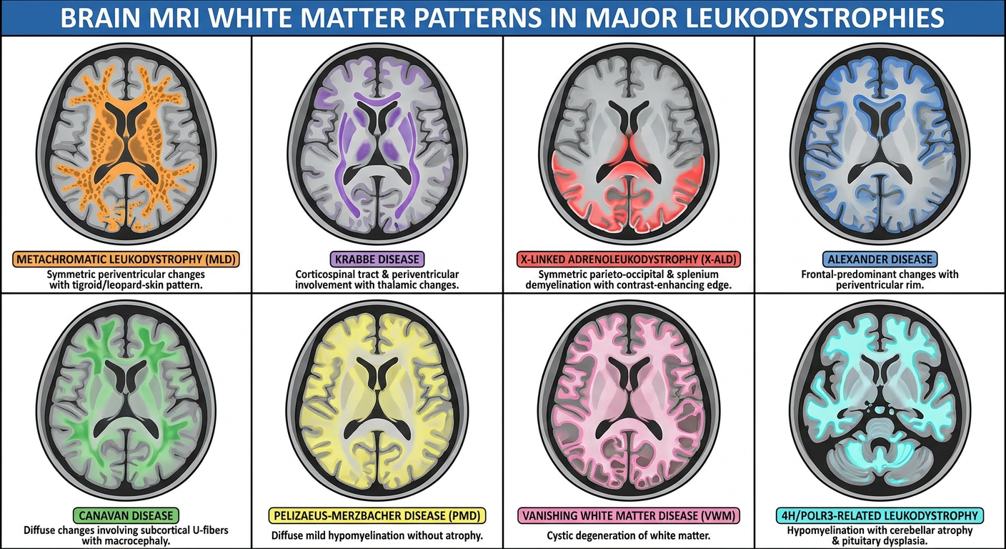
- MLD - symmetric periventricular changes with tigroid/leopard-skin pattern
- Krabbe disease - corticospinal tract and thalamic involvement
- X-ALD - parieto-occipital/splenial demyelination with enhancing leading edge
- Alexander disease - frontal-predominant white matter change
- Canavan disease - diffuse change involving subcortical U-fibers, with macrocephaly
- Pelizaeus-Merzbacher disease - diffuse, non-progressive hypomyelination
- Vanishing white matter disease - cystic degeneration
- 4H/POLR3-related leukodystrophy - hypomyelination with cerebellar atrophy and pituitary dysplasia