
Cushing syndrome
Cushing syndrome clinical features buffalo hump moon face
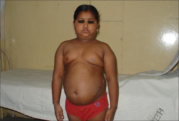
This clinical photograph shows a pediatric patient presenting with characteristic physical features of Cushing syndrome. The face exhibits a rounded, 'moon facies' appearance with associated submental fat deposition (double chin). There is evidence of central adiposity with a protuberant abdomen and fullness in the supraclavicular and upper chest regions, consistent with truncal obesity. A visible dorsocervical fat pad, or 'buffalo hump', is present at the base of the neck. On the patient's left flank, a linear surgical scar is visible, indicating a prior operative intervention. These findings are visually representative of hypercortisolism, which in this case is associated with an ACTH-secreting ganglioneuroma. The image serves as an educational example for identifying the systemic manifestations of glucocorticoid excess in children.
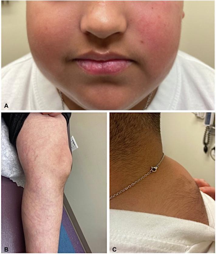
A composite of three clinical photographs demonstrating classic Cushingoid features in a pediatric patient. Panel A: Close-up of the face showing 'moon facies' characterized by rounded cheeks and noticeable facial plethora (erythema). Panel B: View of the left lower extremity showing multiple wide, slightly depressed, erythematous-to-violaceous striae (skin stretch marks) distributed across the proximal thigh and distal leg. Panel C: Lateral view of the upper back and neck region showing a prominent dorsocervical fat pad (buffalo hump) accompanied by significant hypertrichosis (increased hair growth). These visual markers are key clinical indicators of Cushing syndrome, whether endogenous or iatrogenic. The image provides educational value for medical students and clinicians in identifying dermatological and morphological manifestations of hypercortisolism.
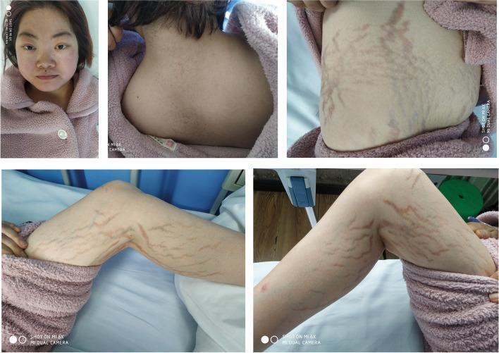
A composite of five clinical photographs illustrating the classic physical manifestations of Cushing's syndrome in a 26-year-old female. The top-left image shows a frontal view of the face, demonstrating a 'moon face' appearance characterized by a rounded facial contour and prominent, plethoric (flushed) cheeks. The top-center photograph displays the posterior cervical and upper thoracic region, showing a significant dorsocervical fat pad, commonly referred to as a 'buffalo hump.' The remaining three images (top-right, bottom-left, and bottom-right) focus on the patient's trunk and limbs, revealing wide, violaceous (purplish) striae distensae across the abdomen and thighs. These striae appear as deep, linear streaks indicating thinning of the dermis. Collectively, these visual findings are hallmark cutaneous and structural signs of chronic hypercortisolism, which in this clinical context was secondary to a left adrenal adenoma. The images serve as educational references for endocrinology and dermatology, highlighting the systemic physical changes associated with cortisol excess.
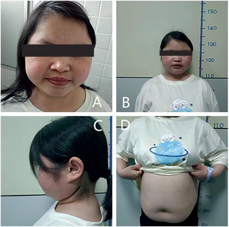
A multi-panel clinical photograph illustrating the classic physical manifestations of hypercortisolism (Cushing's syndrome). Image A provides a close-up of the face, demonstrating a 'moon face' appearance characterized by facial rounding and fullness of the cheeks, accompanied by multiple facial lentigines and mild acne. Image B shows the patient standing against a height scale, highlighting short stature relative to chronological age. Image C is a lateral view of the neck and upper back, showing a dorsocervical fat pad (buffalo hump). Image D displays the torso, revealing centripetal obesity with a protuberant, rounded abdomen; notably, this specific case lacks the characteristic wide, violaceous striae often seen in Cushing's syndrome, though the skin appears thin. This composite clinical image serves as an educational tool for identifying the systemic and dermatologic signs of primary pigmented nodular adrenocortical disease (PPNAD) or other causes of chronic glucocorticoid excess in pediatric or adolescent patients.
"Cushing syndrome"[MeSH Terms] AND diagnosis
Cushing Syndrome
Definition
Epidemiology
- Affects 2-5 per million people per year (endogenous forms)
- Peak age of onset: twenties and thirties
- Women affected 4x more frequently than men in non-iatrogenic cases
- Iatrogenic Cushing syndrome (exogenous steroids) is by far the most common form in the developed world - Quick Compendium of Clinical Pathology, 5th ed.
Pathophysiology - The HPA Axis
- Hypothalamic CRH (also oxytocin, vasopressin) stimulates anterior pituitary corticotrophs
- Pituitary ACTH (cleaved from POMC precursor) stimulates adrenal zona fasciculata to secrete up to 20 mg cortisol/day
- Cortisol feeds back negatively on both hypothalamus (suppresses CRH) and pituitary (suppresses ACTH)
- CRH follows a circadian rhythm - cortisol peaks in the morning, nadirs around 11 PM
Causes / Classification
1. Exogenous (Iatrogenic) - Most Common Overall
- Oral, topical, inhaled, or nasal corticosteroids
- Can occur with low doses; even topical absorption (especially in children) can cause the syndrome
- ACTH is suppressed, and both adrenal glands become atrophic
2. ACTH-Dependent (80-85% of endogenous cases)
| Cause | Proportion | Notes |
|---|---|---|
| Cushing disease (pituitary adenoma) | ~70% of endogenous | Corticotroph microadenoma (<1 cm) in ~95%; female predominance |
| Ectopic ACTH syndrome | ~10% of endogenous | Bronchial carcinoid, small cell lung ca, pancreatic NET, thymic tumors, medullary thyroid ca |
| Ectopic CRH syndrome | Rare | - |
3. ACTH-Independent (15% of endogenous cases)
- Adrenal adenoma or carcinoma (equal incidence in adults; carcinoma > adenoma in children)
- Bilateral adrenal hyperplasia:
- ACTH-independent macronodular adrenal hyperplasia (AIMAH)
- Primary pigmented nodular adrenocortical disease (PPNAD) - associated with Carney complex
- McCune-Albright syndrome, MEN type 1 (rare associations)
- Andrews' Diseases of the Skin; Campbell-Walsh-Wein Urology
Clinical Features
Fat Distribution (Central/Cushingoid Obesity)
- Moon face - wide, round face
- Buffalo hump - dorsocervical fat pad (liposuction can treat)
- Supraclavicular fat pads
- Central/truncal obesity with thin extremities (centripetal pattern)
- Protuberant abdomen
Skin Changes (Most Striking)
- Wide violaceous striae - abdomen, buttocks, back, breasts, upper arms, thighs (dermis atrophy)
- Skin fragility and thinning - "cigarette paper" wrinkling; easy bruising; ecchymoses
- Liddle sign - skin easily peels off when adhesive tape is removed
- Plethora - over cheeks, anterior neck, V of chest
- Hypertrichosis (including facial lanugo in women), scalp hair thinning
- Acne, skin dryness
- Susceptibility to dermatophyte and Pityrosporum (Malassezia) infections; opportunistic fungal infections
- Livedo reticularis, purpura, brownish pigmentation (occasionally)
Systemic Effects
| System | Manifestation |
|---|---|
| Metabolic | Hyperglycemia, glycosuria, frank DM (20% of patients) |
| Electrolyte | Hypokalemia |
| Cardiovascular | Hypertension, generalized arteriosclerosis |
| Musculoskeletal | Proximal muscle weakness, osteoporosis, kyphosis, back/limb pain |
| Reproductive | Loss of libido; menstrual irregularities |
| Protein catabolism | Muscle wasting, thin skin, poor wound healing |
| Psychiatric | Depression, cognitive changes |
- Andrews' Diseases of the Skin, p. 578; Quick Compendium of Clinical Pathology
Clinical Photos
Diagnosis
Step 1 - Confirm Hypercortisolism (Screening)
| Test | Details |
|---|---|
| 24-hour urinary free cortisol (UFC) | Repeated twice; >3-fold elevation strongly suggestive |
| Low-dose dexamethasone suppression test (LDDST) | 1 mg dexamethasone at 11 PM; cortisol >50 nmol/L at 8 AM = abnormal |
| Late-night salivary cortisol | Loss of circadian nadir; >550 ng/dL = probable Cushing |
Step 2 - Determine Source (Localization)
-
Measure plasma ACTH:
- Undetectable/suppressed ACTH → ACTH-independent (adrenal source) → CT adrenals
- Detectable/elevated ACTH → ACTH-dependent (pituitary or ectopic)
-
For ACTH-dependent disease:
- Pituitary MRI + high-dose dexamethasone suppression test (HDDST)
- If no mass identified: Bilateral inferior petrosal sinus sampling (BIPSS) - gold standard for differentiating pituitary vs. ectopic ACTH (petrosal:peripheral ACTH ratio >2 basal or >3 post-CRH = Cushing disease)
- If no petrosal gradient: CT chest/abdomen + somatostatin receptor scintigraphy (for ectopic source)
Diagnostic Algorithm
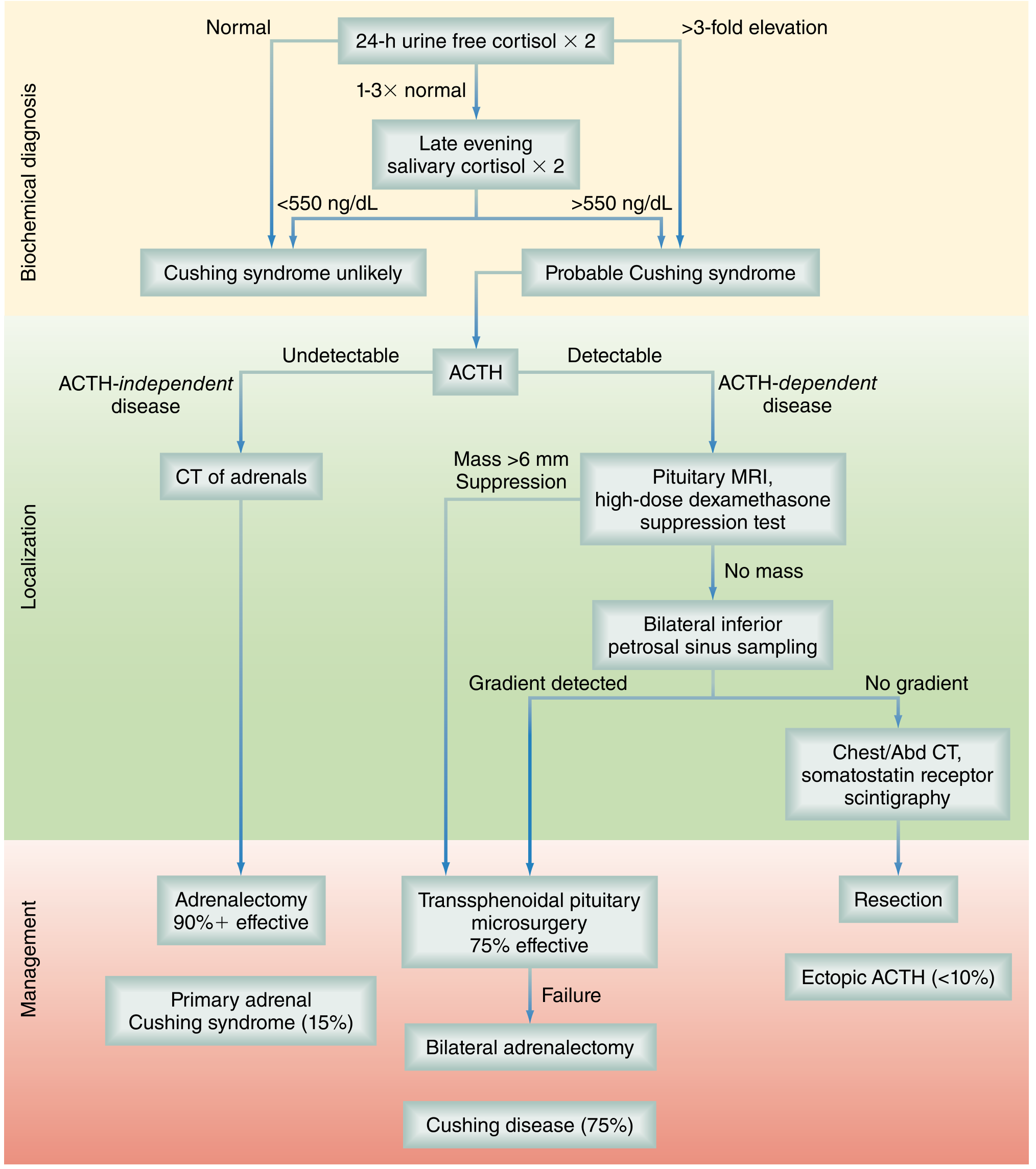
- Quick Compendium of Clinical Pathology, 5th ed.; Campbell-Walsh-Wein Urology; Sabiston Textbook of Surgery
Treatment
| Cause | Treatment | Efficacy |
|---|---|---|
| Cushing disease (pituitary adenoma) | Transsphenoidal pituitary microsurgery | ~75% remission |
| Surgical failure / recurrence | Bilateral adrenalectomy, repeat surgery, or radiation | - |
| Adrenal adenoma/carcinoma | Adrenalectomy (laparoscopic preferred) | 90%+ effective |
| Ectopic ACTH | Resect the primary tumor | Variable |
| Medical therapy (bridge to surgery, inoperable) | Ketoconazole, metyrapone, mitotane, pasireotide (Cushing disease), osilodrostat | Suppresses cortisol |
| Iatrogenic | Taper and discontinue exogenous steroids | - |
- Skin changes, hypertension, and metabolic derangements generally reverse with treatment
- Buffalo hump fat deposits may require liposuction even after cure
- Andrews' Diseases of the Skin; Lippincott Illustrated Reviews: Pharmacology; Smith and Tanagho's General Urology
Key Distinguishing Points
| Feature | Cushing Disease | Ectopic ACTH | Adrenal Cushing |
|---|---|---|---|
| ACTH | Elevated | Very elevated | Suppressed |
| HDDST | Suppresses (usually) | Does not suppress | Does not suppress |
| BIPSS gradient | Present | Absent | N/A |
| Skin pigmentation | Mild | Prominent (high ACTH → MSH) | Absent |
| Adrenal appearance | Bilateral hyperplasia | Bilateral hyperplasia | Unilateral mass |
Recent Evidence (2024-2026)
- A 2024 meta-analysis (PMID 38888685) addressed the diagnostic challenge of distinguishing true Cushing syndrome from non-neoplastic hypercortisolism (pseudo-Cushing) - an ongoing clinical conundrum.
- A 2026 meta-analysis (PMID 42289244) re-examined hypercortisolism screening thresholds in obese/diabetic patients, suggesting current screening strategies may need revision in these populations.