
Explain me 10 common heart diseases with ECG changes and ECG images of those diseases
Reading File
Now let me search the library and for ECG images in parallel:
Finding Sources
Finding Sources
Searching Images
ECG myocardial infarction ST elevation
Good. Now let me search for ECG images for the remaining diseases in parallel:
Searching Images
ECG atrial fibrillation irregular rhythm absent P waves
Searching Images
ECG complete heart block third degree AV block
Searching Images
ECG Wolff-Parkinson-White syndrome delta wave short PR
Searching Images
ECG hypertrophic cardiomyopathy left ventricular hypertrophy
Finding Sources
Finding Sources
Now let me search for ECG images for the remaining diseases:
Searching Images
ECG pericarditis saddle-shaped ST elevation diffuse leads
Searching Images
ECG ventricular tachycardia wide complex tachycardia
Searching Images
ECG pulmonary embolism S1Q3T3 right heart strain
Searching Images
ECG dilated cardiomyopathy left bundle branch block LBBB
Searching Images
ECG Brugada syndrome coved pattern right bundle branch block V1 V2
Excellent! I now have all the images and clinical data needed. Let me compile the comprehensive answer.Here is a comprehensive guide to 10 common heart diseases with their ECG changes and images:
10 Common Heart Diseases: ECG Changes & Images
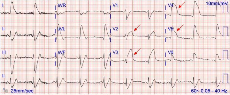
1. ST-Elevation Myocardial Infarction (STEMI)
Pathophysiology: Complete occlusion of a coronary artery causes transmural (full-thickness) myocardial ischemia and injury.
ECG Changes:
- Hyperacute T-waves — earliest sign; tall, peaked T-waves in the affected territory
- ST-segment elevation ≥1 mm in ≥2 contiguous limb leads, or ≥2 mm in ≥2 contiguous precordial leads (convex/tombstone morphology)
- Reciprocal ST depression in opposing leads (e.g., inferior STEMI → ST depression in I, aVL)
- Pathological Q-waves (>40 ms wide, >25% QRS amplitude) — develop over hours, indicate necrosis
- T-wave inversions — evolutionary change as infarction evolves
- Lead localization: Anterior (V1–V4, LAD), Inferior (II, III, aVF, RCA/LCx), Lateral (I, aVL, V5–V6)
2. Non-ST-Elevation Myocardial Infarction (NSTEMI) / Unstable Angina
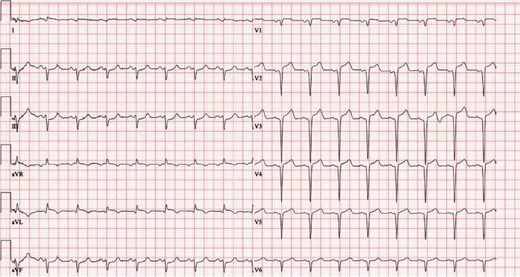
Pathophysiology: Partial occlusion of a coronary artery (or plaque rupture without complete occlusion) causing subendocardial ischemia. NSTEMI: elevated troponin; Unstable Angina: troponin normal.
ECG Changes:
- ST depression ≥0.5–1 mm in ≥2 contiguous leads (horizontal or downsloping)
- T-wave inversions — symmetric, deep inversions, especially in precordial leads
- Transient ST elevation (Prinzmetal/variant angina — vasospasm)
- ECG may be normal in up to 6% of NSTEMI cases — serial ECGs + troponins are essential
- No pathological Q-waves (no full-thickness necrosis)
3. Atrial Fibrillation (AF)
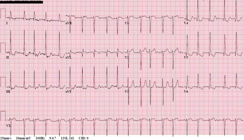
Pathophysiology: Chaotic, disorganized electrical activity in the atria from multiple re-entrant wavelets. The AV node conducts impulses irregularly to the ventricles.
ECG Changes:
- Absent P-waves — replaced by irregular, fibrillatory baseline (best seen in V1)
- Irregularly irregular R-R intervals — hallmark finding
- Narrow QRS complexes (unless aberrant conduction or bundle branch block)
- Rapid ventricular response — rate >100 bpm (uncontrolled AF)
- Fibrillatory baseline — fine undulations at 350–600 cycles/min
4. Complete (Third-Degree) AV Block
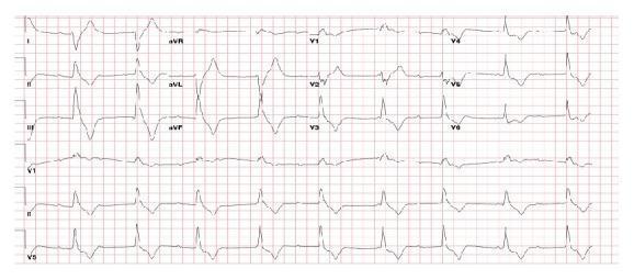
Pathophysiology: Complete failure of conduction through the AV node or His-Purkinje system. Atria and ventricles beat independently. An escape pacemaker (junctional or ventricular) maintains the heartbeat.
ECG Changes:
- Complete AV dissociation — P-waves and QRS complexes march independently with no fixed PR interval
- Regular P-waves at a faster atrial rate (e.g., 70–100 bpm)
- Slow escape rhythm — junctional escape: narrow QRS at 40–60 bpm; ventricular escape: wide QRS at 20–40 bpm
- P-waves "marching through" QRS complexes and T-waves
- Wide QRS if the escape focus is infra-Hisian (ventricular origin)
5. Wolff-Parkinson-White (WPW) Syndrome
Pathophysiology: An accessory pathway (Bundle of Kent) bypasses the AV node, causing ventricular pre-excitation. This predisposes to supraventricular tachycardias and, if AF occurs with the accessory pathway, life-threatening rapid ventricular rates.
ECG Changes (Classic Triad during sinus rhythm):
- Short PR interval < 120 ms (accessory pathway bypasses AV nodal delay)
- Delta wave — slurred, slow upstroke at the onset of QRS (initial pre-excitation of ventricle)
- Widened QRS complex > 120 ms (fusion of pre-excited + normal conduction)
- Secondary ST-T changes — discordant to QRS in pre-excited leads
- "Pseudo-infarction" Q-waves — negative delta waves may mimic Q-waves (e.g., inferior leads if left posteroseptal pathway)
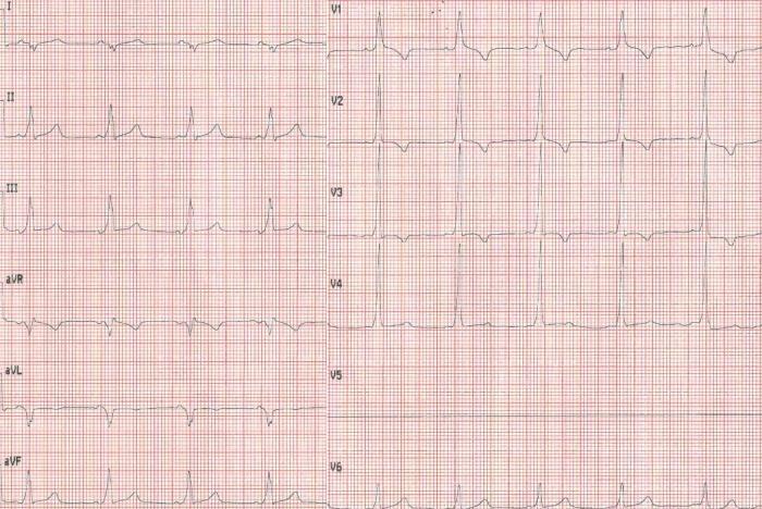
6. Hypertrophic Cardiomyopathy (HCM)
Pathophysiology: Genetic disorder (sarcomere mutations) causing asymmetric LV hypertrophy, diastolic dysfunction, dynamic outflow obstruction, and myofiber disarray — a major cause of sudden cardiac death in young athletes.
ECG Changes:
- Left ventricular hypertrophy (LVH) voltage criteria — Sokolow-Lyon (S in V1 + R in V5/V6 ≥35 mm)
- Giant T-wave inversions — especially in V3–V5 (apical HCM/Yamaguchi variant: deeply inverted "giant" T-waves)
- ST-segment depression (strain pattern) in lateral leads
- Absent septal Q-waves in I, aVL, V5, V6 (septal hypertrophy disrupts normal septal activation)
- Deep Q-waves in inferior and lateral leads (from abnormal septal depolarization)
- Left axis deviation, left atrial enlargement
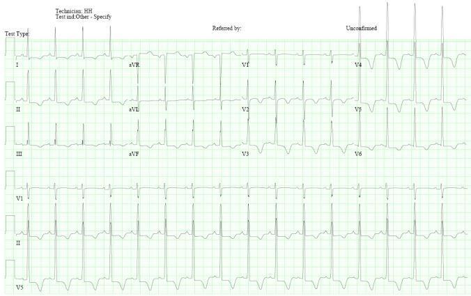
7. Acute Pericarditis
Pathophysiology: Inflammation of the pericardium (viral, autoimmune, post-MI, uremic) causing diffuse superficial myocardial irritation — hence diffuse (not territory-specific) ECG changes.
ECG Changes (4 classic stages):
- Stage 1 (acute): Diffuse concave ("saddle-shaped") ST elevation in most leads (I, II, III, aVF, V2–V6); ST depression in aVR (reciprocal); PR-segment depression (most specific sign) in II, V4–V6 with PR elevation in aVR; Spodick's sign (downsloping TP segment)
- Stage 2: ST normalizes, T-waves flatten
- Stage 3: Diffuse T-wave inversions
- Stage 4: ECG returns to normal
- Key difference from STEMI: Diffuse (not territory-specific), concave morphology, PR depression, no reciprocal changes (except aVR)
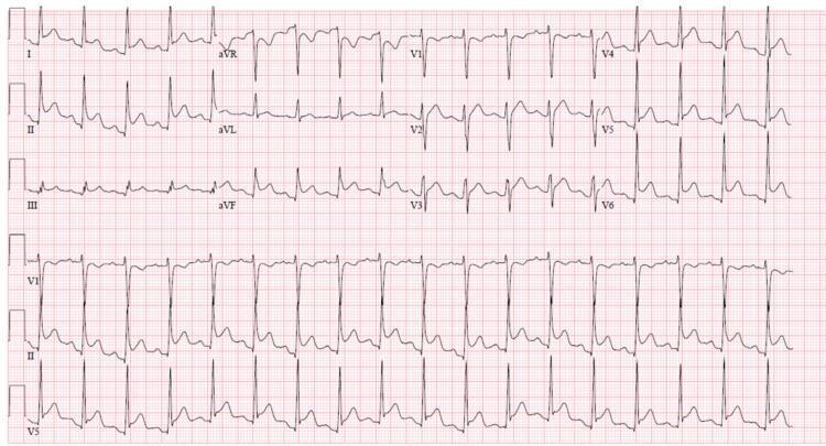
8. Pulmonary Embolism (PE)
Pathophysiology: Massive PE causes acute right heart strain — the RV dilates under elevated pulmonary vascular resistance, shifting the heart's electrical axis rightward and causing characteristic ECG changes.
ECG Changes:
- Sinus tachycardia — most common finding (present in >40%)
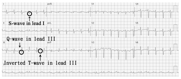
- S1Q3T3 pattern — deep S-wave in lead I, Q-wave in lead III, inverted T-wave in lead III (classic but only in ~20%)
- Right axis deviation (shift toward +90° to +180°)
- Incomplete or complete RBBB — V1 rSR' pattern
- T-wave inversions in V1–V4 (right ventricular strain pattern)
- P pulmonale — peaked P-waves in II, III, aVF (right atrial enlargement)
- Sinus bradycardia or AF in severe PE
9. Dilated Cardiomyopathy (DCM)
Pathophysiology: Systolic dysfunction with LV (and often biventricular) dilation and reduced ejection fraction. Causes include ischemia, alcohol, viral myocarditis, idiopathic, or genetic. Conduction abnormalities are common from fibrosis.
ECG Changes:
- Left Bundle Branch Block (LBBB) — broad notched R in I, aVL, V5–V6; deep S in V1–V3; QRS >120 ms — seen in ~25–30%; indicates dyssynchrony
- Non-specific intraventricular conduction delay (IVCD)
- Poor R-wave progression (V1–V4)
- Prolonged PR interval (first-degree AV block) from fibrotic conduction system
- Left axis deviation
- Low voltage QRS if extensive fibrosis or pericardial effusion
- Atrial fibrillation — common complication
- Frequent PVCs or non-sustained VT
- LVH voltage criteria (early stages)
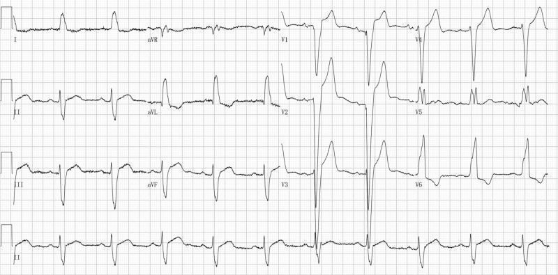
10. Brugada Syndrome
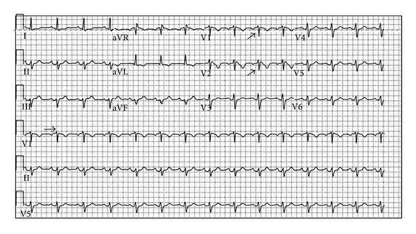
Pathophysiology: Inherited sodium channelopathy (SCN5A mutations, ~20% of cases) causing abnormal depolarization in the right ventricular outflow tract (RVOT). Predisposes to polymorphic VT/VF and sudden cardiac death, especially during fever or at rest/sleep.
ECG Changes:
- Type 1 (Diagnostic — "coved" pattern):
- ST elevation ≥2 mm in V1–V2 (or V1–V3)
- Coved morphology: high-takeoff ST slope descending to inverted T-wave, no isoelectric segment
- Pseudo-RBBB (rSR' in V1)
- Spontaneous or unmasked by Na-channel blockers (ajmaline, flecainide, procainamide)
- Type 2 ("saddle-back"): ≥2 mm J-wave elevation, saddleback ST, positive T-wave — not diagnostic alone
- QRS fragmentation — marker of fibrosis
- PR prolongation (conduction slowing)
- Can be intermittent or concealed (fever unmasks it)
Quick Reference Summary Table
| Disease | Key ECG Features |
|---|---|
| STEMI | ST elevation (convex), Q-waves, reciprocal ST depression, hyperacute T-waves |
| NSTEMI/UA | ST depression, T-wave inversions, may be normal |
| Atrial Fibrillation | No P-waves, irregularly irregular R-R, fibrillatory baseline |
| 3rd-Degree AV Block | AV dissociation, slow escape rhythm, P-waves ≠ QRS |
| WPW Syndrome | Short PR, delta wave, wide QRS, secondary ST-T changes |
| Hypertrophic CM | LVH voltage, giant T-wave inversions, absent septal Q-waves |
| Acute Pericarditis | Diffuse concave ST elevation, PR depression, saddle-shaped |
| Pulmonary Embolism | Sinus tachycardia, S1Q3T3, RBBB, RV strain T inversions V1–V4 |
| Dilated CM | LBBB, poor R progression, AF, prolonged PR, low voltage |
| Brugada Syndrome | Coved ST elevation V1–V2, pseudo-RBBB, inverted T-waves |
Clinical Pearl: The ECG is a snapshot in time — serial ECGs, clinical context, and biomarkers (troponins, BNP) are always needed alongside ECG interpretation for accurate diagnosis.
Sources: Harrison's Principles of Internal Medicine 22E; Tintinalli's Emergency Medicine; Braunwald's Heart Disease; Rosen's Emergency Medicine; Goldman-Cecil MedicineHere's a summary of what was covered — all 10 diseases with ECG images sourced from peer-reviewed medical literature:
- STEMI — Convex ST elevation, tombstone morphology, reciprocal changes, Q-waves
- NSTEMI/Unstable Angina — ST depression, T-wave inversions (subendocardial pattern)
- Atrial Fibrillation — Absent P-waves, irregularly irregular rhythm, fibrillatory baseline
- Complete (3rd-Degree) AV Block — Full AV dissociation, slow escape rhythm independent of P-waves
- WPW Syndrome — Short PR, delta wave, widened QRS (pre-excitation triad)
- Hypertrophic Cardiomyopathy — LVH voltage, giant T-wave inversions (especially apical variant), absent septal Q-waves
- Acute Pericarditis — Diffuse saddle-shaped ST elevation, PR depression in II/V4–V6, PR elevation in aVR
- Pulmonary Embolism — Sinus tachycardia, S1Q3T3, RBBB, right precordial T-wave inversions
- Dilated Cardiomyopathy — LBBB, poor R-wave progression, AF, prolonged PR
- Brugada Syndrome — Coved ST elevation ≥2 mm in V1–V2 with inverted T-wave, pseudo-RBBB
This is a shared conversation. Sign in to Orris to start your own chat.