
Fluorescence In Situ Hybridization (FISH): Principles, types of probes, and its specific utility in detecting microdeletions or oncogenic translocations (e.g., BCR-ABL1). Refer to Robins and cotran
Reading File
Now let me search the Robbins and Cotran textbook for FISH content in parallel:
Finding Sources
Finding Sources
Found the Robbins & Cotran entry. Let me read it along with related sections:
Reading File
Finding Sources
Good. Now let me search for BCR-ABL and microdeletions in Robbins, and also look for the FISH figures referenced in the text:
Finding Sources
Finding Sources
Finding Sources
Finding Sources
Let me look at the figures mentioned in the Robbins text (Fig 5.21 and 5.23) and also search Henry's for FISH probe types:
Finding Sources
Reading File
Excellent content. Let me also read a bit more of the Henry's section and look for BCR-ABL FISH in Robbins specifically:
Reading File
Finding Sources
Now let me fetch key images and read the BCR-ABL1 section from Henry's:
Loading Image
Reading File
I now have comprehensive content from Robbins & Cotran and supporting sources. Let me compile the full answer.
Fluorescence In Situ Hybridization (FISH)
Primary source: Robbins, Cotran & Kumar Pathologic Basis of Disease (10th ed.), Chapter 5, p. 178
1. Principle of FISH
FISH uses labeled DNA probes that correspond to and bind to particular chromosome intervals. The workflow is:
- Probe preparation: Large fragments of cloned genomic DNA - spanning up to 200 kb - are labeled with fluorescent dyes.
- Denaturation: The probe and the sample (metaphase chromosome preparation or interphase nuclei) are both "melted" to yield single-stranded DNA.
- Hybridization: The labeled single-stranded probe is applied to the denatured sample and anneals to its homologous genomic sequence under conditions that favor specific base pairing.
- Visualization: After washing away excess probe, the hybridized region is visualized under a fluorescence microscope, where the fluorescent signal marks the exact chromosomal locus.
FISH can be performed on:
- Prenatal samples (amniotic fluid, chorionic villi)
- Peripheral blood cells
- Touch preparations from cancer biopsies
- Fixed archival (formalin-fixed, paraffin-embedded) tissue sections
Robbins & Cotran, p. 178
2. Types of FISH Probes
(Henry's Clinical Diagnosis and Management by Laboratory Methods, 23rd ed., Chapter 71)
There are three basic probe categories:
A. Chromosome Painting Probes
- A cocktail of many unique DNA fragments spanning the entire length of a chromosome.
- After hybridization, the entire chromosome fluoresces as one color.
- Used to identify structural rearrangements (translocations, insertions) between chromosomes.
B. Repeat Sequence (Centromeric / Enumeration) Probes
- Hybridize to pericentromeric repetitive sequences specific to one chromosome.
- Used for chromosome enumeration - detecting gain or loss (aneuploidy).
- Examples: chromosome-specific pericentromeric probes (chromosomes 13, 18, 21, X, Y in prenatal aneuploidy screening).
C. Unique Sequence (Locus-Specific) Probes
This is the most clinically informative category, and includes three subtypes:
| Subtype | Design | Use |
|---|---|---|
| Locus-specific | Probes flanking a single gene locus | Detects deletions, amplification |
| Subtelomere probes | Target subtelomeric regions | Detects cryptic subtelomeric deletions causing unexplained intellectual disability |
| Break-apart probes | Two probes flanking a translocation breakpoint in different colors | Detects rearrangements with unknown/variable partners (e.g., MYC, ALK, MLL) |
| Dual-fusion (D-FISH) | Probes for each gene in a specific translocation, in two colors | Detects specific fusion genes (e.g., BCR-ABL1); fusion appears as a co-localized yellow signal |
3. Clinical Utility: Microdeletion Detection
One of the most important uses of FISH is detecting microdeletions too small to be seen by conventional karyotyping - Robbins & Cotran explicitly identifies this as a core application.
How it works:
- A locus-specific probe is designed complementary to the critical deleted region.
- In a normal cell: two signals are seen (one per allele).
- In a deletion carrier: only one signal is seen - the other allele's signal is absent.
Classic examples:
| Syndrome | Deletion | FISH finding |
|---|---|---|
| DiGeorge / Velocardiofacial syndrome | 22q11.2 | One red locus signal lost on deleted chromosome 22; control (green) persists |
| Prader-Willi / Angelman syndrome | 15q11-q13 | Loss of one locus signal |
| Williams syndrome | 7q11.23 (elastin gene) | Loss of one signal |
| Cri-du-chat | 5p15 | Loss of one signal |
The image below (Henry's Fig. 71.8) shows the FISH pattern for 22q11.2 microdeletion (velocardiofacial syndrome):
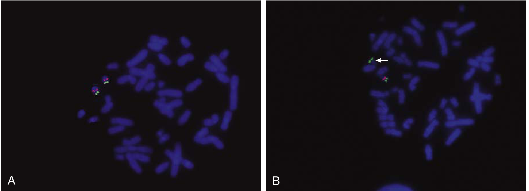
Fig. 71.8 from Henry's: FISH detection of a microdeletion of chromosome 22 (velocardiofacial syndrome). A = normal cell (one red locus + one green control signal per chromosome 22). B = patient cell - the arrow points to a chromosome 22 bearing only the green control signal, confirming hemizygous deletion of the locus.
4. Clinical Utility: Oncogenic Translocations - BCR-ABL1
Robbins & Cotran gives the acute promyelocytic leukemia (APL) translocation as a direct clinical example of FISH-guided therapy: FISH is used to confirm the PML-RARA translocation before initiating retinoic acid (ATRA) therapy, which only works when this specific rearrangement is present.
For BCR-ABL1 (the Philadelphia chromosome, t(9;22)(q34;q11.2)) - the paradigm oncogenic translocation - FISH has distinct advantages over conventional cytogenetics:
Why FISH outperforms standard karyotyping for BCR-ABL1:
- ~5% of CML patients are "Ph-chromosome negative" on cytogenetics, yet harbor the BCR-ABL1 fusion at the molecular level (detectable only by FISH or RT-PCR).
- FISH can be performed on interphase cells - no need to culture cells or obtain dividing metaphase cells.
- FISH works on fixed tissue and archival samples.
Probe strategy - Dual Fusion FISH (D-FISH):
- A red probe labels the ABL1 region on chromosome 9.
- A green probe labels the BCR region on chromosome 22.
- In a normal cell: 2 red + 2 green signals, spatially separated.
- In a CML cell: 1 red + 1 green (residual normal alleles) + 2 yellow fusion signals (co-localization of red and green = BCR-ABL1 and the reciprocal ABL1-BCR on the der(9)).
Break-apart probes are used alternatively when the translocation partner is variable (e.g., MYC rearrangements in lymphoma): the two flanking probes separate when a rearrangement occurs, giving distinct red and green signals instead of the normal fused yellow signal.
5. Other FISH Applications (Robbins, p. 178)
- Aneuploidy detection: e.g., trisomy 21, 18, 13 in rapid prenatal diagnosis
- Gene amplification: e.g., HER2/ERBB2 amplification in breast cancer (more than 6 copies of HER2 signal)
- Variable-partner rearrangements: MYC break-apart probes detect rearrangements even when the fusion partner is unknown
- Rapid diagnosis scenarios: When waiting for karyotype is clinically unacceptable (e.g., suspected APL - start ATRA within hours)
6. Comparison with Array CGH (context from Robbins)
Robbins explicitly notes that FISH requires prior knowledge of the suspected chromosomal region - you must already suspect a locus to probe for it. When no specific region is suspected, array CGH (comparative genomic hybridization) or SNP arrays are preferred for genome-wide copy number analysis. FISH thus remains the method of choice when a specific known locus is suspected clinically.
Summary table:
| Application | Probe type | Signal interpretation |
|---|---|---|
| Aneuploidy (trisomy/monosomy) | Centromeric/enumeration | Count of discrete spots |
| Microdeletion (e.g., 22q11) | Locus-specific | Loss of one signal = deletion |
| Specific translocation (BCR-ABL1) | Dual-fusion | Red + green → yellow fusion dots |
| Unknown-partner rearrangement (MYC) | Break-apart | Separation of fused signals |
| Gene amplification (HER2) | Locus-specific | Numerous red dots per nucleus |
| Chromosome ID / painting | Whole-chromosome paint | Entire chromosome fluoresces |
Robbins, Cotran & Kumar Pathologic Basis of Disease, p. 178 (Chapter 5: Molecular Basis of Disease); Henry's Clinical Diagnosis and Management by Laboratory Methods, Chapter 71
Please teach me in simple language
FISH - Explained Simply 🔬
Let me teach you FISH like you're hearing it for the first time.
The Big Picture: What Problem Does FISH Solve?
Imagine you have a huge library with 46 books (your chromosomes). Something is wrong — maybe a page is missing from one book, or two books got their chapters swapped. You want to find exactly which page is missing or swapped.
A regular microscope (karyotyping) can only show you the rough shape of the books. It cannot spot a missing single page.
FISH is a molecular GPS that lights up a specific page so you can see if it's there or not.
How Does It Work? (Step by Step)
Step 1 - Make a "Glow Stick" Probe
Scientists take a small piece of DNA that matches exactly the region they're looking for. They attach a fluorescent dye to it - think of painting it with glow-in-the-dark paint.
This glowing piece of DNA is called the probe.
Step 2 - "Unzip" the DNA
Your chromosomes are double-stranded (like a zipper). You heat the sample to pull the zipper apart - this is called denaturation. Now both the probe and the chromosome DNA are single-stranded and "sticky."
Step 3 - Let the Probe Find Its Match
You pour the glowing probe onto the chromosome preparation. Like a puzzle piece, it snaps onto its exact matching location on the chromosome. This is called hybridization (hence the name - in situ = in place, hybridization = joining together).
Step 4 - Look Under the Fluorescence Microscope
Wash away any unattached probe. Turn on the special light. Wherever the probe stuck → you see a glowing dot.
Reading the Result - Think of It Like This:
You have 2 copies of every chromosome (one from mom, one from dad). So normally you expect to see 2 glowing dots per cell.
Normal cell:    🟢 🟢   → 2 dots = both copies present ✅

Deletion:       🟢 ⬛   → 1 dot = one copy MISSING ❌

Translocation:  🔴🟢    → colors merge = two genes fused together ⚠️

Amplification:  🟢🟢🟢🟢🟢 → too many dots = gene copied many times ⚠️
Types of Probes (Simple Version)
Think of probes as different types of flashlights:
1. Chromosome Paint Probe 🎨
- Lights up an entire chromosome in one color
- Like painting one whole book bright blue
- Used when: you want to see if a piece of one chromosome got stuck onto another
2. Centromere (Counting) Probe 🔢
- Lights up the center dot of a specific chromosome
- Like putting a sticky note on the spine of each book
- Used when: you want to count how many copies of a chromosome exist
- Example: Is there an extra chromosome 21 (Down syndrome)?
3. Locus-Specific Probe 🎯
- Lights up one very specific gene region
- Like highlighting one paragraph on one page
- Used when: you suspect a specific gene is deleted or amplified
- Example: Is the HER2 gene amplified in this breast cancer?
4. Break-Apart Probe ✂️
- Two probes in two different colors placed on either side of a gene
- Normally they sit close together → you see a merged yellow dot
- If the gene breaks (translocation) → they separate → you see red and green apart
- Used when: you want to detect a rearrangement but don't know the partner
5. Dual Fusion Probe 🤝
- Red probe on Gene A (on one chromosome), green probe on Gene B (on another chromosome)
- Normally → 2 red + 2 green dots, all separate
- After translocation → red and green come together → yellow fusion dot appears
- Used for: BCR-ABL1 in CML
Clinical Use 1 - Microdeletions
A microdeletion is a tiny piece of chromosome that's gone missing - too small to see even with a good microscope during karyotyping.
Example: DiGeorge Syndrome (22q11.2 deletion)
A tiny piece of chromosome 22 is deleted. The child gets heart defects, immune problems, and learning difficulties.
Without FISH? Karyotype looks completely normal. You'd miss it.
With FISH? You use a locus-specific probe for the 22q11.2 region:
- Normal parent: 2 dots (both copies of chromosome 22 are there) ✅
- Affected child: 1 dot (one chromosome 22 is missing that segment) ❌
It's like placing a GPS tracker on a specific page of a book - if that page is torn out, the tracker signal disappears.
Clinical Use 2 - BCR-ABL1 Translocation (CML)
This is the most famous oncogenic translocation. In Chronic Myeloid Leukemia (CML):
- A piece of chromosome 9 (carrying the ABL1 gene) breaks off
- It sticks onto chromosome 22 (which carries BCR)
- This creates the Philadelphia chromosome with the BCR-ABL1 fusion gene
- BCR-ABL1 makes an always-on tyrosine kinase → uncontrolled cell division → leukemia
How FISH detects it (Dual Fusion method):
Normal:                    After translocation:
Chrom 9 → 🔴 🔴           🔴🟢 (fusion on der22 = Ph chromosome)
Chrom 22 → 🟢 🟢           🟢🔴 (reciprocal on der9)
                            🔴   (residual normal chr 9)
                            🟢   (residual normal chr 22)
Result: 2R + 2G            Result: 2 fusion yellows + 1R + 1G
Why FISH matters here over regular karyotype:
- ~5% of CML patients have a "hidden" Philadelphia chromosome - karyotype looks normal but FISH/PCR catches the fusion
- FISH works on interphase cells - no need to grow cells in culture
- Works on fixed tissue and old archived samples
Clinical urgency example from Robbins:
In suspected Acute Promyelocytic Leukemia (APL), you need to confirm the PML-RARA translocation before starting treatment with retinoic acid (ATRA). This treatment only works with this specific translocation. FISH gives you the answer within hours - you don't wait days for a full karyotype.
Quick Summary Table
| What you want to know | Probe used | What you see |
|---|---|---|
| Is there an extra chromosome? | Centromere probe | 3 dots instead of 2 |
| Is a gene segment deleted? | Locus-specific probe | 1 dot instead of 2 |
| Is a gene rearranged (unknown partner)? | Break-apart probe | Red and green separate |
| Is a specific fusion present (BCR-ABL1)? | Dual fusion probe | Yellow merged dots |
| Is a gene over-amplified (HER2)? | Locus-specific probe | 6+ dots per cell |
One Sentence to Remember FISH:
FISH is a glow-in-the-dark GPS for your DNA - you design a probe for exactly what you're looking for, and it either lights up (present) or stays dark (deleted/absent).
This is a shared conversation. Sign in to Orris to start your own chat.