
Action potentials of CNS
Reading File
Now let me search the medical library for action potentials in the CNS.
Finding Sources
Finding Sources
The best sources are Neuroscience: Exploring the Brain (dedicated CNS section) and Guyton & Hall. Let me read them in parallel.
Reading File
Reading File
Now let me read more detailed content from Neuroscience: Exploring the Brain for the actual AP mechanics and CNS-specific content, and also fetch the action potential diagram image.
Reading File
Loading Image
Excellent content. Let me get the next section covering voltage-gated channels, conduction, and CNS-specific firing properties.
Reading File
I now have very rich content. Let me fetch key images and also get the conduction/myelination section content.
Reading File
I now have comprehensive content. Let me fetch one more key image showing the sodium channel states and then compile the full answer.
Loading Image
Reading File
I now have comprehensive content to write a thorough answer. Let me compile it.
Action Potentials of the CNS
What Is an Action Potential?
An action potential (AP) is a rapid, transient reversal of the membrane potential that propagates along a neuronal axon to convey information. It is also called a spike, nerve impulse, or discharge. At rest, the inside of the neuronal membrane is negatively charged relative to the outside (resting membrane potential: approximately -70 mV). An AP temporarily makes the inside positive, then quickly restores the negative resting state.
The number, frequency, and timing of action potentials constitute the code neurons use to transfer information from one location to another.
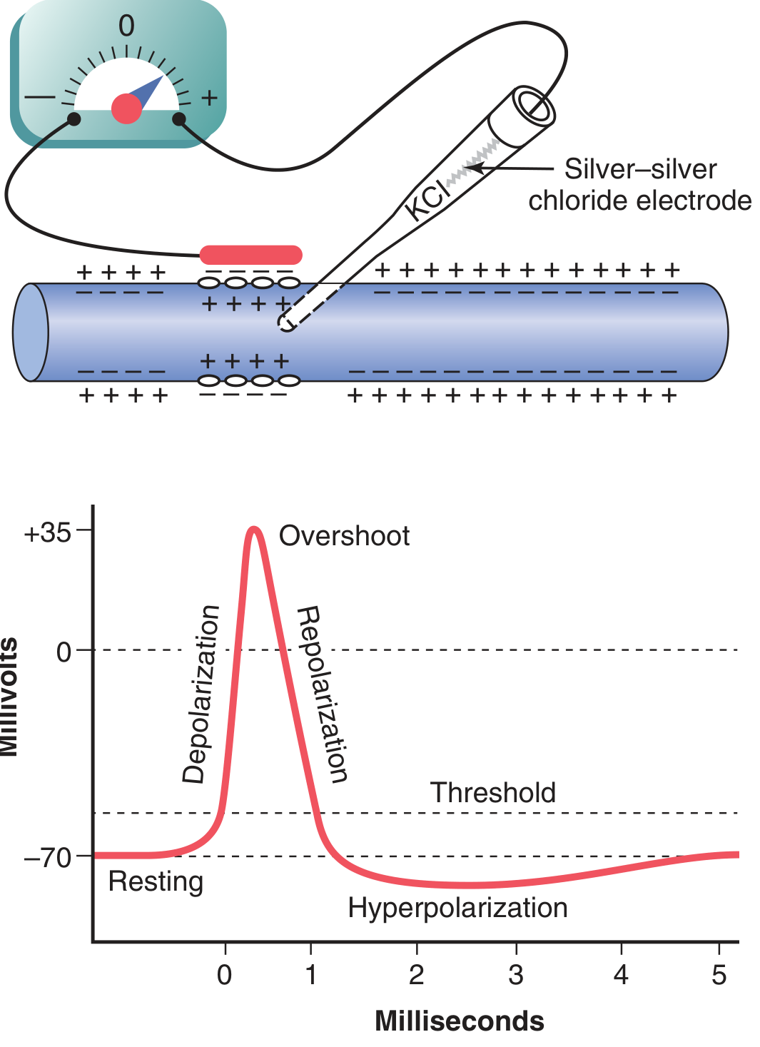
Phases of the Action Potential
1. Resting Stage (-70 mV)
The membrane is polarized - the inside is negative relative to outside. Voltage-gated Na+ and K+ channels are closed. The resting potential is maintained by the Na+/K+ ATPase pump and selective membrane permeability.
2. Depolarization (Rising Phase)
- A stimulus depolarizes the membrane toward threshold (around -55 mV).
- Once threshold is crossed, voltage-gated Na+ channels open rapidly.
- Na+ rushes into the cell (high extracellular [Na+] + electrical gradient both drive it inward).
- Membrane potential shoots from -70 mV up to approximately +35 mV - this positive overshoot is called the overshoot or spike peak.
- In some smaller fibers and many CNS neurons, the potential only approaches zero without fully overshooting to a positive value.
3. Repolarization (Falling Phase)
- Within less than 1 msec, Na+ channels begin to inactivate (close spontaneously).
- Voltage-gated K+ channels open (delayed ~1 msec after depolarization - the "delayed rectifier").
- K+ rushes out of the cell, rapidly restoring the negative membrane potential.
4. Hyperpolarization (Undershoot / After-Hyperpolarization)
- K+ channels stay open slightly longer than needed - K+ continues flowing out.
- Membrane potential dips below resting potential (to around -75 to -80 mV).
- Once K+ channels close, the potential returns to -70 mV.
Voltage-Gated Sodium Channels - The Key Players
The Na+ channel is a large transmembrane protein with four conformational states:
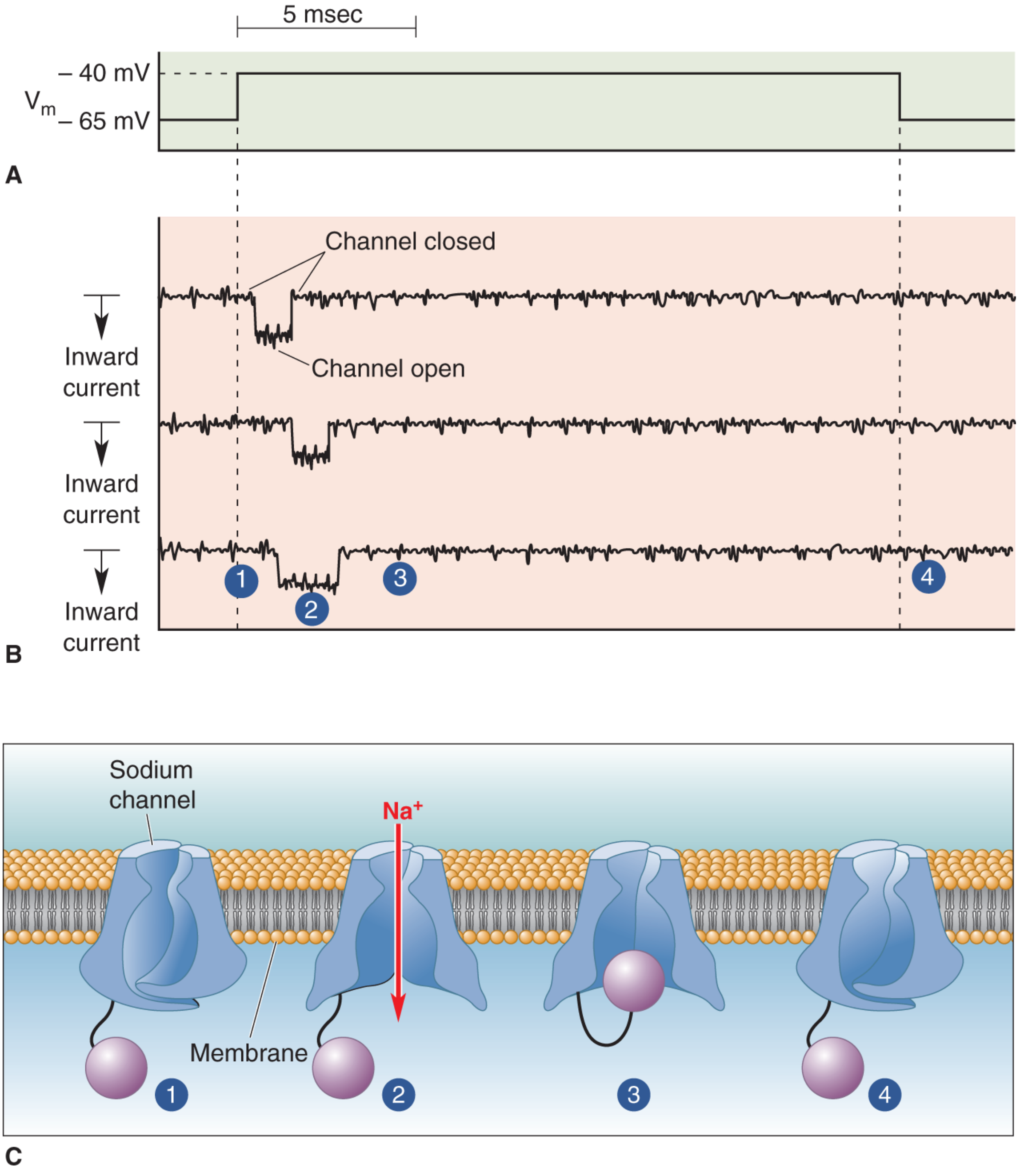
| State | Condition | Na+ Flow |
|---|---|---|
| 1 - Closed (resting) | At rest (~-70 mV) | None |
| 2 - Open (activated) | Depolarization to threshold | Inward rush |
| 3 - Inactivated | Sustained depolarization | None (blocked) |
| 4 - Closed (recovering) | After repolarization | None |
Key properties of voltage-gated Na+ channels (from patch-clamp studies):
- They open with a brief delay after depolarization.
- They stay open for only about 1 msec then spontaneously inactivate.
- They cannot be reopened until the membrane repolarizes to near resting potential - this is the basis of the refractory period.
Voltage-Gated K+ Channels
- ~40 different types exist - the largest ion channel family in the human genome.
- Open more slowly than Na+ channels (delayed rectifier) - ~1 msec lag.
- Four separate polypeptide subunits form the pore.
- Their diversity allows different CNS neuron types to tune their intrinsic excitability for specific functional needs.
The ionic current equation (Ohm's law applied to channels):
I_ion = g_ion (V_m - E_ion)
Where g_ion is the membrane conductance and (V_m - E_ion) is the driving force.
All-or-None Law
Once the membrane reaches threshold, the AP fires with full amplitude regardless of stimulus strength. A stronger stimulus cannot produce a larger AP in a single axon - it can only increase the frequency of APs (rate coding). This is the all-or-none principle.
Refractory Periods
| Period | Mechanism | Significance |
|---|---|---|
| Absolute Refractory Period | Na+ channels are inactivated; cannot open again | No AP can fire, no matter how strong the stimulus |
| Relative Refractory Period | K+ channels still open; membrane hyperpolarized | AP can fire but requires a stronger-than-normal stimulus |
A typical mammalian CNS neuron can fire up to about 500 Hz; once an AP is initiated, it is impossible to trigger another during the absolute refractory period.
Action Potential Conduction
Unmyelinated Axons
- Ionic currents from the active region (where the AP is occurring) spread passively to adjacent membrane, depolarizing it to threshold.
- AP propagates continuously along the entire membrane surface.
- This is slow and energetically costly.
- Conduction velocity depends on axon diameter - larger diameter = faster conduction.
Myelinated Axons (Saltatory Conduction)
- Myelin sheaths (from oligodendrocytes in CNS, Schwann cells in PNS) insulate the axon.
- Voltage-gated Na+ channels are concentrated only at nodes of Ranvier (gaps in myelin).
- The AP "jumps" from node to node - this is saltatory conduction (from Latin saltare = to jump).
- Much faster and more energy efficient than unmyelinated conduction.
- Demyelination (e.g., multiple sclerosis) disrupts this and slows or blocks conduction.
CNS-Specific Features
Initiation Zone
In CNS neurons, the AP is typically initiated at the axon hillock (initial segment), where the density of voltage-gated Na+ channels is highest and the threshold is lowest.
Integration
CNS neurons receive thousands of synaptic inputs simultaneously. EPSPs and IPSPs summate (temporally and spatially) at the axon hillock. If the net depolarization reaches threshold, an AP fires.
Firing Patterns
Different CNS neuron types display characteristic firing patterns based on their ion channel composition:
- Tonic/regular spiking - e.g., pyramidal cortical neurons
- Bursting - clusters of rapid APs followed by silence
- Fast-spiking - interneurons firing at very high frequencies (up to 500 Hz) with little adaptation
- Adapting - firing frequency decreases over time despite sustained stimulus
Role of Ca2+ Channels
In addition to Na+ and K+ channels, CNS neurons express voltage-gated Ca2+ channels. Ca2+ entry during an AP triggers neurotransmitter release at presynaptic terminals and activates Ca2+-dependent K+ channels that contribute to after-hyperpolarization.
Pharmacology - Toxins and Drugs Acting on AP Channels
| Agent | Target | Effect |
|---|---|---|
| Tetrodotoxin (TTX) | Voltage-gated Na+ channel (external) | Blocks Na+ influx; eliminates AP |
| Local anesthetics (lidocaine, procaine) | Na+ channel pore (internal) | Use-dependent block; preferentially blocks small pain fibers |
| Scorpion/sea anemone toxins | Na+ channel inactivation gate | Prolongs Na+ current; sustained depolarization |
| 4-Aminopyridine (4-AP) | K+ channels | Blocks K+ conductance; prolongs AP |
Local anesthetics are use-dependent - neurons that fire more rapidly are blocked more effectively because the anesthetic enters the open channel pore more frequently.
Summary
| Phase | Voltage | Ion Movement | Channel State |
|---|---|---|---|
| Resting | -70 mV | Minimal leak | All closed |
| Depolarization | -70 to +35 mV | Na+ in | Na+ channels open |
| Repolarization | +35 to -70 mV | K+ out, Na+ stops | Na+ inactivates, K+ opens |
| Hyperpolarization | -70 to -80 mV | K+ still out | K+ slowly closes |
| Recovery | -80 to -70 mV | Na+/K+ pump restores | Return to resting |
Sources: Neuroscience: Exploring the Brain, 5th Ed.; Guyton and Hall Textbook of Medical Physiology, 14th Ed.
This is a shared conversation. Sign in to Orris to start your own chat.