attachment
attachment

Identify the pathology specimen

analyse_media
Reading File
Searching Images

cauliflower-like papillary growth gross specimen kidney renal pelvis carcinoma

Searching Images

papillary serous cystadenocarcinoma ovary gross specimen cauliflower excrescences

Finding Sources
query_images_db
Searching Images

Wilms tumor gross specimen cut section kidney
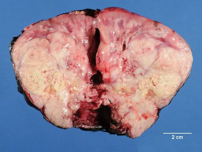
Renal mass, gross pathology photograph of a resected pediatric kidney with an intraparenchymal neoplasm. Modality: Gross pathology; Coronal/transverse cut surface. The kidney shows a large, soft, pale tan to pink mass replacing most of the renal parenchyma with heterogeneous appearance due to hemorrhagic and necrotic foci. The lesion is friable and poorly circumscribed relative to the surrounding cortex, with indistinct margins and occasional satellite nodules evident on the cut surface. The capsule may be variably thinned or breached at the center. Scale bar indicates 2 cm, illustrating a mass several centimeters in greatest dimension. No visible calcification is apparent in this section. The gross appearance aligns with high-grade renal neoplasms in infancy, most characteristically rhabdoid tumor of the kidney (RTK). RTK typically affects children under 5 years, presents as abdominal mass and/or hematuria, and shows aggressive behavior with early metastatic spread. This image emphasizes the gross phenotype: large, soft, fleshy, hemorrhagic, necrotic intrarenal mass with possible satellite nodules. Clinically, RTK requires prompt genetic workup for SMARCB1/INI1 deficiency and multidisciplinary management. This specimen is valuable for educational discussion of differential diagnoses against Wilms tumor, clear cell sarcoma, and other pediatric renal neoplasms. Useful for pathology curricula and radiologic-pathologic correlation.
Renal mass, gross pathology photograph of a resected pediatric kidney with an intraparenchymal neoplasm. Modality: Gross pathology; Coronal/transverse cut surface. The kidney shows a large, soft, pale tan to pink mass replacing most of the renal parenchyma with heterogeneous appearance due to hemorrhagic and necrotic foci. The lesion is friable and poorly circumscribed relative to the surrounding cortex, with indistinct margins and occasional satellite nodules evident on the cut surface. The capsule may be variably thinned or breached at the center. Scale bar indicates 2 cm, illustrating a mass several centimeters in greatest dimension. No visible calcification is apparent in this section. The gross appearance aligns with high-grade renal neoplasms in infancy, most characteristically rhabdoid tumor of the kidney (RTK). RTK typically affects children under 5 years, presents as abdominal mass and/or hematuria, and shows aggressive behavior with early metastatic spread. This image emphasizes the gross phenotype: large, soft, fleshy, hemorrhagic, necrotic intrarenal mass with possible satellite nodules. Clinically, RTK requires prompt genetic workup for SMARCB1/INI1 deficiency and multidisciplinary management. This specimen is valuable for educational discussion of differential diagnoses against Wilms tumor, clear cell sarcoma, and other pediatric renal neoplasms. Useful for pathology curricula and radiologic-pathologic correlation.

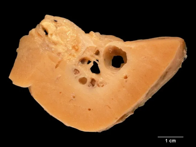
Gross pathology image of a renal tissue specimen showing a cross-section through the renal medullary region. The cut surface reveals numerous round to ovoid cystic-appearing spaces embedded within a tan-orange parenchyma, giving a sponge-like architecture consistent with dilated collecting ducts in the renal medulla. The cortex appears relatively preserved with no large focal necrosis or mass effect. The cavities are irregular in size but show a radial distribution toward the papillary tips, suggestive of medullary collecting duct dilation. The overall impression is compatible with medullary sponge kidney (MSK), a congenital malformation of the collecting system. MSK is usually bilateral, slightly more common in males, and frequently clinically silent; symptoms—when present—include nephrolithiasis, hematuria, and recurrent infection, typically in the third to fifth decades. The condition is often sporadic (~95%), with a genetic predisposition suspected in about 5% of cases. Associations include congenital hemihypertrophy, Beckwith-Wiedemann syndrome, Ehlers-Danlos syndrome, Wilms tumor, and multiple endocrine neoplasia type II. Clinically, recognition of this pattern informs differential diagnoses of medullary calcifications and recurrent stones and guides imaging follow-up with CT urography or MRI. This specimen highlights the distinctive medullary sponge morphology relevant for education, radiology correlation, and surgical planning when symptomatic. Clinical history should be considered globally.

Gross pathology image of a renal tissue specimen showing a cross-section through the renal medullary region. The cut surface reveals numerous round to ovoid cystic-appearing spaces embedded within a tan-orange parenchyma, giving a sponge-like architecture consistent with dilated collecting ducts in the renal medulla. The cortex appears relatively preserved with no large focal necrosis or mass effect. The cavities are irregular in size but show a radial distribution toward the papillary tips, suggestive of medullary collecting duct dilation. The overall impression is compatible with medullary sponge kidney (MSK), a congenital malformation of the collecting system. MSK is usually bilateral, slightly more common in males, and frequently clinically silent; symptoms—when present—include nephrolithiasis, hematuria, and recurrent infection, typically in the third to fifth decades. The condition is often sporadic (~95%), with a genetic predisposition suspected in about 5% of cases. Associations include congenital hemihypertrophy, Beckwith-Wiedemann syndrome, Ehlers-Danlos syndrome, Wilms tumor, and multiple endocrine neoplasia type II. Clinically, recognition of this pattern informs differential diagnoses of medullary calcifications and recurrent stones and guides imaging follow-up with CT urography or MRI. This specimen highlights the distinctive medullary sponge morphology relevant for education, radiology correlation, and surgical planning when symptomatic. Clinical history should be considered globally.

Finding Sources
Searching Images

urinary bladder papillary carcinoma cauliflower growth gross specimen bisected

Searching Images

gallbladder carcinoma papillary cauliflower growth filling lumen gross specimen

Finding Sources
Searching the Web

pathology museum specimen cauliflower growth gross photo jar kidney bean shaped organ

Searching Images

ovary serous papillary cystadenocarcinoma bisected specimen jar

Assessment of the Specimen

Gross description of the jar specimen:
  • An ovoid, bean-shaped organ preserved in formalin (yellow-tinged fixative)
  • A smooth, intact, pale tan-white capsule/wall is preserved around the periphery (top, both lateral margins, and lower pole)
  • The bisected/exposed surface is almost entirely replaced by an extensive, coarsely nodular, cream-to-yellow, friable growth with a fissured, coral-like or "cauliflower" architecture
  • Scattered dark greenish-black areas within the fissures, most likely representing hemorrhage, necrosis, or altered blood pigment
  • No obvious cystic spaces or clear residual normal parenchyma visible on the cut surface
Most likely identification: Papillary ("cauliflower") carcinoma involving the kidney/renal pelvis, or a papillary transitional-cell (urothelial) carcinoma
The exophytic, coarsely nodular, friable, "cauliflower-like" surface with fissuring and hemorrhagic/necrotic discoloration is the classic gross description used in pathology teaching for papillary urothelial (transitional cell) carcinomas of the renal pelvis/bladder, and for papillary variants of renal cell carcinoma. Surgical pathology texts explicitly describe malignant transformation of urothelial papillomas as producing villi that become "stunted and swollen like cauliflower" - Manual on Clinical Surgery (S. Das), Diseases of the Bladder.
Key differentials to consider, given the image alone (no visible label):
  1. Papillary/urothelial carcinoma of renal pelvis or bladder - cauliflower-like friable papillary mass filling the lumen, smooth outer wall/capsule preserved - best fit for this gross pattern.
  2. Renal cell carcinoma (papillary subtype) - can present as a lobulated, friable, tan-yellow mass distorting the kidney with a compressed capsule at the periphery.
  3. Papillary serous cystadenocarcinoma of the ovary - also classically described with "cauliflower-like" papillary excrescences studding a cyst wall; the bean/oval shape and smooth outer capsule are also compatible with a bisected ovarian mass.
Without the original specimen label or clinical history (organ of origin, patient age/sex), I can't give a single certain diagnosis - the morphology (cauliflower/papillary, friable, hemorrhagic-necrotic, encapsulated organ) is shared across these entities. If you have any accompanying label, catalog number, or know which organ this jar is meant to represent (urinary tract vs. kidney vs. ovary), I can narrow this down definitively.
If it would help, I can pull reference gross-pathology images of each of these three entities for side-by-side comparison - let me know which organ system you suspect, or if you have the specimen label/catalog text visible elsewhere in your notes.
This is a shared conversation. Sign in to Orris to start your own chat.