
Diagram of heart
Reading File
The user wants a diagram of the heart - this is a visual/anatomy query. Let me search the medical library for heart anatomy diagrams.
Finding Sources
Finding Sources
I found a heart diagram in Harrison's. Let me read that block to get the image URL.
Reading File
I have the image URL. Let me fetch it.
Loading Image
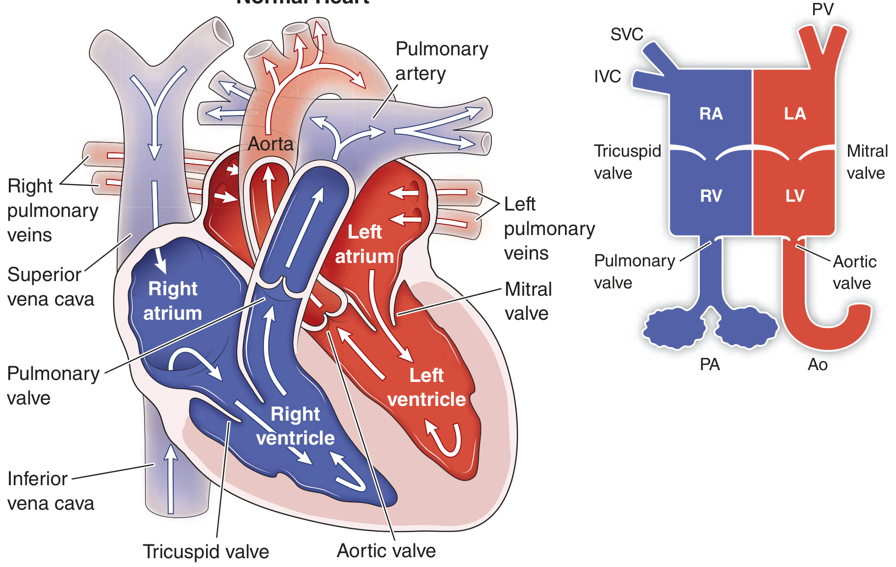
Figure 280-1 - Normal Heart (Harrison's Principles of Internal Medicine, 22E)
The diagram shows both a detailed anatomical cross-section and a simplified box diagram of the heart. Here is a summary of the key structures labeled:
Chambers (4 total):
- Right atrium (RA) - receives deoxygenated blood from the body via the Superior Vena Cava (SVC) and Inferior Vena Cava (IVC)
- Right ventricle (RV) - pumps deoxygenated blood to the lungs via the Pulmonary Artery (PA)
- Left atrium (LA) - receives oxygenated blood from the lungs via the Pulmonary Veins (PV)
- Left ventricle (LV) - pumps oxygenated blood to the body via the Aorta (Ao)
Valves (4 total):
- Tricuspid valve - between RA and RV (right atrioventricular valve)
- Pulmonary valve - between RV and pulmonary artery
- Mitral valve - between LA and LV (left atrioventricular valve)
- Bicuspid/Mitral valve - between LA and LV
- Aortic valve - between LV and aorta
Blood Flow Direction (arrows in diagram):
- Blue = deoxygenated blood (right side of heart + pulmonary artery)
- Red = oxygenated blood (left side of heart + aorta)
- Body → SVC/IVC → RA → Tricuspid valve → RV → Pulmonary valve → Pulmonary artery → Lungs
- Lungs → Pulmonary veins → LA → Mitral valve → LV → Aortic valve → Aorta → Body
Thorax
Finding Sources
Finding Sources
Good results. Let me read the Gray's Anatomy surface anatomy section and fetch the Netter's thorax cross-section image, plus the Gray's thorax surface anatomy image simultaneously.
Reading File
Reading File
Reading File
I have several great image URLs. Let me fetch the most relevant thorax diagrams in parallel - the Netter's cross-section, the dorsal/lateral thorax, and the Gray's chest wall.
Loading Image
Loading Image
Loading Image
Excellent! Three high-quality thorax diagrams retrieved. Here they are with explanations:
Thorax - Anatomical Diagrams
1. Surface Anatomy - Anterior View (Chest Wall)
(Gray's Anatomy for Students)
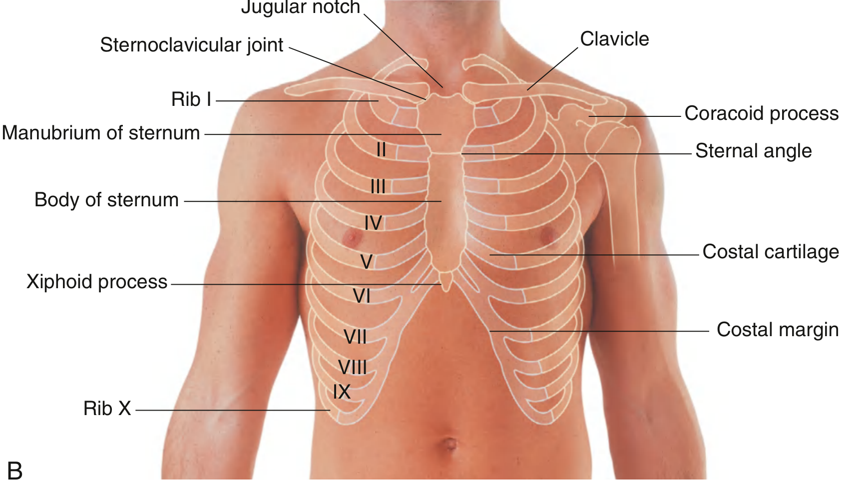
Key surface landmarks:
- Jugular notch - top of the manubrium, used as a reference point
- Sternum - divided into manubrium, body, and xiphoid process
- Sternal angle (angle of Louis) - junction of manubrium and body; marks the level of rib II
- Ribs I-X labeled with costal cartilages
- Costal margin - lower border of the thoracic cage (ribs VII-X)
- Clavicle and coracoid process - superior landmarks
2. Thoracic Cross-Section - Intercostal Muscles & Nerves
(Netter's Atlas of Human Anatomy)
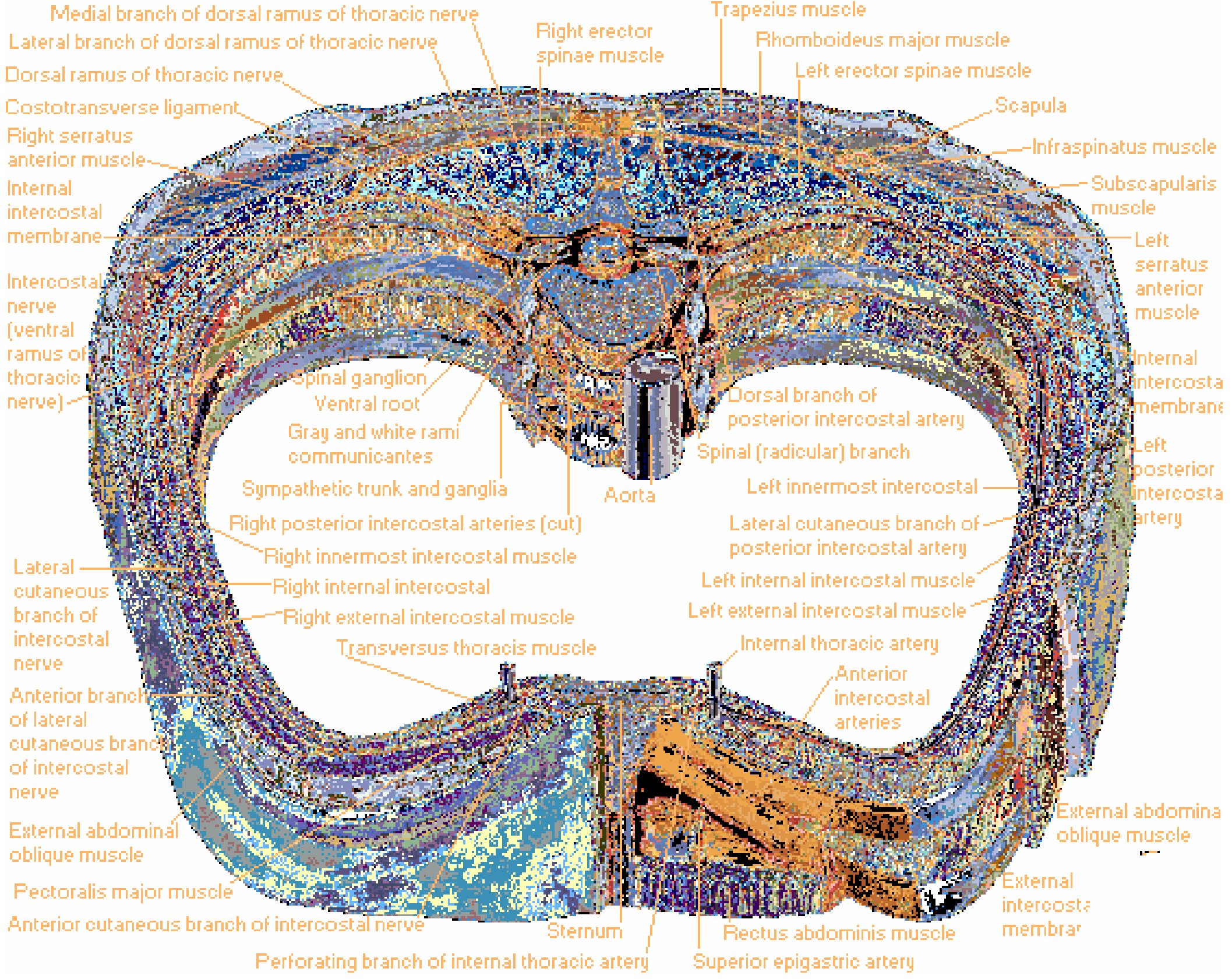
Key structures in this transverse cross-section of the thoracic wall:
- Three intercostal muscle layers: external, internal, and innermost intercostal muscles
- Intercostal nerve (ventral ramus of thoracic nerve) - runs in the costal groove
- Internal thoracic artery and anterior intercostal arteries
- Posterior intercostal arteries and their branches
- Sympathetic trunk and ganglia adjacent to the vertebral column
- Aorta - descending thoracic aorta centrally
- Posterior muscles: trapezius, rhomboideus major, erector spinae, infraspinatus, subscapularis, serratus anterior
3. Thoracic Surface (Inferior View - Diaphragm Level)
(Netter's Atlas of Human Anatomy)
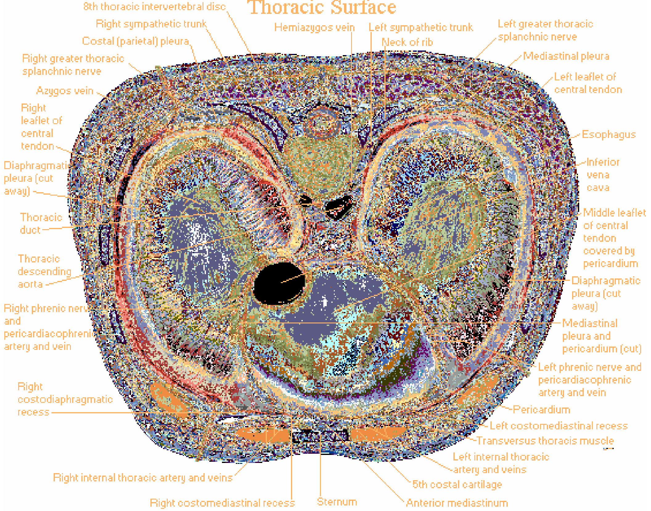
Key structures visible from below:
- Pericardium and heart centrally
- Diaphragmatic pleura (cut away on both sides) revealing the pleural cavities
- Costodiaphragmatic recesses (right and left) - the lowest part of the pleural cavity
- Mediastinal pleura and pericardium
- Azygos vein (right) and hemiazygos vein (left)
- Thoracic descending aorta and esophagus posteriorly
- Sympathetic trunks bilaterally
- Thoracic duct on the left
- Phrenic nerves with pericardiacophrenic arteries and veins
- Transversus thoracis muscle anteriorly
- Internal thoracic arteries and veins running beside the sternum
Summary of thorax contents:
| Region | Structures |
|---|---|
| Thoracic wall | Ribs (12 pairs), sternum, costal cartilages, intercostal muscles & nerves |
| Mediastinum | Heart, great vessels, trachea, esophagus, thoracic duct, vagus & phrenic nerves |
| Pleural cavities | Right and left lungs, enclosed by parietal and visceral pleura |
| Diaphragm | Floor of thorax; openings for aorta (T12), esophagus (T10), IVC (T8) |
This is a shared conversation. Sign in to Orris to start your own chat.