
Polydactaly
Reading File
Polydactyly is the congenital presence of extra digits on the hand or foot. It is one of the most common congenital limb anomalies, classified broadly as preaxial (radial/tibial side), postaxial (ulnar/fibular side), or central.
Searching sources...
Finding Sources
Searching PubMed
polydactyly genetics classification
Reading File
Reading File
Reading File
Reading File
Reading File
Reading File
Reading File
Now let me get the Wassel classification for preaxial (thumb) polydactyly and the dermatology section on rudimentary polydactyly:
Finding Sources
Finding Sources
Reading File
Reading File
Reading File
Now let me fetch the clinical image of polydactyly for inclusion in the answer:
Loading Image
Loading Image
Here is a thorough overview of polydactyly drawn from multiple authoritative sources.
Polydactyly
Polydactyly is the presence of supernumerary (extra) digits on the hands or feet - more than five fingers or toes. It is one of the most common congenital limb anomalies, with an overall incidence of approximately 1-2 per 1,000 live births. The extra digit is often incompletely formed and lacks normal muscular development.
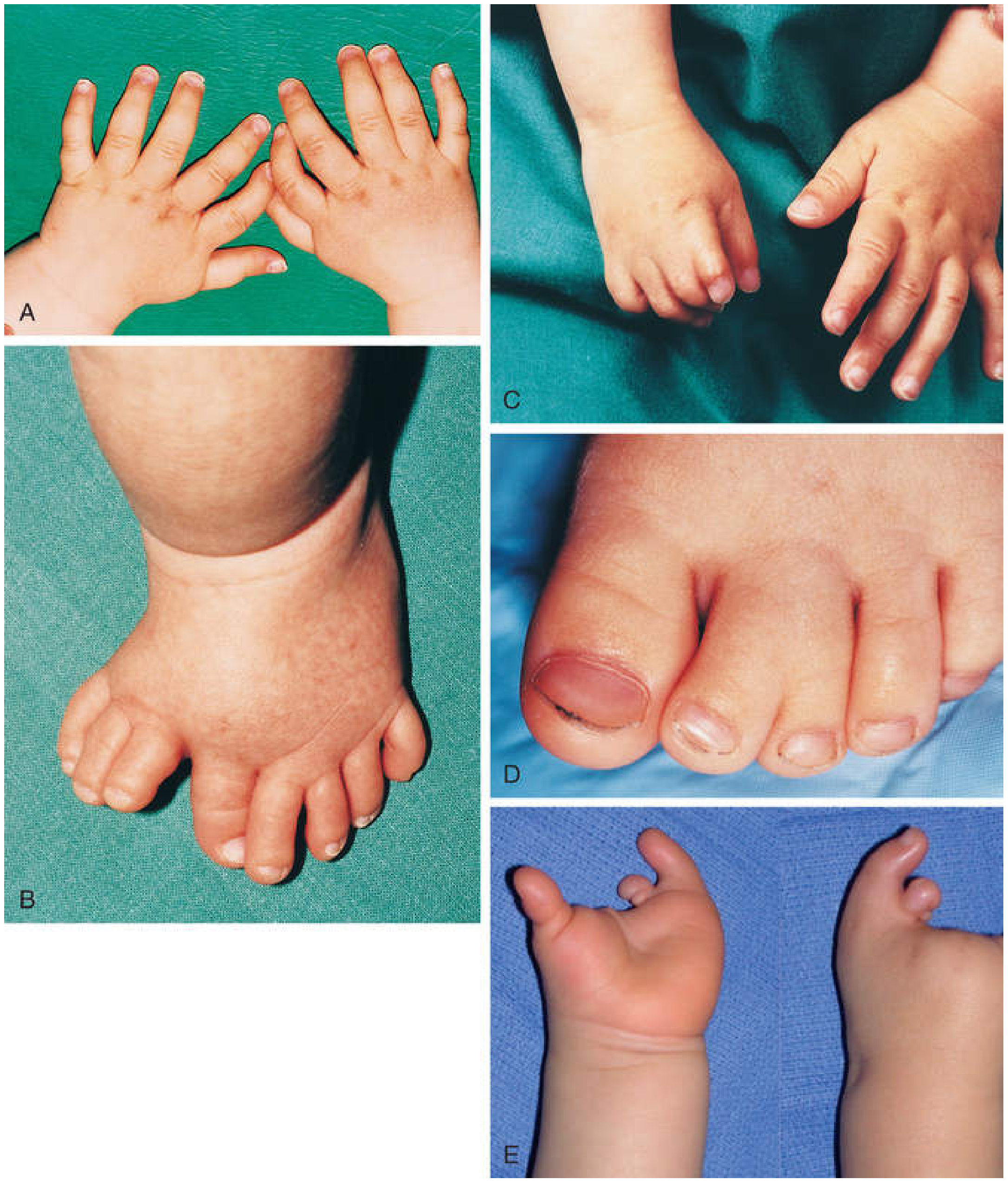
Fig. 16.14 - The Developing Human: Clinically Oriented Embryology. Polydactyly of the hands (A) and feet (B).
Classification by Location
Polydactyly is classified based on which side of the limb is affected:
| Type | Location | Also Known As |
|---|---|---|
| Preaxial | Radial side of hand / tibial side of foot | Thumb or hallux duplication |
| Postaxial | Ulnar side of hand / fibular side of foot | Small finger / 5th toe duplication |
| Central | Index, middle, or ring fingers | Rare; often hidden by syndactyly |
1. Postaxial Polydactyly (Most Common Overall)
Duplication of the small finger (hand) or lateral toe (foot).
- Most common in people of African descent: ~1 in 143 live births (vs. 1 in 1,339 in Caucasians)
- Autosomal dominant inheritance with variable expression
- In the Black population: usually isolated, not syndromic
- In Caucasians: more frequently associated with syndromes and chromosomal abnormalities - warrants a more thorough genetic workup
Temtamy and McKusick Classification:
- Type A - Fully developed, well-formed extra digit; rare; equal incidence across races; may be associated with syndromes (Cornelia de Lange, Chondroectodermal dysplasia, Schinzel syndrome)
- Type B - Rudimentary nubbin attached pedunculated at the proximal phalanx level; much more common in people of African descent (10x); autosomal dominant
Further subclassification (Pritsch et al.):
- Type 1: Soft tissue duplication only
- Type 2: Partial duplication including bone
- Type 3: Complete ray duplication
- Campbell's Operative Orthopaedics 15th Ed 2026, p. 4662
2. Preaxial Polydactyly (Thumb / Hallux Duplication)
- Incidence: ~1 in 3,000 live births
- Usually unilateral and sporadic (not syndromic), except for Wassel Type VII
- Thumb duplication is not a true extra digit but a splitting of a single digit with variable development in each part
Wassel Classification of Thumb Polydactyly
| Type | Description | Frequency |
|---|---|---|
| I | Bifid distal phalanx | 2% |
| II | Duplicated distal phalanx | 15% |
| III | Bifid proximal phalanx | 6% |
| IV | Duplicated proximal phalanx | 43% (most common) |
| V | Bifid metacarpal | 10% |
| VI | Duplicated metacarpal | 4% |
| VII | Triphalangia | 20% |
Type VII is associated with: Holt-Oram syndrome, Fanconi anemia, Blackfan-Diamond anemia, imperforate anus, cleft palate, and tibial defects.
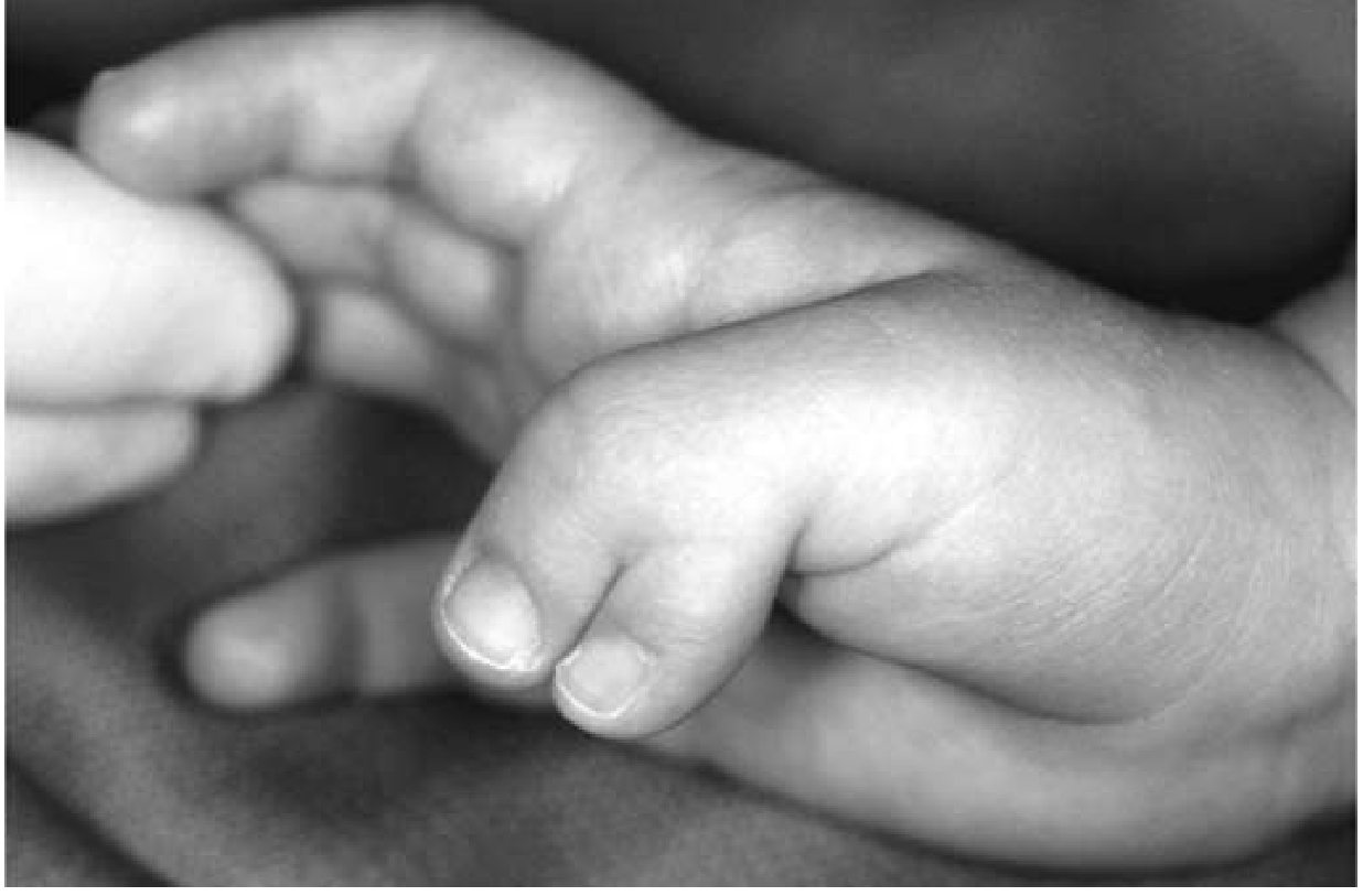
Fig. 7.82 - Miller's Review of Orthopaedics 9th Ed. Type IV duplication.
- Miller's Review of Orthopaedics 9th Edition, p. 2070
3. Central Polydactyly
- Rare - involves index, middle, or ring finger duplication
- Ranges from incomplete to fully formed extra digits
- Commonly associated with syndactyly, cleft hand, and other syndromes
- - Mulholland and Greenfield's Surgery 7e, p. 6046
Associated Syndromes / Genetics
Polydactyly (especially postaxial) is a key feature of the ciliopathies - defects in ciliary proteins produce wide-ranging effects including postaxial polydactyly, retinal degeneration, renal/hepatic cysts, and situs inversus:
| Syndrome | Type of Polydactyly | Inheritance |
|---|---|---|
| Bardet-Biedl syndrome | Postaxial | AR (ciliopathy) |
| Short-rib polydactyly syndromes | Postaxial | AR (ciliopathy) |
| Smith-Lemli-Opitz | Postaxial (~50% of cases) | AR |
| Gorlin syndrome | Postaxial (occasional) | AD |
| Rubinstein-Taybi syndrome | Postaxial (occasional) | AD |
| Simpson-Golabi-Behmel | Postaxial | X-linked |
| Greig cephalopolysyndactyly | Preaxial/mesoaxial | AD (GLI3 variants) |
| Pallister-Hall syndrome | Mesoaxial/postaxial | AD (GLI3) |
| Pfeiffer syndrome | Broad thumbs/halluces | AD (FGFR1/2) |
| Holt-Oram syndrome | Preaxial (thumb) | AD |
- Emery's Elements of Medical Genetics and Genomics
Treatment
Postaxial - Type A
- Surgical excision in the operating room under tourniquet at ~1 year of age due to the complexity of the duplication
- Ulnar digit is removed; soft tissue from ablated digit augments the retained finger
Postaxial - Type B
- Suture ligation / vascular clip in the nursery: fast, no anesthesia needed, cost-effective - BUT 42% revision rate (neuromas, residual polydactyly)
- Surgical excision: preferred - only ~3% complication rate vs. 23.5% for ligation (systematic review data); higher overall patient satisfaction; no revisions needed
- Some centers now perform simple excisions in an office setting under local anesthesia
Preaxial (Thumb)
- Best possible thumb is reconstructed from available anatomic structures (not simple amputation)
- If duplicated thumbs are equal in size: preserve the ulnar thumb to maintain the ulnar collateral ligament (UCL) for pinch
- Soft tissue from the ablated thumb is used to augment the retained one
- Complications: nail deformity, IP joint angulation
- Bilhaut-Cloquet procedure (or modified): used for Wassel types II-III (combines both halves)
Foot Polydactyly
- Preoperative X-rays mandatory before any non-trivial excision
- Extra metatarsal elements must also be removed
- Soft-tissue webspace reconstruction required when central digits are removed
- Optimal age: 6-18 months
- Kirschner wire fixation may be used for 4-6 weeks in preaxial cases after capsule repair
- Campbell's Operative Orthopaedics 15th Ed 2026
Embryological Basis
Polydactyly results from abnormal signaling in the zone of polarizing activity (ZPA) during limb development. It is inherited most often as an autosomal dominant trait with variable expression. The extra digit usually lacks complete muscular development, explaining the importance of selecting the better-developed digit for retention during surgery.
- The Developing Human: Clinically Oriented Embryology, p. 1004
This is a shared conversation. Sign in to Orris to start your own chat.