
boundaries of oral cavity
Reading File
Now let me search the medical library for oral cavity boundaries.
Finding Sources
Finding Sources
Reading File
Reading File
Reading File
Loading Image
Loading Image
Finding Sources
Here is a complete breakdown of the boundaries of the oral cavity, drawn from Gray's Anatomy for Students, Color Atlas of Human Anatomy, and Cummings Otolaryngology.
Boundaries of the Oral Cavity
The oral cavity is the first part of the digestive tract. It lies inferior to the nasal cavities, opens anteriorly onto the face through the oral fissure, and is continuous posteriorly with the pharynx at the oropharyngeal isthmus.
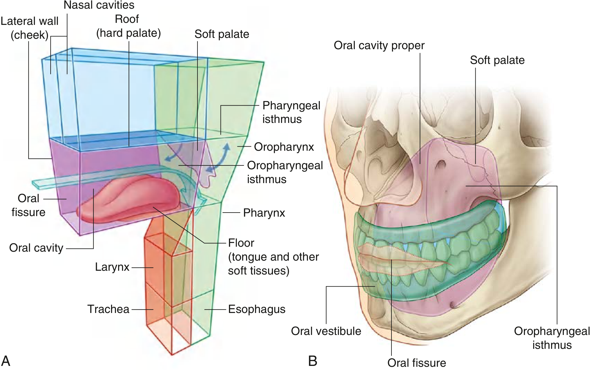
Overall Boundaries
| Wall | Structure |
|---|---|
| Roof | Hard palate anteriorly + soft palate posteriorly (separates it from the nasal cavity above) |
| Floor | Muscular diaphragm of the mouth (mylohyoid + geniohyoid muscles) + tongue resting on it |
| Lateral walls | Cheeks (buccinator muscle lined by buccal mucosa) |
| Anterior opening | Oral fissure (between upper and lower lips) |
| Posterior opening | Oropharyngeal isthmus (leading into the oropharynx) |
Two Subdivisions
The teeth and alveolar processes divide the oral cavity into two regions:
1. Oral Vestibule
- The horseshoe-shaped space between the lips/cheeks externally and the teeth/gingiva/alveolar processes internally
- Opens anteriorly through the oral fissure
- Can be opened and closed by muscles of facial expression and mandibular movements
2. Oral Cavity Proper
- The space enclosed within (medial to) the dental arches
- Anteriorly and laterally: alveolar processes, teeth, and gingiva
- Superiorly (roof): hard palate
- Inferiorly (floor): muscular diaphragm + tongue
- Posteriorly: communicates with the oropharynx via the oropharyngeal isthmus (bounded by the palatoglossal folds/arches on each side)
Key Boundary Landmarks
- Anterior boundary of oral cavity proper: inner surface of teeth and alveolar ridges
- Posterior boundary: the hard palate - soft palate junction superiorly; the palatoglossal arches (anterior faucial pillars) mark the transition to the oropharynx - this boundary is called the oropharyngeal isthmus (isthmus of fauces)
- Superior boundary: hard palate (bony) + soft palate (muscular)
- Inferior boundary: tongue + muscular floor (mylohyoid diaphragm)
- Lateral boundaries: cheeks (buccinator muscle)
Oral Cavity Contents (for context)
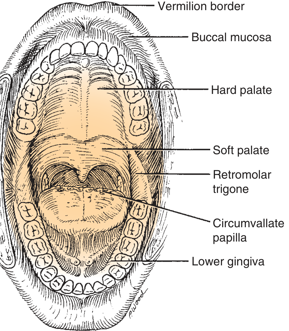
The oral cavity contains:
- Teeth and gingiva (upper and lower)
- Tongue (oral portion = anterior 2/3)
- Hard palate
- Floor of mouth
- Buccal mucosa
- Retromolar trigone (triangular area behind the last lower molar, important in oncology)
Posterior Boundary - Clinical Point
The palatoglossal folds (covering the palatoglossus muscles), one on each side, are the key landmark marking the boundary between the oral cavity and the oropharynx. The oropharynx begins immediately posterior to these folds and includes the soft palate, base of tongue, tonsils, and posterior pharyngeal wall.
Sources: Gray's Anatomy for Students, p. 1227 | Color Atlas of Human Anatomy Vol. 2, p. 245 | Cummings Otolaryngology Head and Neck Surgery, p. 1653
This is a shared conversation. Sign in to Orris to start your own chat.