
Trigeminal neuralgia versus giant cell arteritis.
"trigeminal neuralgia" AND "giant cell arteritis" AND diagnosis
trigeminal neuralgia vascular compression MRI
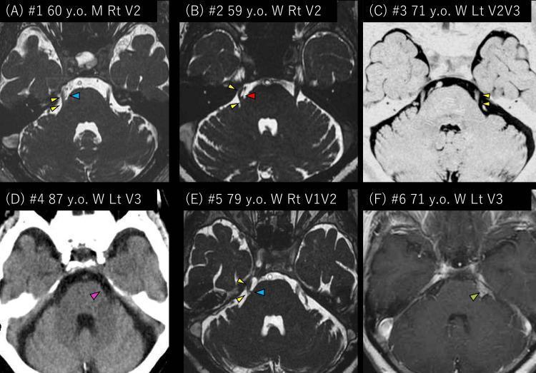
This composite diagnostic image displays radiological findings from six patients with trigeminal neuralgia, primarily using axial Magnetic Resonance Imaging (MRI) and Computed Tomography (CT) focusing on the posterior fossa and cerebellopontine angle (CPA). Panels A, B, and E demonstrate neurovascular compression (NVC) of the trigeminal nerve (yellow arrowheads). Specifically, panels A and E show compression by the petrosal vein (blue arrowheads), while panel B shows compression by the superior cerebellar artery (red arrowhead). Panel C illustrates a left trigeminal nerve without apparent vascular contact. Panel D provides a non-contrast CT view of the left cerebellopontine cistern (pink arrowheads) in a patient where MRI was contraindicated. Panel F displays a T1-weighted gadolinium-enhanced MRI showing a pathological mass/tumor (green arrowhead) within the left cerebellopontine cistern, representing secondary trigeminal neuralgia. The collection serves as an educational comparison between idiopathic, neurovascular, and secondary causes of trigeminal nerve dysfunction, highlighting the anatomical relationship between the nerve root entry zone and adjacent vascular or neoplastic structures.
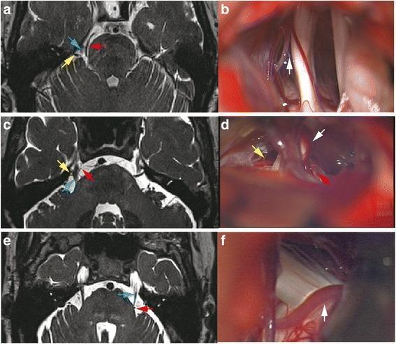
This composite medical image presents a comparison between pre-operative axial MRI scans (a, c, e) and corresponding intra-operative clinical photographs (b, d, f) illustrating neurovascular conflict (NVC) in cases of trigeminal neuralgia. The MRI images demonstrate the spatial relationship between the trigeminal nerve (TGN, blue arrows) and adjacent vascular structures, primarily the superior cerebellar artery (SCA, red arrows) and other vessels (yellow arrows) in the cerebellopontine angle. The intra-operative views provide direct visualization of these structures during microvascular decompression (MVD) surgery. The series highlights clinical discrepancies where routine MRI might suggest arterial compression that is later identified intra-operatively as non-contacting (white arrow gap in b), compression by venous structures (white arrow in d), or arterial compression that was subtle on imaging (white arrow in f). This comparison serves as an educational tool for neurosurgical planning, emphasizing the importance of identifying the Root Entry Zone (REZ) and distinguishing between arterial and venous compression in the management of trigeminal nerve syndromes.
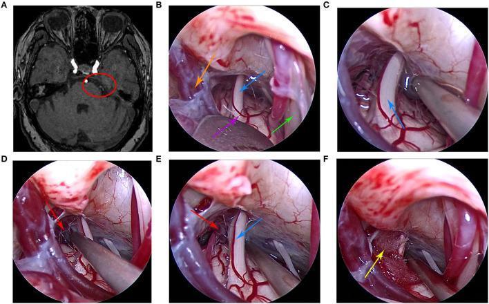
This composite image illustrates the clinical progression and surgical treatment of trigeminal neuralgia using microvascular decompression (MVD). Panel A is a preoperative axial MRI scan of the brain, with a red circle highlighting neurovascular compression at the root entry zone of the right trigeminal nerve in the cerebellopontine angle. Panels B through F provide a sequence of intraoperative endoscopic photographs showing the surgical workflow. Key anatomical structures identified include the trigeminal nerve (blue arrow), brainstem (purple arrow), petrosal vein (orange arrow), and facial nerve (green arrow). The sequence demonstrates the exploration and dissection of arachnoid adhesions (Panels B-D), identifying a petrosal vein branch (red arrow) as the offending vessel compressing the nerve (Panel E). The final panel (F) depicts the therapeutic intervention, where a Teflon pad (yellow arrow) is surgically interposed between the trigeminal nerve and the offending vessel to achieve mechanical decompression. This sequence serves as an educational tool for neurosurgical training, demonstrating the suboccipital retrosigmoid approach and the efficacy of endoscopic visualization in identifying subtle vascular compression.
giant cell arteritis temporal artery biopsy histology granuloma
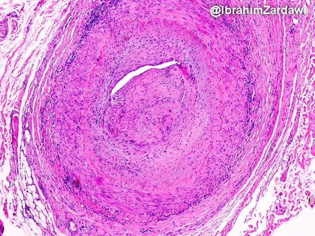
Histology image obtained from a temporal artery biopsy, prepared with Hematoxylin and Eosin stain and viewed under light microscopy at low magnification. The specimen demonstrates classic giant cell arteritis with transmural, granulomatous inflammation encompassing the full thickness of the arterial wall (intima, media, and adventitia) in a segmental distribution. Multinucleated giant cells are interspersed with a dense lymphohistiocytic infiltrate, and fragmentation or disruption of the internal elastic lamina is evident. The inflammatory process extends to the vasa vasorum and small venules within the adventitia, indicating widespread perivascular involvement. Accompanying features include marked intimal hyperplasia producing marked luminal narrowing and, in this field of view, near-occlusive lumen occlusion. The arterial media may show loss of elastic fibers and necrosis is not conspicuous here. These findings are diagnostic of GCA and help differentiate from other large-vessel vasculitides and nonspecific atherosclerotic changes. Clinically, such histology supports a diagnosis of temporal arteritis in patients with new headaches, visual symptoms, jaw claudication, and elevated inflammatory markers, guiding urgent corticosteroid therapy to prevent ischemic complications. This image exemplifies the radiologic-pathologic correlation typical of vasculitic arteritis. The slide highlights diagnostic relevance, research utility, and educational value for trainees and clinicians in practice. For medical education and research.
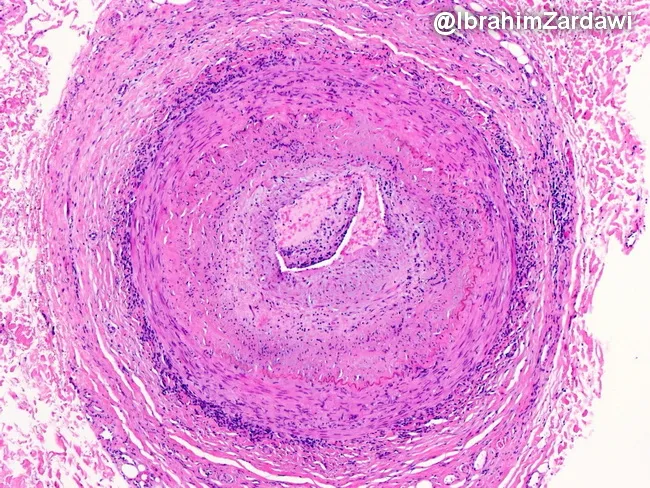
This histology image captures a cross-sectional temporal artery biopsy illustrating the hallmarks of giant cell arteritis. Prepared as a paraffin-embedded tissue section and stained with Hematoxylin and Eosin, the slide reveals a transmural inflammatory infiltrate that traverses all arterial wall layers. At the center, a narrowed lumen is surrounded by concentric intimal hyperplasia with marked medial disruption. Multinucleated giant cells and mononuclear macrophage clusters are present within the media, accompanied by lymphocytic infiltrate along the vessel wall. The elastic lamina appears disrupted, and fragmentation of the media is evident, consistent with robust granulomatous vasculitis. These features produce near-occlusive narrowing of the artery, explaining the ischemic risk associated with temporal arteritis. Clinically, such histopathology confirms a diagnosis of GCA and helps differentiate from atherosclerotic disease, infectious vasculitis, or other large-vessel vasculitides. The image underscores the systemic nature of the disease, as inflammation can extend from cranial vessels to the aorta. The diagnostic significance lies in correlating histology with patient presentation, including age-related cranial symptoms and risk of vision loss; timely immunosuppressive therapy can prevent irreversible ischemic complications. This specimen exemplifies classic angiitis with granulomatous inflammation and intimal hyperplasia driving luminal compromise.
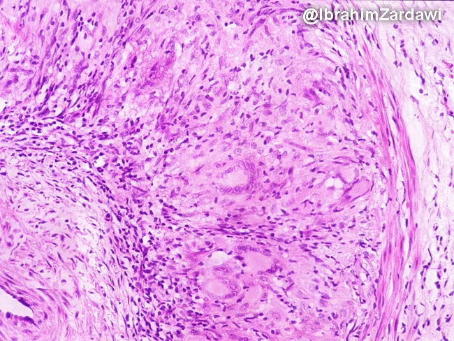
Imaging modality and technique: Brightfield light microscopy of a temporal artery biopsy stained with Hematoxylin and Eosin (H&E). Specimen: arterial wall from the temporal region; cross-sectional vascular biopsy showing inflammatory arteritis. Key features include granulomatous inflammatory infiltrate surrounding the media and adjacent to the internal elastic lamina, composed of lymphocytes, plasma cells, epithelioid histiocytes, and multinucleated giant cells. Giant cells are often present but may be absent; their presence supports diagnosis but is not required. The inflammatory infiltrate disrupts the media and may extend into the adventitia; fragmentation and phagocytosis of the internal elastic lamina are characteristic. Medial necrosis is minimal to absent; there is usually no well-formed granuloma or extensive necrosis. Neointimal hyperplasia and luminal narrowing can be observed as secondary consequences of chronic vasculitis. The image demonstrates a granulomatous arteritis pattern typical of giant cell arteritis. Diagnostic significance: classic histopathology includes granulomatous inflammation of medium-sized arteries with IEL fragmentation; biopsy positivity confirms temporal arteritis; however, skip lesions can yield false negatives; clinical correlation with headache, scalp tenderness, jaw claudication, visual symptoms and elevated inflammatory markers (CRP/ESR) is essential. Differential considerations include Takayasu arteritis, atherosclerosis, infectious vasculitis, and other granulomatous vasculitides. This slide is used in educational settings to illustrate GCA pathology and IEL involvement.
Trigeminal Neuralgia vs. Giant Cell Arteritis
1. Overview
| Feature | Trigeminal Neuralgia (TN) | Giant Cell Arteritis (GCA) |
|---|---|---|
| Mechanism | Neuropathic (demyelination/ectopic firing) | Granulomatous large-vessel vasculitis |
| Primary tissue involved | Trigeminal nerve (CN V) root entry zone | Medium-to-large arteries (temporal, ophthalmic, aortic arch branches) |
| Age of onset | >40 yrs; peak 50-70 yrs | Exclusively >50 yrs (criterion) |
| Sex predominance | Slightly more common in women (1.5:1) | Women more commonly affected |
| Urgency | Urgent (quality of life, weight loss) | Emergency (irreversible blindness risk) |
2. Pathophysiology
3. Clinical Presentation
Trigeminal Neuralgia
- Pain character: Electric shock-like, lancinating, shooting - each paroxysm lasts seconds (up to 2 minutes)
- Distribution: V2 (cheek/upper teeth) and V3 (lower teeth/chin) most common; V2+V3 combination is the classic. V1 (ophthalmic) alone is extremely rare
- Triggers: Touch to the face near the nasolabial fold, chewing, teeth brushing, talking, cool air across the face - a pathognomonic feature
- Refractory period: After a volley, stimuli will not re-trigger the pain for a short time
- No autonomic features: Unlike trigeminal autonomic cephalalgias (cluster, SUNCT)
- Normal examination: No sensory loss, no motor weakness in classical TN; their presence suggests secondary TN/neuropathy
- Interictal pain: Most patients pain-free between attacks; some have a residual dull background ache
- Goldman-Cecil Medicine; Bradley and Daroff's Neurology in Clinical Practice
Giant Cell Arteritis
- Headache: New-onset headache in a patient >50 is the most common presenting complaint - typically temporal, throbbing, or boring; may be continuous
- Jaw claudication: Fatigue and aching of masticatory muscles with chewing - among the most specific signs (LR+ 6.7 alone; combined with headache LR+ 8.0; combined with scalp tenderness LR+ 17.0)
- Scalp/temporal artery tenderness: Erythematous, nodular, tender temporal artery on palpation
- Vision loss: Amaurosis fugax or sudden permanent monocular blindness from arteritic ischemic optic neuropathy - a medical emergency
- Systemic features: Fever, fatigue, anemia, weight loss, markedly elevated ESR/CRP
- Polymyalgia rheumatica: 50% of GCA patients have PMR; 15% of PMR patients have GCA
- Diplopia, stroke: Rare but occur from arteritic involvement of other cranial vessels
- Goldman-Cecil Medicine; Symptom to Diagnosis (Evidence-Based Guide)
4. Key Distinguishing Features
| Feature | Trigeminal Neuralgia | Giant Cell Arteritis |
|---|---|---|
| Pain duration | Seconds (paroxysmal) | Hours-to-days (continuous or persistent) |
| Triggers | Touch, chewing, cold air | None (spontaneous) |
| Jaw pain | Pain while chewing (trigger) | Jaw claudication (fatigue/aching with sustained chewing, stops at rest) |
| Fever/systemic | Absent | Common |
| ESR/CRP | Normal | Markedly elevated (ESR often >50, sensitivity 95%) |
| Temporal artery | Normal | Tender, nodular, beaded |
| Visual symptoms | Absent | Amaurosis fugax, sudden blindness |
| Neurological deficits | Absent (classical TN) | Rare; peripheral neuropathy in ~15% |
| Age | >40 | Strictly >50 |
The jaw pain overlap is clinically important: In TN, chewing is a trigger for brief electric-shock pain. In GCA, jaw claudication is a dull fatigue/ache that develops after sustained chewing and stops with rest - it reflects muscular ischemia, not nerve stimulation.
5. Diagnosis
Trigeminal Neuralgia
- Clinical diagnosis based on ICHD-3 criteria: paroxysmal attacks of intense, shock-like pain lasting 1 second to 2 minutes in ≥1 trigeminal divisions, stereotypical episodes, triggered by innocuous stimuli, no neurological deficit in classical form
- MRI (high-resolution + MRA): Done to exclude secondary causes (MS plaque, pontine lacunar infarct, posterior fossa meningioma/schwannoma, skull base malignancy) and to identify neurovascular compression for surgical planning
- Labs are normal - Bradley and Daroff's Neurology in Clinical Practice
Giant Cell Arteritis
- ACR criteria (3 of 5 required): Age ≥50; new headache; temporal artery abnormality (tenderness/decreased pulse); ESR ≥50 mm/hr; biopsy showing necrotizing arteritis with mononuclear infiltrate or granulomatous inflammation (often with giant cells)
- ESR: Sensitivity 95% for an abnormal result; ESR >100 mm/h has LR+ 1.9
- Temporal artery biopsy: Gold standard - sensitivity 85%, specificity 100% (LR+ infinite, LR- 0.15). Only 30-40% of referred patients are biopsy-positive, reflecting poor clinical predictor performance. Skip lesions can cause false negatives; re-biopsy or treat empirically if suspicion remains high
- Temporal artery ultrasound: Hypoechoic halo sign - present around inflamed arteries, but insufficiently sensitive/specific to replace biopsy
- Do not delay treatment to await biopsy - biopsy can be performed up to ~7 days after starting steroids without significant loss of diagnostic yield - Symptom to Diagnosis (Evidence-Based Guide)
6. Treatment
Trigeminal Neuralgia
- Carbamazepine 400-1200 mg/day (first-line; ~90% initial response rate)
- Oxcarbazepine 300-1800 mg/day (alternative first-line)
- Second-line: baclofen, lamotrigine, gabapentin, phenytoin, clonazepam, valproate, levetiracetam, topiramate
- Botulinum toxin: emerging evidence for refractory cases
- Microvascular decompression (MVD): Suboccipital retrosigmoid craniotomy - separates the offending vessel from the nerve root with a Teflon pad; highest long-term pain-free rate and preserves sensory function; the preferred surgical option for fit patients
- Percutaneous procedures (all ablative - risk of sensory loss): Radiofrequency thermocoagulation, glycerol rhizolysis, balloon compression of the Gasserian ganglion
- Stereotactic radiosurgery (Gamma Knife): Radiation directed at REZ; lower immediate efficacy but non-invasive
- Goldman-Cecil Medicine; Bradley and Daroff's Neurology in Clinical Practice
Giant Cell Arteritis
- High-dose corticosteroids immediately - do not wait for biopsy if clinical suspicion is moderate to high:
- Prednisone 40-60 mg/day orally; if visual symptoms are present or impending, IV methylprednisolone 250-1000 mg/day for 3 days first
- Taper slowly once ESR/CRP normalizes and symptoms remit; total duration 18-24 months
- Monitoring: clinical response + ESR; increase dose if clinical signs of activity appear regardless of ESR result
- Tocilizumab (IL-6 receptor antagonist): FDA-approved corticosteroid-sparing agent; reduces relapse rate and cumulative steroid dose - Bradley and Daroff's Neurology in Clinical Practice
- Methotrexate: An alternative steroid-sparing option for recurrent disease on steroid withdrawal
- Low-dose aspirin: Often added to reduce the risk of visual loss and stroke through antiplatelet effect
7. Prognosis
| Trigeminal Neuralgia | Giant Cell Arteritis | |
|---|---|---|
| Course | Exacerbating-remitting over years | Chronic; requires prolonged treatment |
| Remission | Spontaneous or medication-induced | Uncommon without treatment |
| Best outcome | MVD often curative | Steroids prevent blindness if started early |
| Major risk | Weight loss/malnutrition from pain triggers; depression | Irreversible bilateral blindness; stroke |
Imaging: Trigeminal Neuralgia
Histology: Giant Cell Arteritis (Temporal Artery Biopsy)
Clinical Bottom Line
- A patient >50 with paroxysmal, electric-shock facial pain triggered by touching the face or chewing who has a normal exam and normal labs = trigeminal neuralgia. Start carbamazepine and get MRI.
- A patient >50 with a new persistent temporal headache, jaw claudication (fatigue-type), tender nodular temporal arteries, elevated ESR/CRP, and any visual symptoms = giant cell arteritis until proven otherwise. Start high-dose prednisone immediately, then biopsy.
- The two can occasionally coexist or mimic each other when GCA affects the cavernous sinus or skull base - but the temporal pattern of pain (paroxysmal + triggered vs. persistent + spontaneous) and the presence of systemic inflammation are the key discriminators.
Oxcarbamazepine dose
Oxcarbazepine Dosing - Trigeminal Neuralgia
Starting & Titration (from Tintinalli's Emergency Medicine)
| Step | Dose |
|---|---|
| Starting dose | 300 mg orally once daily |
| After 3 days | Increase to 300 mg twice daily (600 mg/day) |
| Target / typical effective range | 450–1200 mg twice daily (900–2400 mg/day) |
Range Across Sources
| Source | Range |
|---|---|
| Goldman-Cecil Medicine | 300–1800 mg/day |
| Barash's Clinical Anesthesiology | 300–900 mg/day, given twice daily (bid) |
| Tintinalli's Emergency Medicine | Up to 450–1200 mg twice daily |
| Bradley and Daroff's Neurology | Dose titrated to the lowest that controls pain; closely mirrors carbamazepine 600–1200 mg/day |
Key Clinical Notes
- Onset: Pain relief may be noted within 24–48 hours of reaching an effective dose, which is faster than many other agents - Barash's Clinical Anesthesiology
- Compared to carbamazepine: Oxcarbazepine is a 10-keto analogue with a similar sodium-channel blocking mechanism but fewer side effects overall (less hepatotoxicity, fewer drug interactions, no auto-induction)
- Main risk: Hyponatremia - can be prominent and requires monitoring of serum sodium, especially in elderly patients
- Monitoring: Serum sodium, LFTs, CBC at baseline, during the first few months, and annually thereafter - Bradley and Daroff's Neurology in Clinical Practice
- Titrate slowly in elderly patients to minimize dizziness, drowsiness, and ataxia
- Use the lowest effective dose - once pain is controlled, attempt gradual taper to assess for remission
Practical Starting Regimen
300 mg once daily x 3 days → 300 mg twice daily → titrate by 300 mg increments every few days as tolerated, targeting pain control (typical effective dose 600–1800 mg/day in divided doses)