
Why IT band can be reason for back pain, explain me anatomy of IT man how it affecting and how it can be corrected get abstract, the knowledge from textbook as well as from refer articles
iliotibial band low back pain
iliotibial band anatomy tensor fasciae latae hip biomechanics
PMID: 41962100
PMID: 35417568
iliotibial band anatomy lateral thigh hip knee
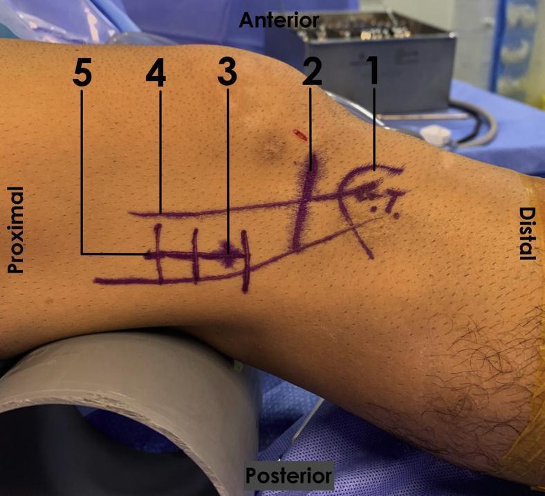
This clinical photograph displays the lateral aspect of a right human knee flexed at approximately 30 degrees, prepared for a surgical procedure (likely iliotibial band lengthening). Preoperative purple skin markers identify key surface anatomy landmarks and surgical planning lines. Numbered labels indicate: (1) Gerdy’s tubercle at the distal insertion of the iliotibial band (ITB); (2) the lateral tibiofemoral joint line; (3) the lateral femoral epicondyle; (4) the longitudinal axis and path of the ITB along the lateral thigh; and (5) the planned 4-cm lateral incision site. Visible distal to the joint line is a 'Z' configuration marking, indicating a planned Z-plasty for ITB lengthening. The image illustrates the anatomical orientation (proximal, distal, anterior, posterior) and the relationship between palpable landmarks and the surgical approach used for treating iliotibial band syndrome in an orthopedic clinical setting.
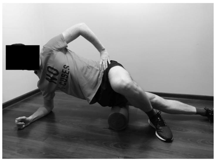
This clinical photograph demonstrates a self-myofascial release (SMFR) technique for the tensor fasciae latae (TFL) and iliotibial (IT) band. The individual is positioned in a lateral side-bridge posture on a wood-laminate floor. Support is maintained by the left forearm and the right foot, which is crossed over the lower leg and planted firmly on the ground to control pressure and movement. A high-density cylindrical foam roller is placed transversely under the lateral aspect of the left thigh, specifically between the iliac crest and the knee joint. The subject’s right hand is placed on the hip to stabilize the torso, which is slightly rotated anteriorly. This exercise is used in physical therapy and sports medicine to improve soft tissue extensibility, reduce myofascial hypertonicity, and manage conditions such as iliotibial band syndrome. The image illustrates the starting position for rolling along the lateral kinetic chain to address muscle tightness in the hip abductors.
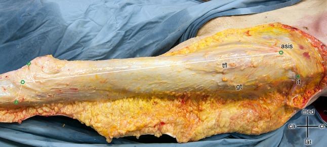
This clinical photograph of a cadaveric specimen illustrates the superficial anatomy and linear measurement of the tractus iliotibialis (iliotibial band, ITB) along the lateral aspect of the right lower limb. The dissection reveals the ITB as a dense, longitudinal band of fibrous connective tissue with a characteristic pale-tan, pearly appearance. A white measurement line extends from the proximal origin at the anterior superior iliac spine (asis) to its distal insertion on the tibia. Key anatomical landmarks visible include the tensor fasciae latae (tfl) muscle, which integrates into the proximal ITB; the iliac tubercle (it); and the greater trochanter (gt) of the femur located deep and posterior to the band. Distally, green points demarcate the tibial plateau level between the fibular head (caput fibulae) and the patellar apex. The orientation is indicated by a directional cross (medial, lateral, cranial, caudal). This image is highly relevant for surgical orthopedics, particularly regarding the Direct Anterior Approach (DAA) to the hip and understanding the biomechanics of the lateral thigh fascia.
This diagnostic image consists of four coronal T2-weighted magnetic resonance imaging (MRI) slices of the left thigh. The imaging demonstrates a localized fluid collection situated in the lateral subcutaneous plane, superficial to the deep fascia and the iliotibial band. This finding is characteristic of a chronic Morel-Lavallée lesion, a post-traumatic internal degloving injury. The collection exhibits high T2 signal intensity with internal septations and a defined pseudocapsule, indicating a chronic stage. In the provided series, the lesion appears reduced in volume compared to previous examinations, as noted in the clinical context. The underlying musculature, including the quadriceps femoris and adductor groups, shows normal signal intensity without evidence of strain or edema. Key anatomical landmarks visible include the femur, hip joint, and knee joint. This image serves as an educational example of the long-term radiological presentation and follow-up of soft tissue shear injuries in orthopedic trauma.
PMID: 19372352
iliotibial band tightness treatment stretching rehabilitation
PMID: 39159925
PMID: 39593624
The IT Band (Iliotibial Tract) and Back Pain: Anatomy, Mechanisms, and Correction
1. Anatomy of the Iliotibial Band (IT Band)
- Proximally: Attaches to the tuberculum of the iliac crest (ASIS region). Here the fascia splits into two layers encasing the tensor fasciae latae (TFL) muscle. It also receives the insertion of the proximal two-thirds of the gluteus maximus muscle.
- Distally: Inserts at Gerdy's tubercle on the anterolateral tibial condyle. Along its course it passes over the lateral femoral epicondyle and parallels the biceps femoris tendon.
- The band also extends deep to connect with the lateral intermuscular septum, anchoring it to the femur.
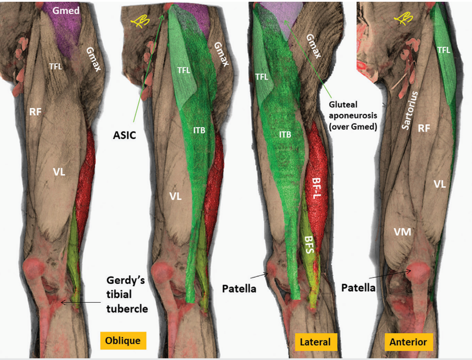
- Tensor fasciae latae (TFL) - arises from the iliac crest between the ASIS and iliac tubercle; innervated by the superior gluteal nerve (L4, L5, S1). Its most important role is postural: steadying the pelvis on the femoral head and stabilizing the femoral condyles on the tibia.
- Gluteus maximus - the proximal two-thirds of its fascicles insert into the ITB. Its proximal origin spans the posterosuperior iliac crest, thoracolumbar fascia (TLF), sacrum, sacroiliac joint region, and sacrotuberous ligament.
2. How the IT Band Causes Back Pain: Mechanisms
2a. The Thoracolumbar Fascia (TLF) Link - Fascial Tension Pathway
- Tension is transmitted retrograde through the gluteus maximus up into the thoracolumbar fascia.
- The TLF is the major posterior fascial structure anchoring the lumbar extensor muscles. Increased tension in it alters loading on the lumbar spine and sacroiliac joint.
- As confirmed by a 2022 review in Clinical Anatomy (Kondrup et al., PMID 35417568), pathological deep fascia (including the ITB) shows increased stiffness, myofibroblast activity, increased nociceptive nerve fiber density, and pro-inflammatory cytokines - all of which propagate pain signals along the fascial continuum.
2b. Gluteus Medius Insufficiency - Pelvic Instability Pathway
- A tight ITB is frequently associated with weakness or inhibition of the gluteus medius.
- Gluteus medius is the primary lateral pelvic stabilizer. When it is insufficient, the pelvis drops on the contralateral side during stance (Trendelenburg pattern).
- To compensate, the lumbar erector spinae and quadratus lumborum on the ipsilateral side hyperactivate to prevent pelvic drop, creating chronic asymmetric loading of the lumbar spine.
- This leads to cumulative stress at the L4-L5 and L5-S1 facet joints and ligaments.
2c. Core Weakness and Movement Pattern Alterations - Kinematic Chain Pathway
- ITB restriction alters hip extension and internal rotation, forcing the lumbar spine to compensate by increasing lordosis or rotating excessively during gait and running.
- Restricted hip mobility (particularly extension) is a well-recognized driver of lumbar hypermobility and segmental overload.
- The ITB also plays a role in stabilizing the hip by preventing lateral displacement of the femoral head from the acetabulum. When it fails this role, altered force vectors travel up the kinematic chain to the pelvis and lumbar region.
2d. Greater Trochanteric Pain Syndrome - Referred Pain Pathway
- A tight ITB rubs over the greater trochanter, contributing to greater trochanteric pain syndrome (GTPS).
- GTPS has a well-documented co-occurrence with low back pain. Williams & Cohen (2009, PMID 19372352) noted that patients with GTPS have a higher prevalence of coexisting LBP, and symptoms can radiate from the lateral hip along the thigh in a pattern that mimics lumbar radiculopathy.
2e. The Sacroiliac Joint Connection
- Because the gluteus maximus attaches both to the ITB and across the sacroiliac joint via the TLF, a tight ITB can produce asymmetric compressive forces at the SIJ.
- Buczek et al. (2026) specifically note that addressing ITB flexibility leads to "substantial reduction in pain of the sacroiliac region."
3. Clinical Presentation
| Feature | Detail |
|---|---|
| Location of pain | Lateral hip, buttock, sacroiliac region, lower lumbar |
| Pattern | Worse with running, prolonged standing, stair climbing |
| Associated findings | Positive Ober test (ITB tightness), weak hip abductors, Trendelenburg sign |
| Common population | Distance runners, cyclists, triathletes, sedentary people with poor hip mechanics |
4. Correction and Treatment
Step 1 - Soft Tissue Release
- Foam rolling (FR) along the lateral thigh and TFL targets myofascial restrictions.
- Instrument-Assisted Soft Tissue Mobilization (IASTM): A 2024 RCT (Unuvar et al., PMID 39159925) in 39 soccer players found both IASTM and foam rolling significantly improved Ober angle, pressure pain threshold, and hip muscle strength compared to exercise alone (P = .001). IASTM and FR produced equivalent results to each other.
- Dry needling: A 2024 clinical trial (Singh et al., PMID 39593624) showed 3 sessions of dry needling added to ITB stretching produced statistically greater reduction in pain (VAS) and disability (LEFS) compared to stretching alone (p = 0.000).
Step 2 - Stretching
- Ober stretch / cross-leg side stretch for the ITB and TFL.
- Hip flexor stretching (Thomas test position) to address concurrent iliopsoas tightness that compounds forward pelvic tilt.
- Consistent daily stretching is foundational before other interventions are added.
Step 3 - Targeted Muscle Strengthening
- Gluteus medius loading - side-lying hip abduction, single-leg stance work, lateral band walks. This is the most important corrective element because gluteus medius insufficiency is identified as a primary driver of the ITB-LBP connection.
- Gluteus maximus activation - hip thrusts, bridges, deadlifts - to normalize ITB tension and restore its normal role in sacroiliac stability.
- Core musculature - transversus abdominis and lumbar multifidus training to reduce compensatory lumbar loading.
Step 4 - Movement Pattern Correction
- Gait retraining to reduce contralateral pelvic drop and hip adduction during running.
- Correction of Trendelenburg gait pattern.
- Addressing training errors (sudden mileage increase, cambered roads, leg length discrepancy) in runners.
Step 5 - In Refractory Cases
- Corticosteroid injection into the trochanteric bursa for GTPS component.
- Surgical ITB lengthening (Z-plasty) is rarely indicated but described in orthopaedic literature when conservative care fails.
Summary
Tight ITB → tension transmitted via gluteus maximus through thoracolumbar fascia → sacroiliac and lumbar loading; PLUS gluteus medius weakness → pelvic instability → lumbar compensatory overload; PLUS altered hip kinematics → lumbar hypermobility.
- Buczek et al. 2026 - "Iliotibial band and lumbar spine pain - a hidden connection" (PMID 41962100)
- Kondrup et al. 2022 - "The deep fascia and its role in chronic pain" (PMID 35417568)
- Unuvar et al. 2024 - "Myofascial Release Techniques in ITB Tightness" (RCT) (PMID 39159925)
- Singh et al. 2024 - "Dry needling in ITB tightness" (PMID 39593624)
- Williams & Cohen 2009 - "Greater trochanteric pain syndrome" (PMID 19372352)
- Gray's Anatomy for Students, pp. 674, 795-796
- Imaging Anatomy, Vol. 3 (Bones, Joints, Vessels and Nerves), pp. 255-257
Why does hip extension and internal rotation gets affected due to IT band tightness?
iliotibial band tightness hip extension internal rotation biomechanics
tensor fasciae latae action hip flexion abduction rotation pelvis anatomy diagram
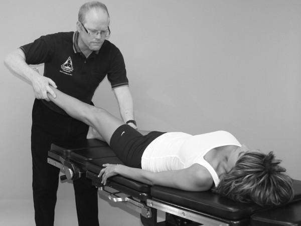
A clinical photograph in black and white demonstrating a Manual Muscle Test (MMT) for the tensor fasciae latae (TFL). The image depicts a patient in a supine position on a clinical examination table. The patient's right leg is extended and elevated at an angle, representing the test position for TFL assessment involving hip flexion, abduction, and internal rotation. A male clinical examiner stands at the foot of the table, applying manual resistance to the patient's distal lower leg near the ankle with both hands. The examiner is positioned with an upright posture, maintaining visual focus on the limb's response. The patient's head is turned away from the side being tested. This visual serves as an educational guide for physical therapy, chiropractic, or orthopedic assessment, illustrating proper practitioner hand placement, patient positioning, and the line of force required to isolate the tensor fasciae latae while minimizing synergist substitution during muscle strength evaluation.
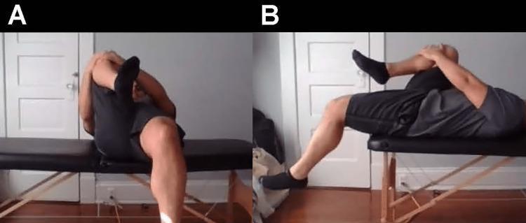
Two clinical photographs demonstrate the performance of the Modified Thomas Test, a physical examination maneuver used to assess hip flexor contractures. Image A shows an anterior view of a patient in a supine position at the edge of an examination table. The patient has one hip and knee fully flexed, held against the chest by the arms to stabilize the pelvis and flatten the lumbar spine. The contralateral leg is allowed to hang freely. This view is used to observe for hip abduction (suggesting tensor fasciae latae involvement) or external rotation (suggesting iliotibial band restriction). Image B provides a lateral view of the same maneuver. This perspective specifically evaluates the degree of hip extension and knee flexion of the hanging leg. A lack of full hip extension indicates psoas or iliopsoas tightness, while an inability to maintain at least 90 degrees of knee flexion suggests rectus femoris contracture. The test is fundamental in musculoskeletal medicine and physical therapy for diagnosing imbalances in the anterior hip and thigh musculature.
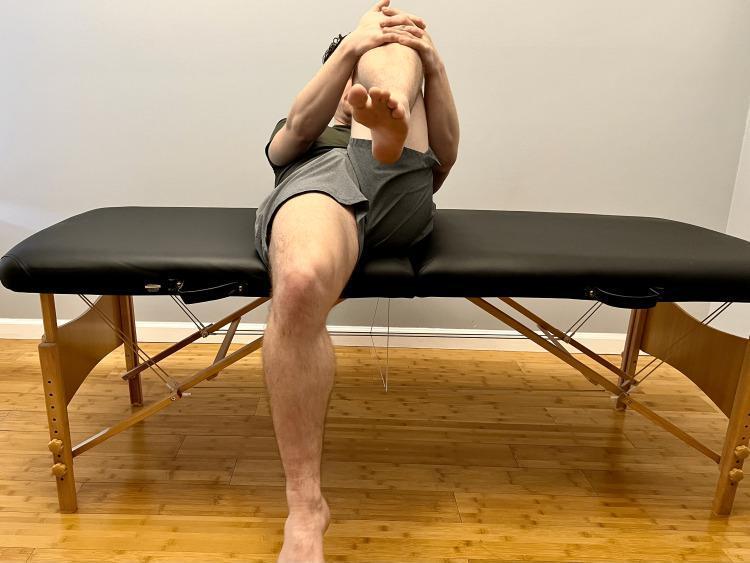
This clinical photograph demonstrates the performance of a Modified Thomas test, a common orthopedic physical examination maneuver used to assess the flexibility of the hip flexors. The patient is positioned supine on a treatment table. One leg (the contralateral limb) is flexed at the hip and knee, with the patient using their hands to pull the knee toward the chest to stabilize the pelvis and flatten the lumbar spine. The test limb is allowed to extend. This visual assessment is used to identify potential muscular tightness or contractures: hip flexion in the test limb suggests psoas involvement, knee extension suggests rectus femoris tightness, and hip abduction or rotation may indicate iliotibial band (ITB) restrictions or tensor fasciae latae (TFL) involvement. This diagnostic maneuver is critical in sports medicine and physical therapy for evaluating hip and knee pathology during a musculoskeletal examination.
iliotibial band hip kinematics gait running
PMID: 36758425
PMID: 31999979
Why IT Band Tightness Restricts Hip Extension and Internal Rotation
Part 1: The TFL - The Root Cause
"Stimulation of the TFL causes extension of the knee, lateral rotation of the leg, and medial rotation of the thigh. Its most important function is its postural role: steadying the pelvis on the head of the femur and the femoral condyles on the tibial condyles."
- Resists hip extension (because it pulls towards flexion)
- Resists hip adduction (because it pulls towards abduction)
- Does NOT restrict internal rotation directly - but the story is more nuanced (see Part 2 below)
Part 2: The IT Band as a Passive Lateral Tether - How it Blocks Extension
2a. Restriction of Hip Extension
- The ITB runs anterior to the hip's axis of flexion-extension in its proximal portion (particularly through the TFL component).
- With the hip in neutral or extended position, a tight ITB + TFL pulls the femur anteriorly and into flexion relative to the pelvis - preventing the hip from reaching full extension.
- Campbell's Operative Orthopaedics (p. 6451-6463) makes this explicit: contracture of the ITB directly produces a "flexion and abduction contracture of the hip" - the hip becomes fixed in flexion and abduction, and the corrective movements required are "extension, adduction, and internal rotation."
"The large expanse of the tensor fasciae latae must be recognized before the deforming possibilities of the iliotibial band can be appreciated... Contracture of the iliotibial band can contribute to flexion and abduction contracture of the hip." - Campbell's Operative Orthopaedics, p. 6452-6460
2b. Restriction of Hip Adduction (and the Link to Apparent Internal Rotation Loss)
- The ITB runs along the lateral side of the thigh. When tight, it acts as a lateral restraint preventing the hip from moving into adduction.
- The Ober test directly tests this: the hip is abducted, then released to fall into adduction under gravity. A tight ITB prevents gravity-driven adduction - the leg stays abducted.
- Per Campbell's: "A positive Ober test with the hip extended past neutral signifies tightness of the iliotibial band."
2c. Why Internal Rotation is Restricted - The Biomechanical Explanation
- It posteriorly rotates the ilium on the affected side due to its pull from the iliac crest downward. This creates an anterior pelvic tilt + ipsilateral posterior iliac rotation pattern.
- The hip compensates into external rotation to unload the tight lateral band - a natural protective posture.
- Over time, the external rotator muscles (piriformis, obturators, gemelli) become adaptively shortened from this habitual external rotation posture.
- The result: the hip is held in slight flexion + abduction + external rotation, and internal rotation range is reduced.
"Female runners with current iliotibial band syndrome exhibited smaller peak hip internal rotation angles and lower isometric hip abductor strength compared to controls... transverse plane hip motion and hip abductor strength weakness may be biomechanical risk factors in female runners with ITBS."
Part 3: The Full Kinematic Chain Summary
Tight TFL / ITB
↓
Hip held in flexion + abduction + (compensatory) external rotation
↓
Hip extension BLOCKED → because TFL spans anterior to flexion axis
Hip adduction BLOCKED → because ITB is a non-elastic lateral tether
↓
External rotators become adaptively shortened
↓
Hip INTERNAL ROTATION BLOCKED → passive tethering + compensatory ER posture
↓
During gait/running: hip cannot fully extend in terminal stance
→ Lumbar spine HYPEREXTENDS or ROTATES to compensate
→ Pelvis tilts anteriorly
→ Increased L4/L5 and sacroiliac joint loading → BACK PAIN
Part 4: Clinical Test Correlation
| Test | What It Detects |
|---|---|
| Ober test | ITB tightness - hip adduction restricted by lateral tether |
| Modified Thomas test | Hip extension restricted - if the hanging leg stays flexed AND abducted, TFL/ITB is implicated (not just iliopsoas) |
| FABER test | Hip abducts + externally rotates easily (the direction of tightness toward) |
| Internal rotation ROM | Reduced passive/active internal rotation on the tight side |
Summary
| Motion Lost | Reason |
|---|---|
| Hip extension | TFL is a hip flexor - when short/tight it resists extension; ITB taut cable prevents posterior femoral glide |
| Hip adduction | ITB is a rigid lateral band - physically prevents the hip from crossing into adduction (Ober test positive) |
| Hip internal rotation | Compensatory external rotation posture adopted to unload the tight lateral band; external rotators shorten; research confirms smaller peak hip internal rotation in ITBS runners (Foch et al. 2023, PMID 36758425) |
Can you say how the special test are performed with picture diagram step-by-step?
Ober test iliotibial band tightness lateral decubitus position clinical examination
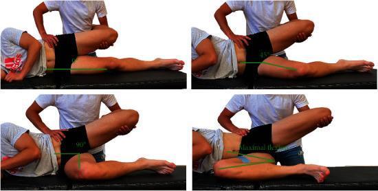
A series of four clinical photographs demonstrating variations of the Ober test, a physical examination maneuver used to assess iliotibial band (ITB) tightness. The patient is positioned in a side-lying (lateral decubitus) posture on an examination table. The examiner stands behind the patient, with one hand stabilizing the superolateral pelvis and the other arm cradling the upper leg with the knee flexed to 90 degrees. The four panels illustrate the test performed with different angles of contralateral (bottom) hip flexion: 0 degrees (extension), 45 degrees of flexion, 90 degrees of flexion, and maximal flexion (stabilized by a belt). Green angular markers indicate the degree of contralateral hip flexion in each scenario. The test focuses on the degree of adduction achievable in the top hip while maintaining the pelvis in a neutral position, illustrating how lumbar and pelvic positioning—influenced by the contralateral hip—can affect clinical measurements of ITB and tensor fasciae latae flexibility.
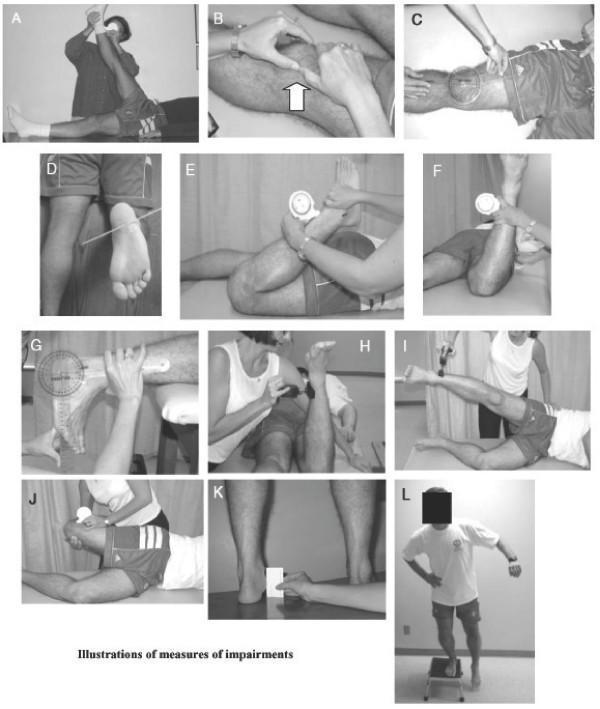
A composite of 12 clinical photographs (labeled A through L) demonstrating physical examination maneuvers and orthopedic tests for evaluating Patellofemoral Pain Syndrome (PFPS). (A) Straight leg raise test for hamstring length. (B) Patellar tilt test assessing lateral retinacular tightness. (C) Measurement of the Q-angle using a universal goniometer. (D) Evaluation of tibial torsion via the intermalleolar axis in a prone position. (E) Quadriceps muscle length measurement using a gravity goniometer during passive knee flexion. (F) Craig’s test for femoral anteversion. (G) Assessment of plantar flexor length (ankle dorsiflexion) with a goniometer. (H) Isometric hip external rotation strength testing. (I) Hip abduction strength testing in a side-lying position. (J) Ober’s test using a gravity goniometer to measure Iliotibial Band (ITB) complex length. (K) Navicular drop test for foot pronation, utilizing an index card and ruler. (L) Functional assessment via the lateral step-down test, observing movement quality and compensation strategies. The collection illustrates diagnostic pathways for biomechanical and musculoskeletal impairments of the lower extremity.
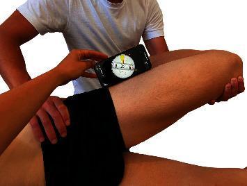
This clinical photograph demonstrates the objective measurement of the Ober test using a smartphone-based digital inclinometer. The patient is positioned in a side-lying posture. The examiner’s left hand is placed on the superolateral aspect of the patient's pelvis to provide stabilization and prevent compensatory pelvic tilt. The examiner’s right arm supports and cradles the patient's flexed knee (at approximately 90 degrees) while guiding the hip through abduction, extension, and into adduction. A mobile device running a clinometer application is positioned on the lateral mid-thigh, aligned between the anterior superior iliac spine (ASIS) and the lateral femoral condyle. The digital interface displays a measurement of 21 degrees, indicating the hip range of motion or degree of adduction at the point of tissue resistance. This procedure is utilized in physical therapy and orthopedics to assess the flexibility or tightness of the iliotibial band (ITB) and tensor fasciae latae (TFL) complex.
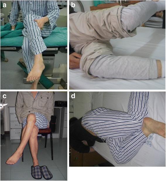
This four-panel clinical photograph illustrates the functional manifestations of bilateral gluteal muscle contracture (GMC) and the results following arthroscopic release. (a) Pre-operative view showing the 'cross-leg sign'; the patient is unable to cross their legs while sitting due to limited hip adduction and flexion. (b) Pre-operative Ober's test demonstrating a positive result, where the upper leg remains abducted and cannot adduct toward the examination table while the patient is in a lateral decubitus position, indicating tight iliotibial bands or gluteal fascia. (c) Post-operative functional assessment three days after arthroscopic release, showing the patient successfully crossing their legs without assistance. (d) Post-operative Ober's test demonstrating a negative result, with the upper leg now able to adduct fully toward the table. The series demonstrates the clinical efficacy of surgical release in restoring hip range of motion, specifically adduction and flexion, which are pathognomonic limitations in GMC.
modified Thomas test hip flexor contracture TFL iliotibial band clinical examination
Two clinical photographs demonstrate the performance of the Modified Thomas Test, a physical examination maneuver used to assess hip flexor contractures. Image A shows an anterior view of a patient in a supine position at the edge of an examination table. The patient has one hip and knee fully flexed, held against the chest by the arms to stabilize the pelvis and flatten the lumbar spine. The contralateral leg is allowed to hang freely. This view is used to observe for hip abduction (suggesting tensor fasciae latae involvement) or external rotation (suggesting iliotibial band restriction). Image B provides a lateral view of the same maneuver. This perspective specifically evaluates the degree of hip extension and knee flexion of the hanging leg. A lack of full hip extension indicates psoas or iliopsoas tightness, while an inability to maintain at least 90 degrees of knee flexion suggests rectus femoris contracture. The test is fundamental in musculoskeletal medicine and physical therapy for diagnosing imbalances in the anterior hip and thigh musculature.
This clinical photograph demonstrates the performance of a Modified Thomas test, a common orthopedic physical examination maneuver used to assess the flexibility of the hip flexors. The patient is positioned supine on a treatment table. One leg (the contralateral limb) is flexed at the hip and knee, with the patient using their hands to pull the knee toward the chest to stabilize the pelvis and flatten the lumbar spine. The test limb is allowed to extend. This visual assessment is used to identify potential muscular tightness or contractures: hip flexion in the test limb suggests psoas involvement, knee extension suggests rectus femoris tightness, and hip abduction or rotation may indicate iliotibial band (ITB) restrictions or tensor fasciae latae (TFL) involvement. This diagnostic maneuver is critical in sports medicine and physical therapy for evaluating hip and knee pathology during a musculoskeletal examination.
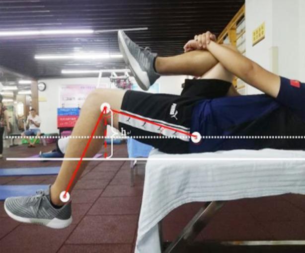
This clinical photograph demonstrates the performance of a Thomas test, a physical examination maneuver used to assess hip flexor contracture and psoas tightness. The subject is positioned supine on an examination table with the sacrum at the table's edge. One hip is maximally flexed toward the chest and held in place by the subject's arms to flatten the lumbar spine. The contralateral leg hangs freely off the table. The image is overlaid with quantitative diagnostic annotations: a horizontal white dotted line represents the pelvic baseline, solid white lines indicate the table level, and red lines delineate the femur and tibia segments. A vertical indicator labeled 'h' measures the elevation of the thigh from the table surface. This visual focuses on the biomechanical assessment of the iliopsoas and rectus femoris muscles, with the failure of the extended thigh to touch the table surface indicating a positive test for hip flexor tightness.
FABER test FADIR test hip flexion abduction external rotation internal rotation clinical
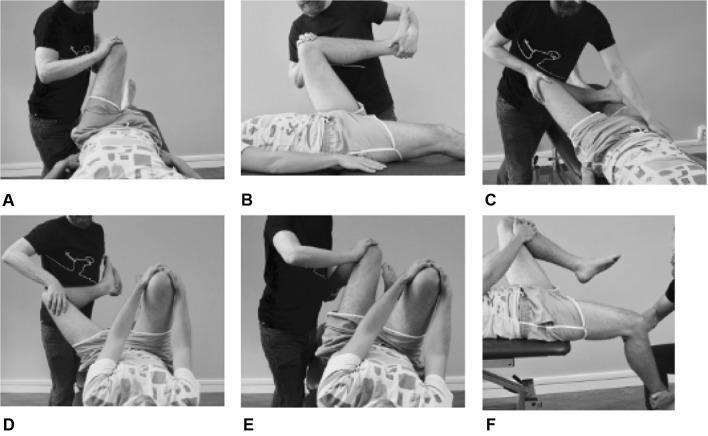
A composite of six clinical photographs (a–f) demonstrating a systematic orthopedic physical examination of the hip joint for femoroacetabular impingement (FAI) and labral pathology. The images show a patient in the supine position while an examiner performs specific provocative maneuvers. (a) AIMT (Anterior Impingement Test): hip at 90° flexion with internal rotation and adduction. (b) FADIR test: maximal hip flexion, adduction, and internal rotation to assess for anterosuperior impingement. (c) FABER (Patrick’s) test: the examined leg is in flexion, abduction, and external rotation with the foot placed proximal to the contralateral knee; the examiner stabilizes the contralateral pelvis. (d) DEXRIT and (e) DIRIT: Dynamic External Rotation and Internal Rotation Impingement Tests involving wide arcs of motion while the patient holds the contralateral hip in >90° flexion to stabilize the lumbar spine. (f) PRIMT (Posterior Rib-Impingement Test): the patient is positioned at the edge of the table with the contralateral hip flexed while the tested hip is moved into extension, abduction, and external rotation to assess posterior impingement.
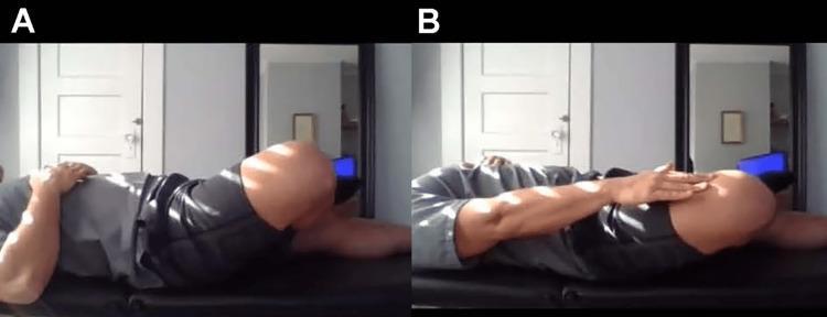
This clinical photograph demonstrates the FABER (Flexion, Abduction, and External Rotation) test, also known as Patrick's test, used to evaluate hip and sacroiliac joint pathology. Part A shows a patient in the supine position on an examination table with the right leg placed in a 'figure-four' configuration: the hip is flexed, abducted, and externally rotated so that the lateral malleolus rests on the contralateral knee. Part B illustrates the progression of the maneuver, where the patient applies downward pressure onto the medial aspect of the flexed knee. This movement increases the degree of abduction and external rotation. Clinically, pain elicited during the initial positioning (A) may indicate intra-articular hip joint pathology, such as osteoarthritis or labral tears. Pain elicited during the application of overpressure (B) is more suggestive of sacroiliac joint dysfunction or posterior hip involvement. The image serves as a guide for physical examination techniques in orthopedics and primary care.
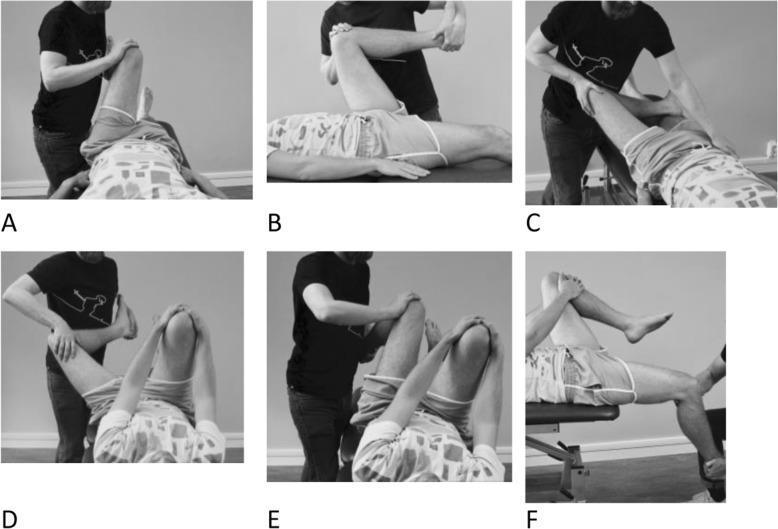
A series of six clinical photographs (A-F) demonstrating common orthopedic physical examination maneuvers for diagnosing hip impingement syndromes. All images show a patient in a supine position on an examination table with an examiner performing passive movements. (A) Anterior Impingement Test (AIMT) with 90° flexion, medial rotation, and adduction. (B) FADIR test showing maximal flexion, medial rotation, and adduction. (C) FABER (Patrick’s) test with the foot placed proximal to the contralateral knee, involving combined flexion, abduction, and external rotation while stabilizing the pelvis. (D) DEXRIT and (E) DIRIT involve the patient holding the contralateral hip in >90° flexion while the examiner moves the tested hip through arcs of extension/abduction/external rotation or extension/adduction/internal rotation. (F) Posterior Rim Impingement Test (PRIMT) with the patient at the table's edge, keeping the contralateral hip flexed while the tested hip undergoes extension, abduction, and lateral rotation. These tests are essential for evaluating femoroacetabular impingement (FAI) and labral pathology in sports medicine and orthopedics.
Trendelenburg test hip abductor weakness gluteus medius single leg stance
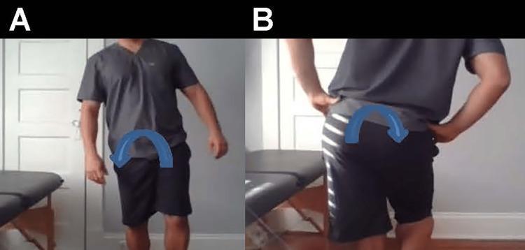
Clinical photograph sequence illustrating a positive Trendelenburg sign and compensated Trendelenburg gait. Image A presents an anterior view of an individual during the stance phase on the left leg. There is a visible lateral trunk lean (compensated gait) toward the stance side, indicated by a blue curved arrow, to maintain pelvic stability despite hip abductor weakness. Image B provides a posterior view demonstrating a significant pelvic drop on the contralateral (right) side while standing on the left leg, indicated by a blue downward-curving arrow. This pelvic tilt reflects insufficiency of the gluteus medius and minimus muscles on the weight-bearing side. The sequence highlights key physical examination findings for hip abductor weakness, typically assessed in sports medicine, orthopedics, and physical therapy to diagnose conditions like gluteal tendinopathy or superior gluteal nerve palsy.
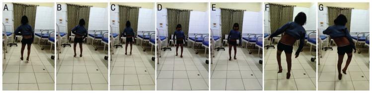
A series of clinical photographs of a 13-year-old female patient demonstrating gait abnormalities and physical examination findings indicative of hip abductor weakness. Panels A through E capture the patient's gait from posterior and anterior views, showing a waddling pattern characterized by lateral trunk oscillations and pelvic instability, consistent with bilateral Trendelenburg gait. Panels F and G display a bilateral positive Trendelenburg test. In these clinical frames, the patient is standing on a single leg; the contralateral side of the pelvis drops (sags) rather than remaining level or rising. To maintain balance and compensate for the pelvic tilt, the patient exhibits a characteristic lateral lean of the torso toward the weight-bearing side. This visual documentation serves as a classic educational representation of gluteus medius and minimus insufficiency, often associated with neuromuscular disorders, proximal myopathies, or hip pathologies. The images clearly illustrate the biomechanical failure of the hip abductor mechanism and the resulting compensatory movements of the upper body and pelvis.
Special Tests for ITB Tightness and Related Hip Dysfunction - Step-by-Step with Pictures
TEST 1: The Ober Test
What it assesses
Step-by-Step Procedure
- Negative (normal): The leg drops down passively toward or below the table level - ITB is flexible enough
- Positive: The leg remains elevated - unable to adduct to table level - indicating ITB/TFL contracture
Interpretation nuance (from Campbell's Operative Orthopaedics, p. 3134-3135):
| Positive Ober position | Meaning |
|---|---|
| Hip extended past neutral | ITB tightness |
| Hip in neutral flexion/extension | Gluteus medius contracture or tendinopathy |
TEST 2: Modified Thomas Test
What it assesses
Step-by-Step Procedure
Reading the result - what each position means:
| Observation of the hanging leg | Tight structure | Explanation |
|---|---|---|
| Thigh stays elevated (hip stays flexed) | Iliopsoas | Primary hip flexor contracture |
| Thigh abducts laterally away from midline | TFL / ITB | The lateral band pulls it into abduction |
| Knee extends (straightens out) | Rectus femoris | Anterior thigh tightness |
| Hip externally rotates | ITB restriction pattern | Compensatory external rotation |
The ITB/TFL component is specifically identified when the leg abducts rather than just failing to reach the table. This is the key finding distinguishing TFL from pure iliopsoas tightness.
TEST 3: FADIR Test (Flexion-Adduction-Internal Rotation)
Step-by-Step Procedure
- Adducts the hip (brings knee across midline)
- Internally rotates the hip (lower leg moves laterally)
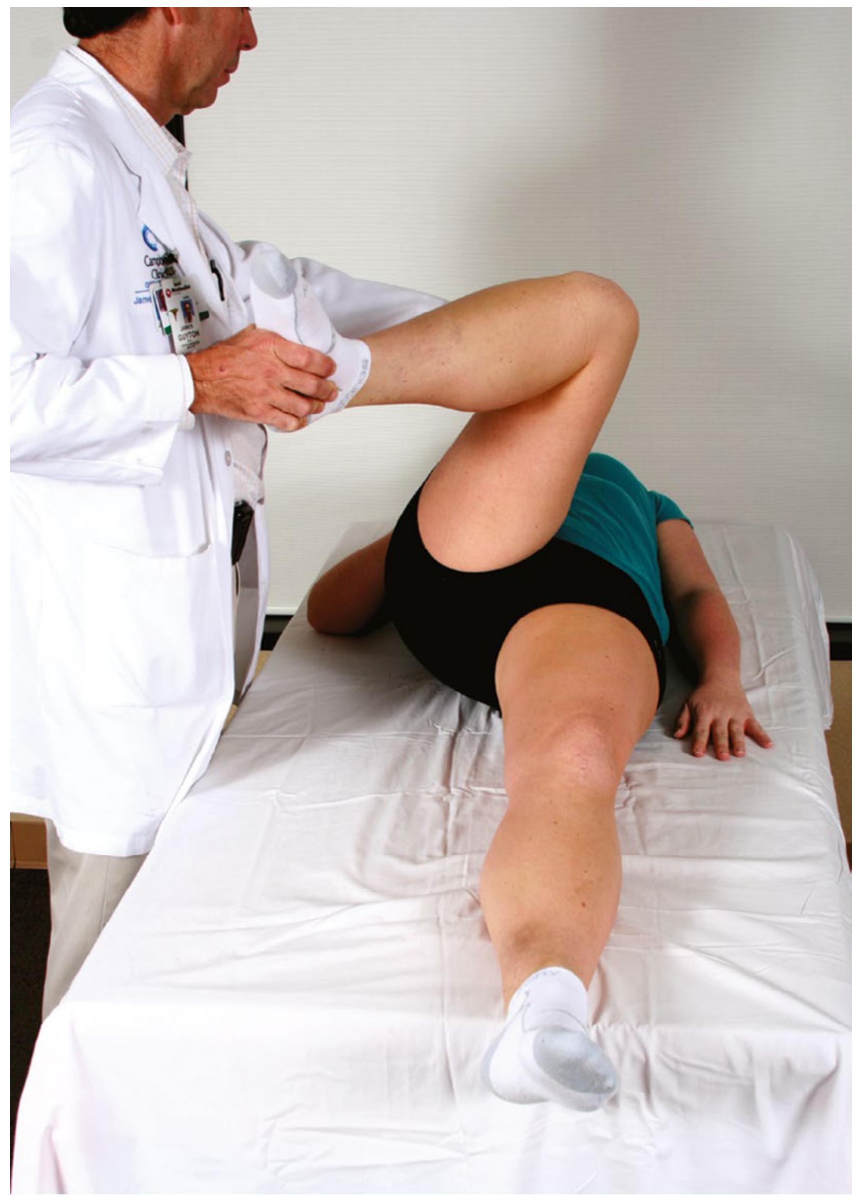
Relevance to ITB:
TEST 4: FABER Test (Flexion-Abduction-External Rotation) / Patrick's Test
Step-by-Step Procedure
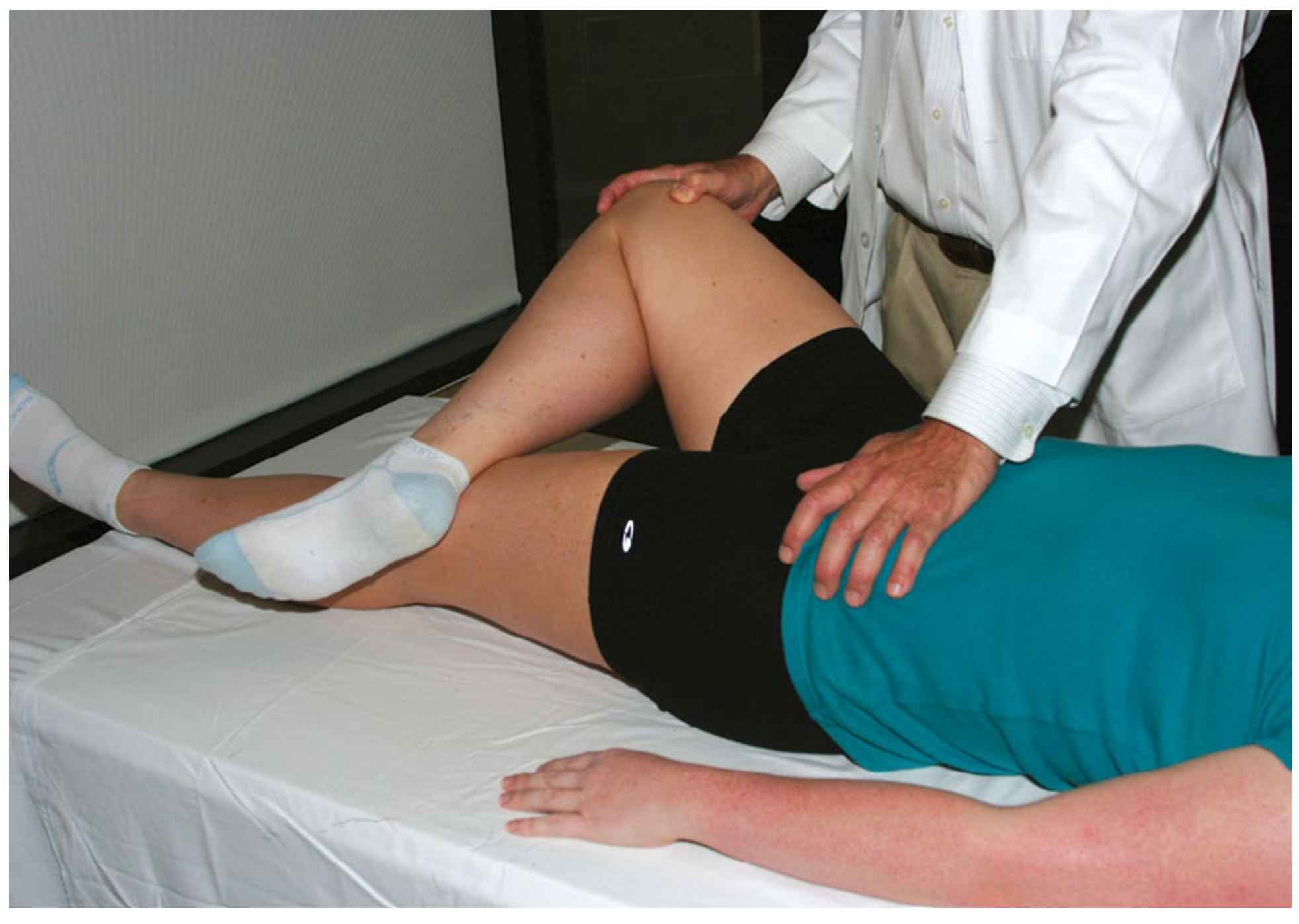
Reading the result:
| Finding | Suggests |
|---|---|
| Groin pain with positioning | Intra-articular hip pathology (OA, labral tear) |
| Posterior hip / SIJ pain with overpressure | Sacroiliac joint dysfunction |
| Lateral hip / greater trochanter pain | ITB tightness, trochanteric bursitis |
| Knee falls easily to table | Normal hip external rotation and abduction |
TEST 5: Trendelenburg Test
Step-by-Step Procedure
Reading the result:
| Finding | Meaning |
|---|---|
| Pelvis drops on the lifted side | Positive Trendelenburg - gluteus medius weakness on stance side |
| Trunk leans toward the stance leg | Compensated Trendelenburg - patient uses lateral trunk lean to maintain balance |
| Pelvis stays level | Negative - gluteus medius functioning normally |
Connection to ITB back pain:
Quick Reference Summary Table
| Test | Position | Key Action | Positive Finding | What it Means |
|---|---|---|---|---|
| Ober | Side-lying | Abduct + extend hip, release | Leg stays abducted | ITB/TFL contracture |
| Modified Thomas | Supine, leg off table edge | Lower test leg freely | Leg abducts (not just stays up) | TFL/ITB tightness |
| FADIR | Supine | Flex + Adduct + Int. Rotate | Pain / restricted motion | Hip impingement; restricted IR confirms ITB pattern |
| FABER | Supine | Flex + Abduct + Ext. Rotate | Lateral hip pain | ITB / trochanteric involvement |
| Trendelenburg | Standing, single leg | Observe pelvis | Contralateral pelvic drop | Gluteus medius weakness |