
amniotic fluid and its disorders in obs
Reading File
Now let me search the medical library for amniotic fluid content in parallel.
Finding Sources
Finding Sources
Searching PubMed
"amniotic fluid" AND "disorders" AND obstetrics
Excellent - found rich content in Creasy & Resnik's Maternal-Fetal Medicine. Let me read multiple sections in parallel.
Reading File
Reading File
Finding Sources
Finding Sources
Reading File
Reading File
Finding Sources
Finding Sources
Reading File
Finding Sources
Reading File
Finding Sources
Reading File
Reading File
Now I have comprehensive information from Creasy & Resnik's. Let me also fetch the amniotic fluid volume image and search for polyhydramnios/oligohydramnios clinical details.
Loading Image
Finding Sources
I now have comprehensive data from Creasy & Resnik's Maternal-Fetal Medicine. Let me compile a thorough, well-structured answer.
Amniotic Fluid and Its Disorders in Obstetrics
Source: Creasy & Resnik's Maternal-Fetal Medicine: Principles and Practice
1. What Is Amniotic Fluid?
Amniotic fluid (AF) is essential for normal fetal growth and development. Its key roles include:
- Cushioning - protects the fetus from mechanical trauma
- Bacteriostatic properties - helps maintain a sterile intrauterine environment
- Space for movement - allows fetal mobility and supports normal development of lungs and limbs
- Diagnostic access - provides fetal cells and metabolic byproducts; used in amniocentesis (first performed in 1956 for sex determination)
2. Volume of Amniotic Fluid
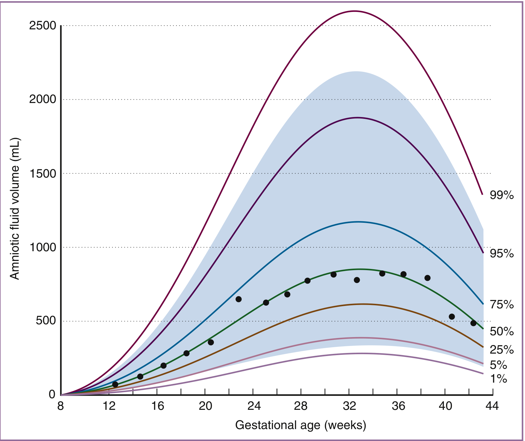
AF volume changes predictably throughout pregnancy:
| Gestational Age | AF Volume |
|---|---|
| 8 weeks | < 10 mL |
| 22 weeks | ~630 mL |
| 28 weeks | ~770 mL |
| 34-36 weeks | Maximum (~800 mL) |
| 41 weeks | ~515 mL |
| Post-term | Falls ~33% per week |
Key dynamics:
- Volume increases at 10 mL/week early in fetal period, peaks at 50-60 mL/week at 19-25 weeks
- After 34 weeks, volume falls at 60-70 mL/week at 40 weeks
- The late-pregnancy decline is a physiologic progression, not an aberration
3. Composition of Amniotic Fluid
First trimester:
- Isotonic with maternal/fetal plasma
- Minimal protein
- Very low oxygen tension
- High concentration of sugar alcohols (product of anaerobic metabolism)
- Arises as a transudate of plasma - through nonkeratinized fetal skin or across uterine decidua/placenta
Second half of pregnancy (mid-trimester onward):
- Fetal urine becomes the major component (fetus produces dilute urine)
- AF osmolality falls to ~85-90% of maternal serum osmolality (drops by 20-30 mOsm/kg with advancing gestation)
- AF urea, creatinine, and uric acid increase to 2-3x fetal plasma levels
- The entire volume of AF turns over on a daily basis - a highly dynamic system
4. Production and Resorption - Sources and Sinks
| Source/Sink | Role |
|---|---|
| Fetal urine | Major source in second half of pregnancy |
| Fetal lung fluid | Secreted into AF |
| Fetal swallowing | Major route of resorption (~500-1000 mL/day near term) |
| Intramembranous (IM) flow | Water crosses amnion into fetal vasculature |
| Transmembranous flow | Across uterine wall to maternal circulation |
The amnion is a "leaky" epithelium with significant water flux potential, comparable to renal tubular epithelium. IM flow can be modulated to maintain AF volume homeostasis - for example, when fetal swallowing is experimentally blocked, IM flow increases to normalize volume.
5. Ultrasound Assessment of AF Volume
Two main methods are used clinically:
- Maximum Vertical Pocket (MVP) - single deepest pocket
- Amniotic Fluid Index (AFI) - sum of MVP in four uterine quadrants
Both can be used; subjective assessment is also acceptable. Per ISUOG guidelines, AFI and MVP are used to classify volume as normal, reduced, or excessive.
6. Disorders of Amniotic Fluid
A. Polyhydramnios (Hydramnios) - Excess AF
Definition: AFI > 24-25 cm or MVP > 8 cm
Classification by AFI:
| Grade | AFI |
|---|---|
| Mild | 25-30 cm |
| Moderate | 30.1-35 cm |
| Severe | > 35.1 cm |
Causes:
- Impaired fetal swallowing - neurologic abnormalities, obstructions
- GI anomalies - esophageal atresia, tracheoesophageal fistula
- Respiratory anomalies - congenital diaphragmatic hernia
- Maternal diabetes mellitus - fetal hyperglycemia leads to osmotic diuresis
- Fetal anemia - increased cardiac output and urine flow
- Twin-to-twin transfusion syndrome (TTTS) - recipient twin
- Idiopathic - no identifiable cause in many cases
- Hypoproteinemia with decreased maternal plasma oncotic pressure
Consequences / Complications:
- Preterm birth (PTB): 18.5% with mild, 21.8% with moderate, 14.3% with severe polyhydramnios
- Uterine overdistension triggers inflammatory cytokines (IL-1β, TNF-α, IL-6, IL-8, CCL2, PGE2, PGF2α) - causing preterm labor
- The underlying cause (congenital malformations, diabetes) determines PTB risk more than the fluid volume itself
- Maternal discomfort, dyspnea
- Malpresentation
- Cord prolapse
- Placental abruption (after rapid decompression)
B. Oligohydramnios - Reduced AF
Definition: AFI < 5 cm or MVP < 2 cm
Causes:
- Renal agenesis / bilateral renal anomalies - no urine production (Potter sequence)
- Posterior urethral valves / lower urinary tract obstruction - leads to oligohydramnios, pulmonary hypoplasia, renal dysplasia, Potter facies, clubfeet
- Placental insufficiency / uteroplacental insufficiency - reduced fetal perfusion reduces urine output
- Post-term pregnancy - AF falls 33%/week after 41 weeks (physiologic)
- Rupture of membranes (PPROM/PROM) - loss of AF
- Maternal dehydration - osmotic mechanism; rehydration can reverse it
Consequences:
- Pulmonary hypoplasia - AF is required for normal lung development; severe oligohydramnios early in pregnancy is particularly dangerous
- Limb deformities - restricted fetal movement
- Potter sequence - renal agenesis → oligohydramnios → pulmonary hypoplasia, Potter facies (flat nose, low-set ears, epicanthal folds), limb contractures
- Fetal distress - cord compression, fetal hypoxia
- Perinatal mortality - higher with severe oligohydramnios
- Intrauterine growth restriction (IUGR) association
C. Amniotic Fluid Embolism (AFE)
AFE is a rare but potentially fatal obstetric emergency:
Nature: A "spectrum disorder" - not precisely an embolism nor strictly amniotic fluid-related. It reflects a breach in the normal physiologic barrier between mother and fetus, with an abnormal maternal response to fetal tissue exposure - similar to systemic inflammatory response syndrome (SIRS) and anaphylaxis.
Diagnostic Triad (SMFM/AFE Foundation criteria):
- Hemodynamic compromise (cardiovascular collapse)
- Respiratory compromise
- Disseminated intravascular coagulopathy (DIC)
Clinical features:
- Sudden cardiovascular collapse and cardiac arrest
- Severe hypoxia
- DIC with hemorrhage
- Often occurs during labor, delivery, or within 30 minutes postpartum
Management (supportive - no specific treatment):
- Early intubation + 100% O2 + PEEP
- High-quality CPR immediately for cardiac arrest
- If AFE occurs before delivery → emergent cesarean delivery to improve maternal resuscitation and fetal survival
- Large-bore IV access + arterial line
- Transesophageal echocardiography after intubation - to manage pulmonary hypertension and cardiac failure
- Pressors, inotropes, pulmonary vasodilators as needed
- ECMO - consider after prolonged arrest or severe ventricular dysfunction unresponsive to medical management
- Massive transfusion: Packed RBCs : FFP : Platelets in 1:1:1 ratio
- TEG / ROTEM point-of-care testing for coagulopathy guidance
- Note: Recombinant activated Factor VIIa is controversial - a systematic review found it may worsen outcomes in AFE
Cannot be predicted or prevented.
7. Summary Table
| Feature | Polyhydramnios | Oligohydramnios |
|---|---|---|
| Definition | AFI > 24-25 cm / MVP > 8 cm | AFI < 5 cm / MVP < 2 cm |
| Key causes | GI obstruction, diabetes, fetal anemia, TTTS, idiopathic | Renal agenesis, LUTO, placental insufficiency, PROM, post-term |
| Complications | PTB, malpresentation, cord prolapse, abruption | Pulmonary hypoplasia, limb deformities, Potter sequence, IUGR |
| Mechanism of PTB | Uterine overdistension → inflammatory cytokines | - |
| Key diagnostic tool | Ultrasound (AFI/MVP) | Ultrasound (AFI/MVP) |
References: Creasy & Resnik's Maternal-Fetal Medicine: Principles and Practice - Chapter 4 (Amniotic Fluid Dynamics, Ross & Beall) and Chapter 18 (Amniotic Fluid Embolism, p. 6137-6146).

169f91eb-5fc9-4ff5-83a0-0611c44032a0.jpeg
breast
Reading File
The image shows a surgery/pathology question paper covering: A) Classification of breast tumors, B) Carcinoma Breast - etiopathogenesis, clinical features, TNM staging, management, spread, prognostic factors, and breast reconstruction & MRM(SM), C) ANDI of breast, and VSAQ on fibroadenoma. Let me now search the library in parallel for all these topics.
Finding Sources
Finding Sources
Finding Sources
Reading File
Finding Sources
Finding Sources
Reading File
Finding Sources
Reading File
Reading File
Reading File
Reading File
Reading File
Reading File
Reading File
I now have all the key content. Let me compile a comprehensive answer covering everything in the question paper.
Breast - Complete Surgery Notes
Source: Bailey and Love's Short Practice of Surgery, 28th Edition; Robbins & Cotran Pathologic Basis of Disease
A) Classification of Breast Tumors
I. Benign Tumors
- Fibroadenoma - most common benign lump in young women
- Cystosarcoma phyllodes (Phyllodes tumor) - benign/borderline/malignant
- Lipoma
- Duct papilloma - solitary or multiple (papillomatosis)
- Adenoma (tubular, lactating)
- Granular cell tumor
II. Malignant Tumors (Carcinoma)
Non-invasive (In Situ):
- Ductal Carcinoma In Situ (DCIS)
- Lobular Carcinoma In Situ (LCIS) - high-risk benign lesion per 8th AJCC edition
Invasive:
- Invasive Ductal Carcinoma (IDC / No Special Type) - most common (70-80%)
- Invasive Lobular Carcinoma (ILC)
- Mucinous (colloid) carcinoma
- Tubular carcinoma
- Medullary carcinoma
- Papillary carcinoma
- Inflammatory carcinoma (T4d)
- Paget's disease of nipple
Molecular Subtypes (IHC-based):
| Subtype | ER | PR | HER2 | Notes |
|---|---|---|---|---|
| Luminal A | + | + | - | Best prognosis |
| Luminal B | + | +/- | +/- | Intermediate |
| HER2-enriched | - | - | + | Targeted therapy |
| Triple Negative (TNBC) | - | - | - | Poorest prognosis; common in BRCA1 |
B) Carcinoma Breast
Etiopathogenesis (Risk Factors)
Non-modifiable:
- Female sex, increasing age
- Early menarche, late menopause (prolonged estrogen exposure)
- Nulliparity, first pregnancy after age 35
- Family history / BRCA1 (chr 17q21) or BRCA2 (chr 13q12.3) mutation
- BRCA1: 50-85% lifetime breast cancer risk + 40% ovarian cancer risk
- BRCA2: 50-60% lifetime risk + 20% ovarian cancer risk
- Previous breast cancer, atypical ductal/lobular hyperplasia (ADH/ALH)
- Dense breast tissue
Modifiable:
| Factor | Relative Risk |
|---|---|
| Obesity (BMI >30) | RR 1.29 (postmenopausal) |
| HRT >10 years | RR 1.2 |
| Heavy alcohol (>4 drinks/day) | RR 1.46 |
| Radiation exposure | RR 6 |
| Tobacco (>25 cig/day) | RR 1.14 |
| Late first childbirth (>35 yr) | Higher risk |
| Breastfeeding >12 months | Protective |
Pathogenesis: Prolonged estrogenic exposure → epithelial proliferation → atypical hyperplasia → carcinoma in situ → invasive carcinoma. Growth factors (FGF, TGFα, TGFβ, VEGF) promote tumor growth and invasion.
Clinical Features
Symptoms:
- Painless lump in the breast (most common presentation) - usually upper outer quadrant
- Nipple discharge (bloody)
- Nipple retraction / inversion
- Skin changes: dimpling, puckering, peau d'orange
- Axillary lump (lymph node metastasis)
Signs:
- Hard, irregular, non-tender, fixed mass
- Skin dimpling (shortened Cooper's ligaments due to desmoplastic reaction)
- Peau d'orange (orange peel appearance) - dermal lymphatic obstruction
- Nipple retraction / Paget's disease of nipple (eczematous lesion)
- Inflammatory carcinoma: diffuse erythema + skin edema >1/3 of breast (T4d)
- Palpable axillary or supraclavicular nodes
Spread of Breast Carcinoma
1. Local Spread:
- Invades adjacent breast parenchyma
- Skin → ulceration, satellite nodules
- Deep → pectoralis major, serratus anterior, chest wall
- FGF → desmoplastic reaction → collagen shortening → Cooper's ligament retraction → dimpling/nipple retraction
2. Lymphatic Spread (most common):
- Axillary nodes (primary drainage - especially outer quadrant tumors)
- Level I: lateral to pectoralis minor
- Level II: behind pectoralis minor
- Level III: medial to pectoralis minor (apical nodes)
- Internal mammary nodes - especially inner quadrant tumors
- Contralateral axillary nodes (via subdermal lymphatics or internal mammary interconnections)
- Supraclavicular nodes
3. Haematogenous Spread:
- Bones (most common - spine, pelvis, femur, skull)
- Lungs (nodules, pleural effusion)
- Liver
- Brain
- Adrenals
TNM Staging (UICC/AJCC 8th Edition)
T - Primary Tumor:
| Stage | Criteria |
|---|---|
| Tis | DCIS or Paget's (without invasive Ca) |
| T1mi | ≤1 mm |
| T1a | >1 mm but ≤5 mm |
| T1b | >5 mm but ≤10 mm |
| T1c | >10 mm but ≤20 mm |
| T2 | >20 mm but ≤50 mm |
| T3 | >50 mm |
| T4a | Extension to chest wall |
| T4b | Ulceration / satellite nodules / peau d'orange |
| T4c | T4a + T4b |
| T4d | Inflammatory carcinoma |
N - Regional Lymph Nodes:
| Stage | Criteria |
|---|---|
| cN0 | No nodal metastasis |
| cN1 | Movable ipsilateral axillary nodes (Level I/II) |
| cN2 | Fixed/matted axillary nodes OR internal mammary nodes (clinical) |
| cN3 | Infraclavicular, supraclavicular, or combined internal mammary + axillary |
M - Distant Metastasis:
- M0: No distant metastasis
- M1: Distant metastasis present
Stage Groups:
| Stage | Summary |
|---|---|
| Stage 0 | Tis N0 M0 |
| Stage I | T1 N0 M0 |
| Stage IIA | T0-1 N1 or T2 N0 M0 |
| Stage IIB | T2 N1 or T3 N0 M0 |
| Stage IIIA | T0-3 N2 or T3 N1 M0 |
| Stage IIIB | T4 any N M0 |
| Stage IIIC | Any T, N3 M0 |
| Stage IV | Any T, Any N, M1 |
8th edition additions: includes ER/PR/HER2/Ki-67, Oncotype DX, histologic grade, and response to neoadjuvant therapy
Prognostic Factors
Good prognosis:
- Small tumor size (T1)
- Node-negative (N0)
- Low histologic grade (Grade 1)
- ER/PR positive
- HER2 negative
- Low Ki-67 (<20%)
- Luminal A subtype
- No LVI (lymphovascular invasion)
Poor prognosis:
- Large tumor, N2/N3
- High grade (Grade 3)
- TNBC (Triple Negative)
- HER2 positive (before targeted therapy era)
- High Ki-67 (>30%)
- Presence of LVI
- Young age at presentation
- BRCA1 mutation
Management of Breast Cancer
Treatment is multimodal requiring a multidisciplinary team (MDT).
Surgery:
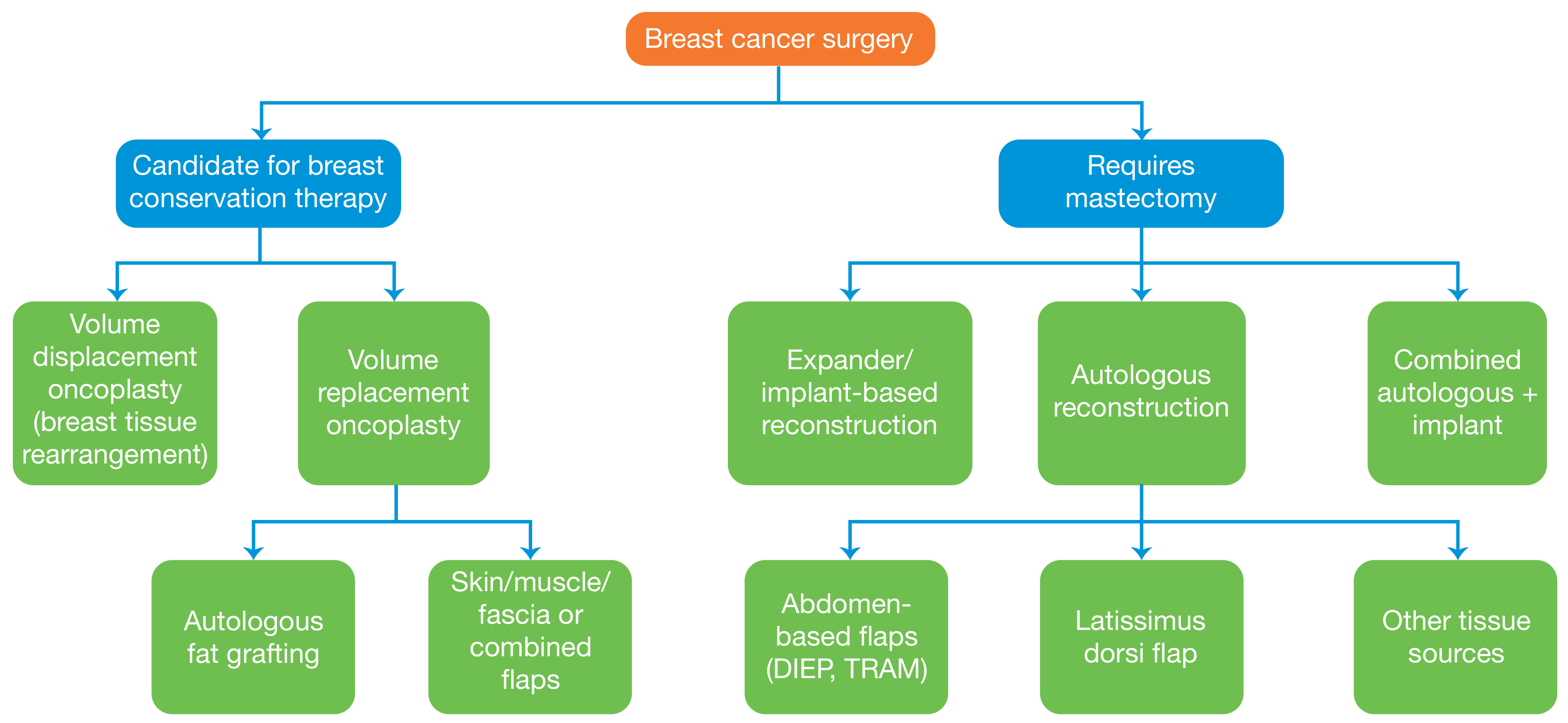
A. Breast-Conserving Surgery (BCS):
- Wide Local Excision (WLE) - up to 20% breast volume
- Oncoplasty - for >20% volume loss:
- Level 1: Volume displacement (local tissue rearrangement)
- Level 2: Volume replacement (local/distant flap)
- Must always be followed by adjuvant radiotherapy
Contraindications to BCS: Previous breast/chest wall radiation, SLE/collagen vascular disease, ankylosing spondylitis, severe orthopnoea
B. Mastectomy:
- Simple mastectomy
- Modified Radical Mastectomy (MRM/Patey's):
- Removal of breast + pectoralis minor + axillary node dissection (Levels I-III)
- Pectoralis major is preserved
- Radical mastectomy (Halsted's): Now rarely performed; removes pectoralis major too
C. Axillary Surgery:
- Sentinel Lymph Node Biopsy (SLNB): First-line for clinically node-negative patients; sentinel node = first echelon of axillary nodes, identified with blue dye and/or radioisotope
- Axillary Lymph Node Dissection (ALND): For SLNB-positive patients
Breast Reconstruction (after MRM)
Timing:
- Immediate (at time of mastectomy)
- Delayed (months to years later)
Methods:
1. Implant-based:
- Direct implant (silicone/saline)
- Tissue expander followed by permanent implant
2. Autologous Flaps:
| Flap | Description |
|---|---|
| TRAM flap | Transverse Rectus Abdominis Myocutaneous - based on superior epigastric artery |
| DIEP flap | Deep Inferior Epigastric Perforator - muscle-sparing TRAM |
| LD flap | Latissimus Dorsi - used with or without implant |
| SGAP/IGAP | Gluteal artery perforator flaps |
3. Nipple-Areola Complex (NAC) reconstruction:
- Local flap for nipple, tattooing for areola
Radiotherapy:
- Mandatory after BCS
- After mastectomy if: tumor ≥5 cm, skin/chest wall involvement, LVI, Grade 3, node-positive
- Dose: 45-50.4 Gy in 25 fractions OR hypofractionation 40-42.5 Gy in 15-16 fractions
- Axilla NOT irradiated after ALND (risk of lymphedema)
Adjuvant Systemic Therapy:
| Type | Indication |
|---|---|
| Chemotherapy | Node-positive, TNBC, HER2+, high grade; improves RFS by ~30%, OS by 10% (EBCCTCG) |
| Hormone therapy (Tamoxifen) | ER/PR positive, pre-menopausal |
| Aromatase inhibitors | ER/PR positive, post-menopausal |
| Trastuzumab (Herceptin) | HER2 positive |
| CDK4/6 inhibitors | Advanced ER+/HER2- disease |
C) ANDI - Aberrations of Normal Development and Involution
Developed by the Cardiff Breast Clinic to replace confusing older terms (fibrosis, adenosis, epitheliosis, fibrocystic disease).
Concept: Most benign breast disorders are minor aberrations of the normal processes of:
- Development (age 15-25 years)
- Cyclical hormone-related change (age 15-50 years)
- Involution (age 35-55 years)
The spectrum ranges from normal → aberration → disease.
ANDI Classification Table:
| Phase | Normal Process | Aberration | Disease |
|---|---|---|---|
| Development (15-25 yr) | Lobule formation, duct development | Fibroadenoma | Giant fibroadenoma |
| Nipple eversion | Nipple inversion | Subareolar abscess | |
| Cyclical changes (15-50 yr) | Cyclical hormonal effect | Cyclical mastalgia, nodularity | Severe mastalgia |
| Epithelial proliferation | Epithelial hyperplasia | Atypical hyperplasia (ADH/ALH) | |
| Involution (35-55 yr) | Lobular involution | Macrocysts, sclerosing adenosis | |
| Ductal involution | Duct ectasia, nipple discharge | Periductal mastitis | |
| Stromal involution | Fibrosis |
Pathology:
- Hyperplasia: >2 cell layers in duct/acini lining; may be typical or atypical
- ADH → DCIS: If ADH involves >2 ducts or >2 mm - reclassified as DCIS
- Papilloma: Fibrovascular core + papillary projections; solitary (RR 1.5-2) or papillomatosis (RR 3)
- Cysts: Kinking of ductules during involution → microcyst → macrocyst
Clinical Features of ANDI:
- Cyclical breast pain (mastalgia) - peaks day 14, worst day 27-28, relieved with menses
- Nodularity (lumpiness) - upper outer quadrant, bilateral, cyclical
- Discrete lump - fibroadenoma in young, cyst in middle-aged
- Nipple discharge
Treatment:
- Reassurance (most cases)
- Mastalgia: Evening primrose oil, Tamoxifen, Danazol, LHRH analogues
- ADH/ALH: Close surveillance (RR 4-5x for developing cancer)
VSAQ - Fibroadenoma of Breast
- Definition: Benign tumor of the breast derived from the terminal duct lobular unit (TDLU); represents an aberration of lobular development (ANDI)
- Epidemiology: Most common benign breast lump in women aged 15-35 years; accounts for ~13% of all palpable breast masses
- Cause: Lobular proliferation in response to estrogen
Types:
- Simple fibroadenoma - <5 cm, common
- Giant/Juvenile fibroadenoma - >5 cm, in adolescents
- Complex fibroadenoma - contains cysts, sclerosing adenosis, epithelial calcifications (slight ↑ cancer risk)
- Multiple fibroadenomas - bilateral, familial
Clinical Features:
- Smooth, rubbery, well-defined, mobile lump ("breast mouse")
- Non-tender
- Usually in upper outer quadrant
- No skin changes, no node involvement
Investigations (Triple Assessment):
- Clinical examination
- Ultrasonography - smooth, hypoechoic, well-defined oval mass
- FNAC or core biopsy
Management:
- Reassurance if <3 cm, young patient, confirmed benign on triple assessment
- Surgical excision if:
-
3-4 cm
- Growing on serial USS
- Patient anxiety
- Not typical on imaging
- Age >35 years
-
Prognosis: Slight increase in cancer risk only with complex type or in patients with family history.
References: Bailey and Love's Short Practice of Surgery, 28th Edition, Chapter 58; Robbins & Cotran Pathologic Basis of Disease
This is a shared conversation. Sign in to Orris to start your own chat.