
Thalamus neurophysiology
Reading File
The thalamus is the brain's main sensory relay station, sitting at the center of the diencephalon. It acts as a "gateway" to the cortex - nearly all sensory information (except olfaction) passes through specific thalamic nuclei before reaching conscious awareness. It also plays key roles in motor control, arousal, memory, and consciousness.
Searching sources...
Finding Sources
Finding Sources
Searching PubMed
"thalamus"[MeSH Terms] AND "neurophysiology"
Good results. Now let me read the key sections in parallel - Bradley's Neurology (most neurophysiology-focused), Costanzo Physiology, and Neuroanatomy through Clinical Cases.
Reading File
Reading File
Reading File
Excellent content. Let me now fetch the thalamic nuclei diagram and read more of the Neuroanatomy text (which was truncated but had great content) and also search for specific neurophysiology topics.
Loading Image
Reading File
Finding Sources
Excellent content. Let me now read more of the Neuroanatomy text (for the reticular nucleus section) and Ganong's (best for neurophysiology), plus the EEG/thalamic oscillations section from Plum & Posner.
Reading File
Reading File
Reading File
The first read_file got the wrong offset (it went into spinal cord content). Let me get the Kaplan & Sadock section on thalamus (which had 5 paragraphs on the thalamus) and search for thalamic oscillations/sleep spindles.
Reading File
Finding Sources
Now I have rich content from multiple authoritative textbooks. Let me compile the comprehensive answer.
Thalamus: Neurophysiology
1. Overview and Location
The thalamus (Greek: "inner chamber" or "bedroom") is the largest component of the diencephalon, located between the cerebral hemispheres and the brainstem, just rostral to the midbrain. Together with the hypothalamus and epithalamus, it forms the diencephalon. In horizontal sections the two thalami appear as egg-shaped gray matter structures forming an inverted V.
Nearly all pathways projecting to the cerebral cortex synapse first in the thalamus. It conveys:
- All sensory modalities (except olfaction)
- Motor outputs from the cerebellum and basal ganglia
- Limbic inputs
- Modulatory inputs governing arousal and sleep-wake cycles
Corticothalamic projections actually outnumber thalamocortical projections - the thalamus is not a passive relay but an active, bidirectionally regulated processing station.
- Neuroanatomy through Clinical Cases 3rd Edition, p. 306
2. Structural Organization
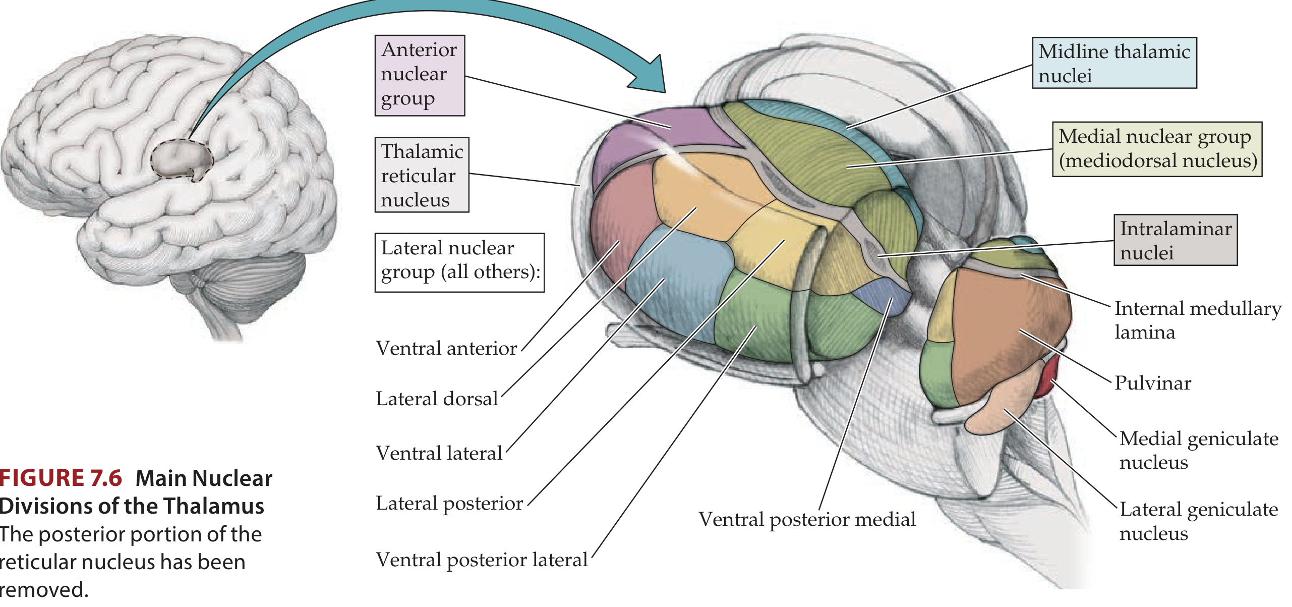
The thalamus is divided into nuclear groups by a Y-shaped white matter structure called the internal medullary lamina:
-
Anterior nuclear group - single anterior nucleus
-
Medial nuclear group - primarily the mediodorsal (MD) nucleus
-
Lateral nuclear group - dorsal tier (lateral dorsal, lateral posterior, pulvinar) and ventral tier (VA, VL, VPL, VPM)
-
Intralaminar nuclei - within the lamina itself; largest is the centromedian nucleus
-
Midline nuclei - thin nuclei adjacent to the third ventricle; may fuse to form the interthalamic adhesion
-
Thalamic reticular nucleus (TRN) - a thin sheet of inhibitory neurons enveloping the lateral surface of the thalamus
-
Kaplan & Sadock's Comprehensive Textbook of Psychiatry, p. 223
3. Three Functional Classes of Thalamic Nuclei
A. Relay Nuclei (Specific Projection)
These form most of the thalamic volume and have localized, topographic projections to specific cortical regions. They are primarily glutamatergic (excitatory).
| Nucleus | Main Inputs | Cortical Target | Function |
|---|---|---|---|
| VPL (Ventral Posterior Lateral) | Medial lemniscus, spinothalamic tract | Primary somatosensory cortex (postcentral gyrus) | Body touch, pain, temperature, proprioception |
| VPM (Ventral Posteromedial) | Trigeminal lemniscus, taste afferents | Somatosensory + taste cortex | Head/face sensation, taste |
| LGN (Lateral Geniculate Nucleus) | Retina (optic tract) | Primary visual cortex (V1) | Vision relay |
| MGN (Medial Geniculate Nucleus) | Inferior colliculus | Primary auditory cortex | Auditory relay |
| VL (Ventral Lateral) | Deep cerebellar nuclei, globus pallidus interna | Motor, premotor, SMA cortex | Cerebellar/basal ganglia motor relay |
| VA (Ventral Anterior) | Substantia nigra pars reticulata, GPi | Widespread frontal lobe | Basal ganglia motor relay |
| Pulvinar | Superior colliculus, extrageniculate visual pathway | Parietotemporo-occipital association cortex | Behavioral orientation toward relevant stimuli |
| Anterior nucleus | Mammillary bodies (via mammillothalamic tract) | Limbic/cingulate cortex | Memory, emotion (Papez circuit) |
| Mediodorsal (MD) | Hypothalamus, amygdala, prefrontal inputs | Prefrontal and temporal cortex | Cognition, affect, working memory |
Specific relay nuclei project to cortical layer IV; nonspecific nuclei project more broadly to layers I-IV.
- Ganong's Review of Medical Physiology, p. 278; Neuroanatomy through Clinical Cases, p. 308
B. Intralaminar Nuclei (Diffuse Projection)
- Located within the internal medullary lamina
- Receive input from ascending reticular activating system (ARAS), spinal cord, basal ganglia, and cerebellum
- Project diffusely to widespread cortical and striatal areas
- Function: regulate global cortical excitability and arousal
- The centromedian nucleus is the largest and receives substantial input from motor cortex
C. Thalamic Reticular Nucleus (TRN)
This is the master modulator of thalamic output:
- A thin shell of GABAergic (inhibitory) interneurons that wraps the lateral thalamus
- Does not project to the cortex - only back to other thalamic nuclei
- Receives collaterals from both thalamocortical and corticothalamic axons as they pass through it
- When activated, it inhibits thalamic relay nuclei, hyperpolarizing them into burst firing mode
- This is the mechanism underlying sleep spindles and thalamic gating of sensory input
"Neurons in the reticular nucleus provide GABAergic inputs to the thalamic relay nuclei, which hyperpolarize them and set them into bursting mode."
- Plum and Posner's Diagnosis and Treatment of Stupor and Coma, p. 43
4. Thalamic Firing Modes
Thalamic relay neurons operate in two distinct electrophysiological modes:
Tonic (Transfer) Mode
- Occurs during wakefulness and alert states
- Neurons fire in a steady, graded fashion in response to incoming signals
- Faithfully transmits sensory information to the cortex with high fidelity
- Maintained by depolarizing monoamine and cholinergic inputs from the brainstem
Burst (Oscillatory) Mode
- Occurs during sleep and drowsy states
- Triggered by hyperpolarization from the TRN (via GABA) or from inhibitory interneurons
- Activates a low-threshold calcium current (I_T, T-type Ca²⁺ channels)
- Produces high-frequency action potential bursts on a slow oscillatory background
- Responsible for thalamic spindles (8-13 Hz) during NREM sleep
- In this mode, sensory information is blocked from reaching the cortex - the thalamus "gates" out incoming signals
5. Thalamocortical Oscillations and EEG
The thalamus is a major pacemaker of cortical EEG rhythms through the thalamocortical loop:
- TRN activation hyperpolarizes relay nuclei → burst firing → synchronizes cortical activity
- Sleep spindles (8-13 Hz): waxing and waning oscillations in light NREM sleep; generated by the TRN-relay nucleus circuit
- Delta waves (0.5-4 Hz): deep slow-wave sleep; thalamic relay neurons in sustained burst mode
- Alpha rhythm (8-12 Hz): resting wakefulness, partially thalamic in origin
However, large thalamic lesions mainly suppress spindles and high-frequency activity - mid-range EEG oscillations appear partly dependent on corticocortical connections, not thalamus alone.
- Plum and Posner's Diagnosis and Treatment of Stupor and Coma, p. 43
The cholinergic pedunculopontine and laterodorsal tegmental nuclei (brainstem ARAS) can hyperpolarize the TRN via GABA_B receptors, thereby releasing the relay nuclei from inhibition and switching them into tonic (waking) mode.
6. The Thalamus as a Sensory Gate
The thalamus actively modulates what information reaches the cortex:
- Corticothalamic feedback (layer VI pyramidal neurons projecting back to thalamus) allows the cortex to selectively amplify or suppress thalamic input - the basis of selective attention
- Under conditions of pain, fear, or attention, the corticothalamic loop can either enhance (amplify signal) or suppress (gate out noise) incoming signals
- The posterior nuclear group (VPL/VPM area) receives nociceptive input from the spinothalamic tract and projects to the secondary somatosensory cortex and insular cortex - important in the affective dimension of pain
7. Thalamus and Consciousness
The thalamus plays a fundamental role in consciousness and arousal:
- The intralaminar nuclei are considered part of the consciousness-generating network; bilateral thalamic lesions cause profound disorders of consciousness
- The concept of the thalamus as the "content of consciousness" vs. the basal forebrain as the "arousal" driver reflects the complementary roles of these structures
- Bilateral thalamic hemorrhage or infarction (particularly paramedian) can cause akinetic mutism or persistent vegetative state
- A clinical case from Plum & Posner illustrates: bilateral thalamic hemorrhage produced vegetative state with continuous 6-7 Hz theta EEG, absent sleep spindles and higher frequencies, reflecting loss of thalamic oscillatory activity
8. Key Neurophysiological Summary Table
| Property | Specific Relay Nuclei | Intralaminar Nuclei | Reticular Nucleus |
|---|---|---|---|
| Transmitter | Glutamate (excitatory) | Glutamate | GABA (inhibitory) |
| Cortical projection | Focal (layer IV) | Diffuse (layers I-IV) | None (intra-thalamic) |
| Function | Sensory/motor relay | Arousal/attention | Thalamic gating/oscillations |
| EEG role | Spindle generation (burst mode) | Global activation | Synchronizes relay nuclei |
Sources used:
- Neuroanatomy through Clinical Cases, 3rd Edition (Blumenfeld) - primary neurophysiology source
- Kaplan & Sadock's Comprehensive Textbook of Psychiatry - structural detail
- Ganong's Review of Medical Physiology, 26th Edition - cortical layer projections, transmitters
- Plum and Posner's Diagnosis and Treatment of Stupor and Coma - oscillations, EEG, consciousness
- Bradley and Daroff's Neurology in Clinical Practice - VPL/VPM sensory relay detail
- Costanzo Physiology, 7th Edition - overview
This is a shared conversation. Sign in to Orris to start your own chat.