
Track of fasciculus gracilis & cuneatus
Reading File
The fasciculus gracilis and cuneatus are the two main ascending tracts of the dorsal column-medial lemniscal (DCML) system, carrying fine touch, proprioception, and vibration sense from the body to the cortex.
Searching sources...
Finding Sources
Finding Sources
Searching Images
fasciculus gracilis cuneatus dorsal column tract spinal cord anatomy
Good results found. Now let me read the detailed content from the most relevant sources.
Reading File
Reading File
Reading File
Excellent content retrieved. Let me also fetch the key diagram image and read a bit more of the Neuroanatomy textbook content.
Loading Image
Reading File
I now have comprehensive information to give a thorough, well-structured answer.
Fasciculus Gracilis & Fasciculus Cuneatus - Complete Track
These are the two subdivisions of the posterior (dorsal) columns of the spinal cord, forming the first leg of the Posterior Column - Medial Lemniscal (DCML) pathway.
Overview Table
| Feature | Fasciculus Gracilis | Fasciculus Cuneatus |
|---|---|---|
| Other name | Column of Goll | Column of Burdach |
| Position in cord | Medial (next to midline) | Lateral (between gracilis & dorsal horn) |
| Spinal levels contributing | Sacral + Lumbar + Lower 6 Thoracic (T7-T12) | Upper 6 Thoracic (T1-T6) + All Cervical |
| Body region served | Lower limbs & lower trunk | Upper limbs, upper trunk, neck |
| Terminates in | Nucleus gracilis (caudal medulla) | Nucleus cuneatus (caudal medulla) |
| Present throughout cord? | Entire spinal cord | Only above T6 (mid-thoracic) |
Pathway - Neuron by Neuron
1st Order Neuron (Receptor → Dorsal Column Nuclei)
- Cell body: Dorsal root ganglion (DRG)
- Receptor types: Meissner's corpuscles, Merkel's discs, Pacinian corpuscles, Ruffini endings, muscle spindles, Golgi tendon organs - all large-diameter, heavily myelinated Aα (Ia) and Aβ (II) fibers
- Entry: Medial bundle of the dorsal root entry zone
- Collaterals: Some collaterals synapse on dorsal horn neurons (laminae III-IV), but the main branch ascends ipsilaterally without synapsing all the way to the medulla
- Somatotopic lamination: Fibers are added in a medial-to-lateral sequence - sacral fibers are most medial, and as you ascend, each new level's fibers push existing fibers medially, so cervical fibers end up most lateral
"Fibers from the lowermost part of the body (sacral, lumbar, and the lowest six thoracic levels) are located more medially and constitute the fasciculus gracilis (Goll column), whereas those coming from the upper part of the body (upper six thoracic and all cervical levels) occupy a more lateral position and constitute the fasciculus cuneatus (Burdach column)."
- Localization in Clinical Neurology, 8e
Important exception for proprioception from lower limbs
Most proprioceptive fibers from the lower limbs do NOT travel all the way up in the fasciculus gracilis. They leave at the upper lumbar spinal cord and synapse in Clarke's column (Clarke's nucleus, C8-L2). Clarke's column neurons then project to Nucleus Z in the medulla via the dorsal spinocerebellar tract pathway. Nucleus Z fibers then join the medial lemniscus. The fibers that remain in fasciculus gracilis up to the medulla mainly carry tactile sensation.
In contrast, upper limb proprioceptive fibers in the fasciculus cuneatus travel all the way to the nucleus cuneatus, from which an accessory cuneate nucleus projects to the cerebellum (cuneocerebellar tract).
2nd Order Neuron (Dorsal Column Nuclei → VPL Thalamus)
- Cell bodies: Nucleus gracilis and nucleus cuneatus of the caudal medulla
- Axons cross midline via the internal arcuate fibers (decussation in the lower medulla)
- Form the medial lemniscus, which ascends through:
- Medulla (posterior midline region)
- Pons (moves laterally)
- Midbrain (lateral tegmentum)
- Terminate: Ventral posterolateral (VPL) nucleus of the thalamus
- Somatotopy in medial lemniscus: Lower body fibers are ventral, upper body fibers are dorsal (inverted compared to cord)
3rd Order Neuron (Thalamus → Cortex)
- Cell bodies: VPL nucleus of thalamus
- Axons travel via the posterior limb of the internal capsule
- Terminate: Primary somatosensory cortex (S1) - postcentral gyrus (Brodmann areas 3, 1, 2)
- Somatotopic map (homunculus) maintained throughout
Sensory Modalities Carried
Both tracts together carry the discriminative sensations:
- Fine/discriminative touch
- Two-point discrimination
- Vibration sense
- Proprioception (conscious joint position sense)
- Pressure touch
- Stereognosis (recognizing objects by touch)
- Graphesthesia (writing on skin recognition)
- Weight perception
- Shape and movement awareness
Summary Diagram
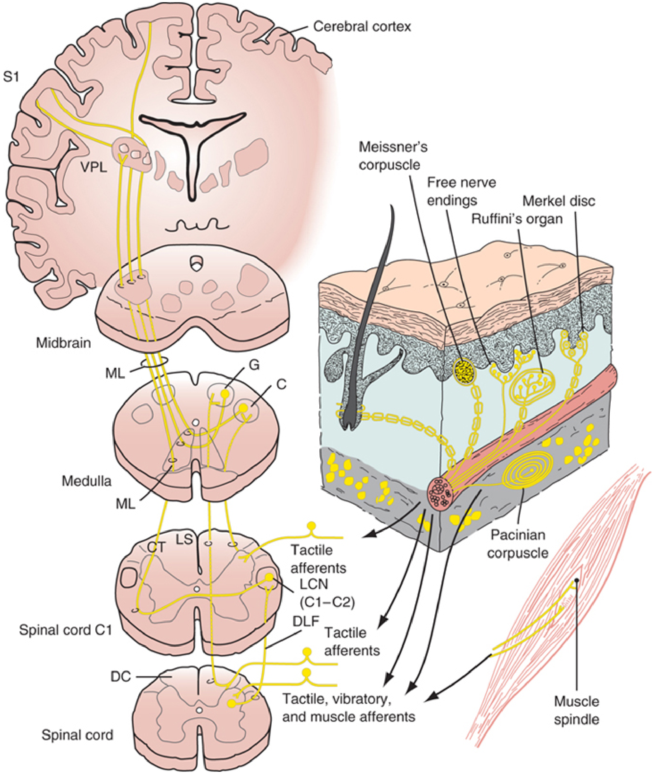
Diagram shows the full DCML pathway: spinal cord dorsal columns → posterior column nuclei (G = gracilis, C = cuneatus) in medulla → medial lemniscus (ML) crossing → midbrain → VPL thalamus → S1 cortex. Also shows the tactile afferents going through the lateral cervical nucleus (LCN) at C1-C2 via the dorsolateral funiculus (DLF).
Lateral Cervical Nucleus (Accessory Pathway)
An accessory route also exists: some tactile afferents synapse in the dorsal horn, and their 2nd-order axons ascend the ipsilateral dorsolateral funiculus to synapse in the lateral cervical nucleus (LCN) at C1-C2. LCN neurons then cross and join the medial lemniscus. This pathway runs in parallel to the main dorsal columns.
Clinical Correlates
| Lesion | Effect |
|---|---|
| Unilateral posterior column damage | Ipsilateral loss of fine touch, vibration, proprioception below the level |
| Subacute combined degeneration (Vit B12 deficiency) | Bilateral dorsal column (+ lateral column) degeneration - loss of proprioception + UMN signs |
| Tabes dorsalis (neurosyphilis) | Posterior column degeneration → Romberg's sign, sensory ataxia, Charcot joints |
| Brown-Séquard syndrome (hemisection) | Ipsilateral dorsal column loss (proprioception/vibration) + ipsilateral UMN signs + contralateral pain/temp loss below lesion |
| Posterior cord syndrome | Dorsal column loss only |
Sources:
- Localization in Clinical Neurology, 8e, pp. 44-45
- Neuroanatomy through Clinical Cases, 3rd ed., Chapter 7
- Gray's Anatomy for Students
This is a shared conversation. Sign in to Orris to start your own chat.