
let's start detail of blood pressure discuss everything like it's equation and effect of it's factor and under what circumstances it increase or decrease also explain it's factor physiologically and also discuss it's pathological conditions
Reading File
Reading File
Reading File
Reading File
Reading File
Blood pressure is a broad physiology and clinical topic. We can build it in focused sections:
- Definition and measurement: systolic, diastolic, pulse pressure, mean arterial pressure, technique and units
- Core equations: cardiac output, total peripheral resistance, MAP, flow and resistance relationships
- Physiological regulation: autonomic nervous system, baroreceptors, kidneys, renin-angiotensin-aldosterone system, ADH, natriuretic peptides, local vascular control
- Factors that raise or lower BP: posture, exercise, sleep, stress, age, temperature, meals, salt, volume status, pregnancy, drugs
- Pathology: primary and secondary hypertension, hypertensive emergency, orthostatic hypotension, shock, and abnormal pulse pressures
- Interpretation and clinical patterns: isolated systolic hypertension, white-coat/masked hypertension, circadian variation, common medication effects.
At its simplest, arterial pressure depends mainly on how much blood the heart pumps and how much resistance the systemic vessels provide:
[
\text{MAP} \approx \text{CO} \times \text{SVR}
]
where cardiac output (CO) = heart rate × stroke volume, and systemic vascular resistance (SVR) is determined largely by arteriolar diameter. The kidneys provide the dominant long-term control by adjusting body sodium, water, and therefore circulating volume.
Which section should we study in depth first?
Asking for Preferences
Choose where to begin the detailed lesson.
Blood pressure is a broad physiology and clinical topic. We can build it in focused sections:
- Definition and measurement: systolic, diastolic, pulse pressure, mean arterial pressure, technique and units
- Core equations: cardiac output, total peripheral resistance, MAP, flow and resistance relationships
- Physiological regulation: autonomic nervous system, baroreceptors, kidneys, renin-angiotensin-aldosterone system, ADH, natriuretic peptides, local vascular control
- Factors that raise or lower BP: posture, exercise, sleep, stress, age, temperature, meals, salt, volume status, pregnancy, drugs
- Pathology: primary and secondary hypertension, hypertensive emergency, orthostatic hypotension, shock, and abnormal pulse pressures
- Interpretation and clinical patterns: isolated systolic hypertension, white-coat/masked hypertension, circadian variation, common medication effects.
At its simplest, arterial pressure depends mainly on how much blood the heart pumps and how much resistance the systemic vessels provide:
[
\text{MAP} \approx \text{CO} \times \text{SVR}
]
where cardiac output (CO) = heart rate × stroke volume, and systemic vascular resistance (SVR) is determined largely by arteriolar diameter. The kidneys provide the dominant long-term control by adjusting body sodium, water, and therefore circulating volume.
Which section should we study in depth first?
1-4 section
Finding Sources
Finding Sources
Finding Sources
Searching PubMed
"blood pressure" AND physiology
Searching the Web
American Heart Association blood pressure readings physiology blood pressure regulation
Reading File
Reading File
Finding Sources
Finding Sources
Finding Sources
Reading File
Reading File
Reading File
Reading File
Blood Pressure: Sections 1 to 4
1. Definition, components, and measurement
Blood pressure (BP) is the lateral pressure exerted by flowing blood on arterial walls. It is conventionally recorded in millimetres of mercury (mmHg) as:
[
\text{BP} = \frac{\text{Systolic BP}}{\text{Diastolic BP}}
]
Example: 120/80 mmHg
- Systolic blood pressure (SBP): maximum arterial pressure during left-ventricular ejection.
- Diastolic blood pressure (DBP): lowest arterial pressure, reached during ventricular relaxation and filling.
- Pulse pressure (PP):
[
PP = SBP - DBP
]
For 120/80 mmHg:
[
PP = 120 - 80 = 40 \text{ mmHg}
]
- Mean arterial pressure (MAP): the average pressure that drives systemic tissue perfusion over one cardiac cycle. It is not the simple numerical average of SBP and DBP because diastole lasts longer than systole at normal heart rates.
[
MAP \approx DBP + \frac{1}{3}(PP)
]
or:
[
MAP \approx \frac{SBP + 2(DBP)}{3}
]
For 120/80 mmHg:
[
MAP \approx 80 + \frac{40}{3} = 93 \text{ mmHg}
]
This approximation becomes less accurate with marked tachycardia because the duration of diastole shortens.
MAP is physiologically important because it is the major pressure gradient driving organ perfusion. Organ blood flow can be represented as:
[
Q = \frac{P_{arterial}-P_{venous}}{R}
]
Since venous pressure is normally low relative to arterial pressure:
[
Q \approx \frac{MAP}{R}
]
Thus, a fall in MAP can reduce cerebral, renal, coronary, and other organ perfusion if local autoregulation cannot compensate. Costanzo Physiology, 7th ed., p. 168.
Accurate measurement matters
A single reading is not necessarily a person's usual BP. BP rises transiently with anxiety, recent exercise, nicotine, caffeine, pain, talking, or a full bladder. For a reliable clinic reading, rest quietly, support the back and arm at heart level, keep feet on the floor, and use a correctly sized cuff. The American Heart Association measurement guidance also advises avoiding exercise, smoking, and caffeine for at least 30 minutes beforehand.
2. The equations of blood pressure
A. Central equation
The main haemodynamic relationship is:
[
\boxed{MAP = CO \times SVR}
]
where:
- CO = cardiac output
- SVR = systemic vascular resistance, also called total peripheral resistance, TPR
[
CO = HR \times SV
]
where:
- HR = heart rate
- SV = stroke volume
Therefore:
[
\boxed{MAP = HR \times SV \times SVR}
]
This means BP can rise if one or more of the following increase:
- Heart rate
- Stroke volume
- Systemic vascular resistance
And BP falls if these decrease.
Costanzo Physiology, 7th ed., p. 168.
B. Important limitation of the equation
The variables are interdependent, not independent.
For example, if SVR rises abruptly because of arteriolar constriction:
- MAP initially rises.
- The left ventricle must pump against higher afterload.
- Stroke volume may fall.
- Cardiac output may fall.
- Therefore, MAP usually does not rise as much as a simplistic calculation suggests.
Conversely, if SVR falls substantially:
- BP initially falls.
- Afterload falls, so the heart can eject blood more easily.
- Cardiac output may rise.
- The increase in CO partly offsets the fall in BP.
Costanzo Physiology, 7th ed., p. 168.
C. Resistance and vessel radius: Poiseuille relationship
For laminar flow in a vessel:
[
R = \frac{8\eta L}{\pi r^4}
]
where:
- (R) = resistance
- (\eta) = blood viscosity
- (L) = vessel length
- (r) = vessel radius
The key component is:
[
R \propto \frac{1}{r^4}
]
A small fall in arteriolar radius produces a large increase in resistance. Arterioles are therefore called the resistance vessels.
Example
If the radius becomes half:
[
R \propto \frac{1}{(1/2)^4} = 16
]
Resistance rises approximately 16-fold, assuming other factors remain constant.
This is why sympathetic arteriolar vasoconstriction can quickly raise SVR and BP, and why vasodilation in sepsis, anaphylaxis, or some drugs can abruptly lower BP.
D. Stroke volume and cardiac output
Stroke volume is influenced by:
[
SV = f(\text{preload, contractility, afterload})
]
1. Preload
Preload reflects ventricular filling before contraction, related clinically to venous return and end-diastolic volume.
- Increased blood volume, venoconstriction, leg elevation, or fluid infusion increase venous return and preload.
- By the Frank-Starling mechanism, increased preload usually raises stroke volume and CO.
- Therefore BP may increase.
2. Contractility
Contractility is the intrinsic force of myocardial contraction at a given preload and afterload.
- Sympathetic stimulation and circulating catecholamines increase contractility through cardiac (\beta_1)-receptors.
- Myocardial infarction, severe hypoxia, acidosis, and some drugs can reduce contractility.
Lower contractility reduces SV, CO, and potentially MAP.
3. Afterload
Afterload is the pressure/resistance against which the ventricle ejects.
- Higher SVR increases afterload.
- Severe hypertension and aortic stenosis increase left-ventricular afterload.
- An abrupt major afterload increase can lower stroke volume.
3. Physiological control of blood pressure
BP regulation occurs over several time scales.
| Time scale | Main mechanisms | Main purpose |
|---|---|---|
| Seconds to minutes | Baroreceptor reflex, autonomic nervous system, chemoreceptors | Stabilize BP rapidly |
| Minutes to hours | Capillary fluid shifts, circulating catecholamines, angiotensin II, vasopressin | Support circulating volume and vascular tone |
| Hours to days or longer | Kidneys, sodium balance, RAAS, thirst, aldosterone, ANP | Long-term BP control |
A. Baroreceptor reflex: the rapid controller
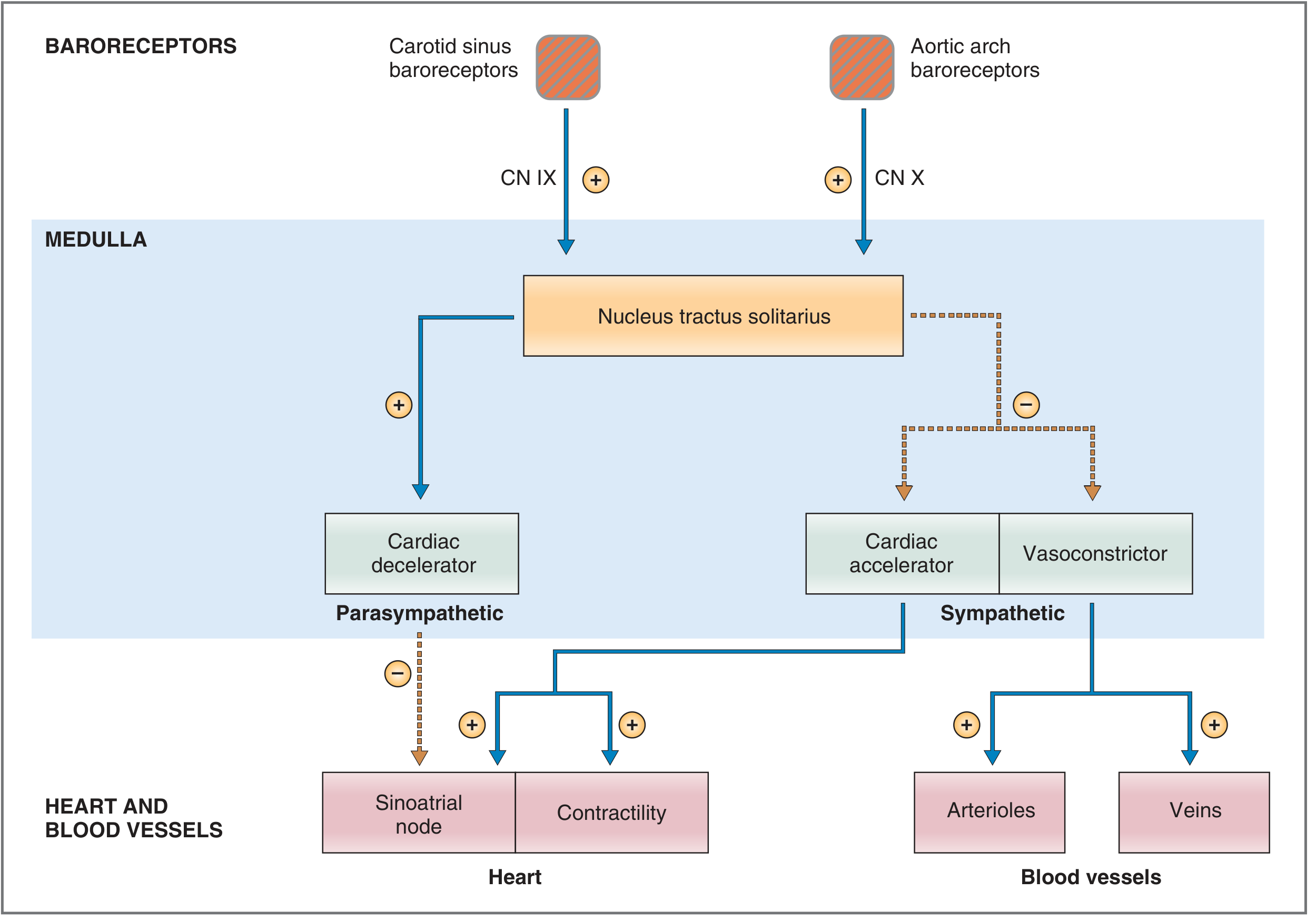
Baroreceptors are stretch-sensitive mechanoreceptors in:
- Carotid sinus: at the bifurcation of the common carotid artery
- Aortic arch
Their afferent pathways are:
- Carotid sinus to medulla via glossopharyngeal nerve, CN IX
- Aortic arch to medulla via vagus nerve, CN X
When arterial pressure increases, arterial walls stretch more, and baroreceptor firing increases. When pressure falls, stretch and firing decrease. Costanzo Physiology, 7th ed., pp. 169-170.
When BP rises
[
\uparrow BP \rightarrow \uparrow \text{baroreceptor firing}
]
The medulla responds by:
- Decreasing sympathetic outflow
- Increasing parasympathetic, vagal outflow
Consequences:
- ↓ Heart rate
- ↓ Contractility
- Arteriolar vasodilation, so ↓ SVR
- Venodilation, so ↓ venous return and preload
- ↓ CO and ↓ MAP toward normal
When BP falls
[
\downarrow BP \rightarrow \downarrow \text{baroreceptor firing}
]
The medulla responds by:
- Increasing sympathetic outflow
- Decreasing vagal outflow
Consequences:
- (\beta_1) stimulation at heart: ↑ heart rate and ↑ contractility
- (\alpha_1) stimulation of arterioles: vasoconstriction, therefore ↑ SVR
- Venoconstriction: ↑ venous return and preload
- Increased CO and MAP toward normal
This reflex is rapid, particularly useful during standing, haemorrhage, pain, fear, or acute exercise. It is less effective for long-term BP regulation because baroreceptors can reset to a higher operating pressure in chronic hypertension.
B. Response to standing
When a person suddenly stands:
- Gravity causes blood to pool in leg and splanchnic veins.
- Venous return falls.
- Preload falls.
- Stroke volume falls.
- Cardiac output and MAP transiently fall.
- Baroreceptor firing falls.
- Sympathetic activity increases.
- Heart rate, contractility, venous tone, and SVR rise.
This restores cerebral perfusion.
A simplified chain is:
[
\text{Standing} \rightarrow \downarrow VR \rightarrow \downarrow SV \rightarrow \downarrow CO \rightarrow \downarrow MAP
]
[
\downarrow MAP \rightarrow \uparrow sympathetic\ activity \rightarrow \uparrow HR,\ \uparrow contractility,\ \uparrow SVR
]
Costanzo Physiology, 7th ed., p. 190.
C. Sympathetic nervous system
The sympathetic system raises BP through several pathways.
| Site | Receptor | Effect | BP consequence |
|---|---|---|---|
| SA node | (\beta_1) | Increased HR | ↑ CO |
| Ventricular myocardium | (\beta_1) | Increased contractility | ↑ SV and CO |
| Arterioles | (\alpha_1) | Vasoconstriction | ↑ SVR |
| Veins | predominantly (\alpha_1) | Venoconstriction | ↑ venous return and preload |
| Kidney | (\beta_1) | Increased renin release | Activates RAAS |
The parasympathetic system acts mainly through the vagus nerve to slow the heart. It has relatively little direct effect on most systemic blood vessels.
D. Renin-angiotensin-aldosterone system: RAAS
The RAAS is especially important for longer-term BP and extracellular-fluid volume regulation.
Stimuli for renin release
Renin release from renal juxtaglomerular cells increases with:
- Reduced renal perfusion pressure
- Reduced sodium chloride delivery to macula densa
- Increased renal sympathetic activity via (\beta_1)-receptors
Sequence:
[
\text{Renin} \rightarrow \text{Angiotensin I} \rightarrow \text{Angiotensin II}
]
Angiotensin II raises BP by:
- Direct arteriolar vasoconstriction: raises SVR.
- Aldosterone release from adrenal cortex: raises distal sodium reabsorption.
- Thirst stimulation: increases water intake.
- ADH release: increases water retention.
- Facilitating sympathetic activity.
Sodium retention leads to water retention:
[
\uparrow Na^+ \text{ retention} \rightarrow \uparrow ECF\ volume \rightarrow \uparrow venous\ return \rightarrow \uparrow CO \rightarrow \uparrow BP
]
RAAS has a major role in salt-sensitive hypertension and in BP maintenance during volume depletion. Guyton and Hall Textbook of Medical Physiology, Chapter 19.
E. Kidney and pressure natriuresis: long-term controller
The kidneys are the dominant long-term regulators of arterial pressure because they determine the amount of sodium and water in the body.
When arterial pressure rises:
[
\uparrow BP \rightarrow \uparrow renal\ Na^+\ and\ water\ excretion
]
This is pressure natriuresis and pressure diuresis.
The result is:
[
\downarrow ECF\ volume \rightarrow \downarrow venous\ return \rightarrow \downarrow CO \rightarrow \downarrow BP
]
When BP falls, the kidney tends to retain sodium and water, partly through RAAS and sympathetic activation. This increases extracellular volume and supports BP.
For sustained hypertension to exist, there is generally some abnormality in the renal-pressure natriuresis relationship, excess sodium retention, or both.
F. Vasopressin, also called ADH
Antidiuretic hormone, ADH, is released particularly when:
- Plasma osmolality rises
- Blood volume or arterial pressure falls substantially
ADH raises BP by:
- Increasing water reabsorption in collecting ducts through V2 receptors
- Causing vasoconstriction through V1 receptors at higher concentrations
It is especially important during severe dehydration, blood loss, and shock.
G. Atrial and brain natriuretic peptides
ANP is released when the atria are stretched by increased circulating volume. BNP is released largely from ventricles under increased wall stress.
Their overall effects oppose RAAS:
- Promote sodium excretion and water loss
- Suppress renin and aldosterone
- Promote vasodilation
- Reduce effective circulating volume and BP
H. Local vascular control
Each tissue can alter arteriolar tone according to its own metabolic needs.
Metabolites that usually cause local vasodilation
- ↓ Oxygen
- ↑ Carbon dioxide
- ↑ Hydrogen ion concentration
- ↑ Potassium
- Adenosine
- Lactate
- Nitric oxide
This increases local flow during exercise or hypoxia. Systemic BP may not fall much during exercise because sympathetic vasoconstriction in nonessential vascular beds offsets vasodilation in active muscle.
Endothelial mediators
| Mediator | Main action |
|---|---|
| Nitric oxide | Vasodilation |
| Prostacyclin | Vasodilation and reduced platelet aggregation |
| Endothelin | Potent vasoconstriction |
| Angiotensin II | Vasoconstriction |
| Thromboxane A2 | Vasoconstriction and platelet activation |
Endothelial dysfunction, often associated with hypertension, diabetes, smoking, and atherosclerosis, can reduce nitric-oxide-mediated dilation and contribute to high SVR.
4. Factors that increase or decrease blood pressure
A. Physiological circumstances that increase BP
| Situation | Main physiological explanation |
|---|---|
| Exercise | ↑ Sympathetic activity, HR, contractility, and CO. Dynamic exercise usually raises SBP more than DBP because active muscles vasodilate. |
| Fear, anxiety, pain, anger | Sympathetic discharge causes tachycardia, increased contractility, and vasoconstriction. |
| Cold exposure | Cutaneous vasoconstriction increases SVR. |
| Standing initially | CO falls first, then sympathetic compensation raises HR and SVR. |
| Caffeine and nicotine | Sympathetic stimulation and vasoconstriction can cause temporary BP elevation. |
| High salt intake in salt-sensitive people | Sodium retention increases extracellular volume and CO. |
| Obesity | Increased blood volume, sympathetic activation, insulin resistance, renal sodium retention, and often sleep apnea. |
| Sleep apnea | Recurrent hypoxia, arousal, sympathetic surges, RAAS activation. |
| Pregnancy complications such as pre-eclampsia | Endothelial dysfunction, vasoconstriction, and altered placental vascular signalling. |
| Aging | Arterial stiffening raises SBP and pulse pressure. |
| Full urinary bladder | Sympathetic activation may transiently raise BP. |
Exercise: SBP versus DBP
During dynamic exercise:
- HR and stroke volume increase.
- CO rises greatly.
- Arterioles in active skeletal muscle dilate.
- SBP rises.
- DBP often changes little or may decrease slightly because SVR falls.
During heavy static or resistance exercise, marked sympathetic activation and mechanical vascular compression can raise both SBP and DBP transiently.
A recent exercise systematic review found that BP responses vary substantially by exercise type and an individual's health and training status (2025 systematic review).
B. Physiological circumstances that decrease BP
| Situation | Main physiological explanation |
|---|---|
| Sleep | Lower sympathetic activity and lower metabolic demand reduce HR, CO, and vascular tone. |
| Warm environment | Skin vasodilation reduces SVR; dehydration can compound the fall. |
| Rest after exercise | Persistent vasodilation in skeletal muscle can cause post-exercise hypotension. |
| Standing with volume depletion | Venous pooling plus reduced circulating volume lowers venous return, SV, CO, and MAP. |
| Haemorrhage | Reduced blood volume lowers preload, SV, and CO. |
| Vomiting, diarrhoea, poor intake, sweating | Extracellular-volume depletion reduces venous return and CO. |
| Anaphylaxis | Vasodilation and capillary leak reduce SVR and effective circulating volume. |
| Sepsis | Widespread vasodilation, capillary leak, and sometimes myocardial depression. |
| Drugs | Vasodilators, diuretics, alpha-blockers, nitrates, some antidepressants, anaesthetic agents, and excessive antihypertensive treatment can lower BP. |
Pathological conditions related to BP
1. Primary, or essential, hypertension
Primary hypertension means sustained BP elevation without one single identifiable cause. It accounts for most adult hypertension.
It results from interacting factors:
- Genetic susceptibility
- Obesity and insulin resistance
- Excess dietary sodium in susceptible individuals
- Reduced renal sodium excretion
- Excess sympathetic activity
- RAAS activity
- Endothelial dysfunction
- Vascular remodelling and arterial stiffness
- Alcohol excess, smoking, inactivity, and poor sleep
- Obstructive sleep apnea
Hypertension often has no symptoms but gradually injures target organs, including brain, heart, kidneys, retina, and arteries. The AHA overview summarizes its associations with stroke, myocardial infarction, heart failure, kidney disease, and vision loss.
2. Secondary hypertension
Secondary hypertension has an identifiable cause. Consider it especially in abrupt, severe, resistant, or early-onset hypertension.
Common causes include:
| Category | Examples | Mechanism |
|---|---|---|
| Renal parenchymal disease | Chronic kidney disease, glomerulonephritis | Sodium retention, impaired pressure natriuresis, RAAS activation |
| Renovascular disease | Renal artery stenosis | Reduced renal perfusion causes renin release and angiotensin II excess |
| Endocrine disorders | Primary aldosteronism, pheochromocytoma, Cushing syndrome, thyroid disease | Mineralocorticoid excess, catecholamine excess, cortisol effects, altered vascular tone |
| Sleep disorder | Obstructive sleep apnea | Hypoxia and recurrent sympathetic activation |
| Vascular cause | Coarctation of aorta | Elevated proximal arterial pressure |
| Drugs/substances | NSAIDs, glucocorticoids, oral contraceptives, stimulants, cocaine, alcohol, decongestants, licorice | Sodium retention, sympathetic stimulation, vasoconstriction, hormonal effects |
Primary aldosteronism
Excess aldosterone causes:
[
\uparrow Na^+\ retention \rightarrow \uparrow ECF\ volume \rightarrow hypertension
]
It may produce hypokalaemia, although normal potassium does not exclude it.
Renal artery stenosis
Reduced pressure at the affected kidney is interpreted as low effective arterial pressure:
[
\downarrow renal\ perfusion \rightarrow \uparrow renin \rightarrow \uparrow Ang\ II + \uparrow aldosterone \rightarrow hypertension
]
Pheochromocytoma
Catecholamine-secreting tumours can cause episodic or sustained hypertension through alpha-mediated vasoconstriction and beta-mediated cardiac stimulation.
3. Isolated systolic hypertension and wide pulse pressure
Isolated systolic hypertension means elevated SBP with normal or relatively normal DBP, commonly in older adults.
Mechanism:
- Large elastic arteries become stiff.
- They cannot expand effectively during systole.
- SBP rises.
- Reduced elastic recoil may lower DBP.
- Pulse pressure widens.
[
PP = SBP - DBP
]
A wide pulse pressure may occur with:
- Aging and arterial stiffness
- Aortic regurgitation
- Hyperthyroidism
- Severe anaemia
- High-output states
4. Narrow pulse pressure
A narrow pulse pressure can occur when stroke volume is low, such as:
- Severe heart failure
- Hypovolaemia
- Cardiac tamponade
- Cardiogenic shock
- Severe aortic stenosis
This is often a warning that forward cardiac output is poor.
5. Orthostatic hypotension
Orthostatic hypotension occurs when BP falls abnormally after standing, producing dizziness, blurred vision, weakness, presyncope, or syncope.
Common mechanisms:
- Dehydration or blood loss
- Medications, especially diuretics, vasodilators, alpha-blockers, nitrates, and some antidepressants
- Autonomic neuropathy, such as diabetic autonomic neuropathy
- Parkinson disease and other neurodegenerative disorders
- Prolonged bed rest
- Adrenal insufficiency
The core physiology is:
[
\text{Standing} \rightarrow \text{venous pooling} \rightarrow \downarrow VR \rightarrow \downarrow CO \rightarrow \downarrow MAP
]
If baroreceptor-mediated sympathetic compensation is inadequate, cerebral perfusion falls and symptoms develop. Costanzo Physiology, 7th ed., p. 190.
6. Shock
Shock is not simply “low BP.” It is a state of inadequate tissue perfusion and oxygen delivery. A patient can be in compensated shock with a near-normal BP initially.
Major categories:
| Type | Primary defect | BP mechanism |
|---|---|---|
| Hypovolaemic | Reduced blood volume | ↓ preload, ↓ SV, ↓ CO |
| Cardiogenic | Pump failure | ↓ contractility, ↓ SV, ↓ CO |
| Distributive | Pathological vasodilation | ↓ SVR, often capillary leak |
| Obstructive | Blocked filling or outflow | ↓ venous return or impaired ejection |
Examples:
- Hypovolaemic shock: haemorrhage, severe dehydration, burns
- Cardiogenic shock: large myocardial infarction, severe arrhythmia, acute severe valve failure
- Distributive shock: sepsis, anaphylaxis, neurogenic shock
- Obstructive shock: pulmonary embolism, cardiac tamponade, tension pneumothorax
In haemorrhage, the early response includes tachycardia, vasoconstriction, venoconstriction, sodium and water retention, and reduced skin blood flow. With worsening blood loss, these compensations fail, tissue hypoxia develops, lactate rises, and hypotension becomes evident. Ganong’s Review of Medical Physiology, 26th ed., Clinical Box 30-2.
7. Hypertensive emergency
A hypertensive emergency is severe BP elevation accompanied by acute target-organ injury, for example:
- Hypertensive encephalopathy
- Acute pulmonary oedema
- Acute kidney injury
- Aortic dissection
- Acute myocardial ischaemia
- Intracranial haemorrhage
- Eclampsia
- Retinal haemorrhages or papilloedema
The emergency is defined by organ damage, not a BP number alone. It requires urgent in-person assessment and carefully controlled BP reduction, because lowering pressure too abruptly can reduce cerebral, renal, or coronary perfusion.
Seek emergency care for markedly elevated BP with chest pain, breathlessness, new neurological symptoms, fainting, severe headache with confusion, weakness/numbness, visual loss, or severe back pain.
A useful summary chain
BP rises when:
[
\uparrow HR,\ \uparrow SV,\ \uparrow CO,\ \uparrow SVR,\ \uparrow blood\ volume
]
Examples: sympathetic activation, vasoconstriction, sodium/water retention, RAAS activation, arterial stiffness.
BP falls when:
[
\downarrow CO \quad \text{and/or} \quad \downarrow SVR \quad \text{and/or} \quad \downarrow circulating\ volume
]
Examples: haemorrhage, dehydration, heart failure, vasodilation from sepsis/anaphylaxis, drugs, autonomic failure.
The long-term principle:
[
\boxed{\text{Kidney sodium and water handling sets long-term arterial pressure}}
]
The autonomic system saves BP in seconds. The kidneys determine where BP settles over days to weeks.
This is a shared conversation. Sign in to Orris to start your own chat.