
Amyloidosis
amyloidosis Congo red staining apple green birefringence histology
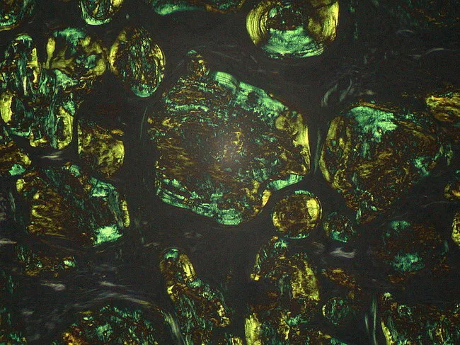
Light microscopy image of a lymph node biopsy stained with Congo Red and examined under polarized light. The tissue reveals extracellular, amorphous eosinophilic deposits distributed throughout nodal architecture consistent with amyloid. The deposits assume a dense, granular to plaque-like appearance on routine brightfield, and demonstrate distinctive apple-green birefringence when illuminated with polarized light, confirming the beta-pleated sheet rich configuration of amyloid fibrils. Congo Red positivity with birefringence distinguishes amyloid from other PAS-positive glycoprotein material and helps differentiate from non-amyloid nodal deposits. The beta-pleated sheet conformation accounts for the characteristic staining behavior and optical properties used in diagnosis. Clinically, identification of nodal amyloid supports systemic or localized amyloidosis and prompts workup for light-chain (AL) or serum amyloid A (AA) subtypes, including serum free light chains, immunofixation, and mass spectrometry-based typing. This image is relevant for educational illustrations of amyloid histology, pathophysiology, and diagnostic pathology workflows, including differential diagnosis with glycoprotein-rich PAS-positive deposits that are Congo Red negative. Potential clinical scenarios include workup of lymphadenopathy in patients with chronic inflammatory states, plasma cell dyscrasias, and lymphoproliferative disorders. High quality images like this support interdepartmental communication, teaching, and reproducible reporting of amyloid histology for junior pathologists and medical trainees in pathology education programs.
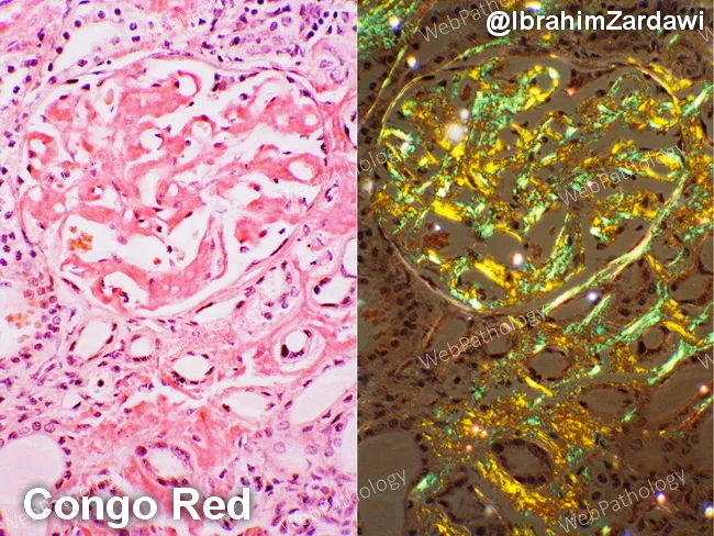
This renal cortical histology specimen demonstrates AL-type amyloidosis with kidney involvement in a patient with multiple myeloma. The left panel shows hematoxylin and eosin–stained tissue with amorphous, eosinophilic extracellular deposits within the glomerular mesangium and extending into the tubulointerstitium, causing mesangial expansion and variable basement membrane thickening. The right panel uses Congo Red staining to highlight the amyloid deposits, which appear salmon-pink on bright-field examination. Under polarized light, the same deposits display the diagnostic apple-green birefringence, a hallmark of Congo Red–positive amyloid. The deposits are diffuse, involving multiple glomeruli and extending into the tubulointerstitium, sometimes outlining capillary walls. In AL amyloidosis associated with plasma cell dyscrasia, the deposited light-chain–derived proteins disrupt normal renal architecture and contribute to proteinuria and progressive renal impairment. Differential considerations include AA (secondary) amyloidosis and other renal amyloidoses, but the combination of a known plasma cell disorder, Congo Red positivity, and apple-green birefringence strongly supports AL-type amyloidosis. This image serves as a valuable teaching reference for renal pathology, nephrology, and hematology-oncology, illustrating classic Congo Red histology, amyloid distribution patterns within glomeruli and interstitium, and the polarizing light signature. Correlates with proteinuria and renal dysfunction in many myeloma patients, guiding targeted therapy and monitoring of clonal plasma cell burden.
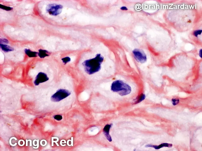
This histology image depicts human cardiac tissue (myocardium) stained with Congo Red and examined under bright-field microscopy. The image highlights extracellular, eosinophilic amyloid deposits that encircle and encroach upon individual cardiomyocytes (perimyocytic/interstitial deposition). The deposits appear as amorphous, orange to salmon-pink material on Congo Red, consistent with amyloid. When the same tissue is examined under polarized light, Congo Red–positive material exhibits characteristic apple-green birefringence, a pathognomonic feature of amyloidosis. The prevailing pattern is perimyocytic, with amyloid accumulating in the interstitium around myocytes and near small vessels, potentially contributing to myocardial stiffness and diastolic impairment. Histologic architecture shows preserved cellular morphology of myocytes with basophilic nuclei contrasted against the pink-red amyloid matrix. This image is diagnostic for cardiac amyloidosis on biopsy and would typically prompt typing (AL vs ATTR) by immunohistochemistry or mass spectrometry, as well as correlation with serum free light chains or transthyretin testing. Clinically, such infiltration predisposes to restrictive cardiomyopathy, diastolic dysfunction, arrhythmias, and heart failure symptoms. This slide underscores the utility of Congo Red staining in identifying myocardial amyloid deposits and the diagnostic value of polarized light birefringence for definitive confirmation. Useful for teaching, differential diagnosis, and research into infiltrative cardiomyopathies and treatment monitoring.
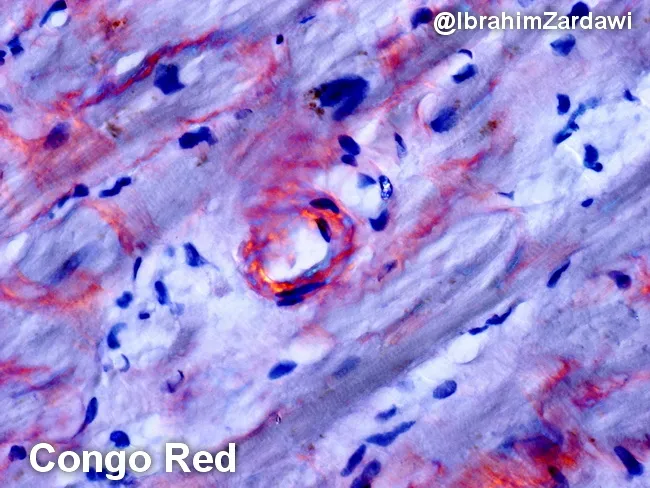
Imaging modality and technique: histopathology using light microscopy with Congo red staining of cardiac tissue, demonstrating characteristic amyloid deposition in the perimyocytic (pericellular) region of the myocardium. The deposited material appears orange to salmon pink on conventional hematoxylin-eosin–like chromogen, and exhibits classic apple-green birefringence under polarized light, confirming amyloid protein with a beta-pleated sheet configuration. The pattern is typically diffuse to pericellular around individual myocytes, producing a subtle, eosinophilic, amorphous coating that can obscure underlying cardiomyocyte detail. In this specimen, the amyloid is evident as an acellular, amorphous, hyaline-like material hugging muscle fibers, with irregular but cohesive contours and occasional nodular clusters. Congo red positivity combined with birefringence is diagnostic of systemic or cardiac amyloidosis when correlated with clinical data. Typing by immunohistochemistry, immunofluorescence, or mass spectrometry is often required to distinguish AL (light-chain) from ATTR (transthyretin) amyloidosis, which has implications for prognosis and treatment. Clinically, these deposits stiffen the myocardium, contributing to restrictive physiology and heart failure with preserved ejection fraction. The image illustrates foundational histopathologic criteria used in diagnosing cardiac amyloidosis and serves as educational material for pathology residents, cardiology fellows, and medical students studying protein misfolding diseases and Congo red histology. This image supports diagnostic confidence globally.
amyloidosis diagnosis treatment
cardiac amyloidosis echocardiogram restrictive cardiomyopathy
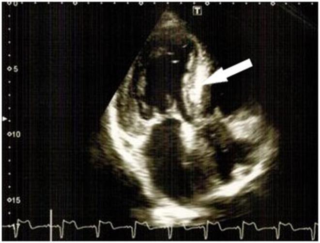
This diagnostic image is a 2D transthoracic echocardiogram in an apical four-chamber view, demonstrating hallmark features of restrictive cardiomyopathy, specifically cardiac amyloidosis. The visual highlights significant concentric left ventricular hypertrophy (LVH) with a markedly thickened interventricular septum. A white arrow points to the 'characteristic luminescence' or 'sparkling' appearance of the myocardial tissue, which indicates increased echogenicity due to amyloid protein deposition. Both atria appear moderately dilated, which is consistent with the restrictive filling pattern and diastolic dysfunction typical of infiltrative heart diseases. The image also captures a concurrent ECG strip at the bottom for cardiac cycle timing. Key educational concepts illustrated include the visualization of myocardial texture changes, wall thickening (hypertrophy), and chamber enlargement as diagnostic indicators for amyloid-related cardiac involvement.
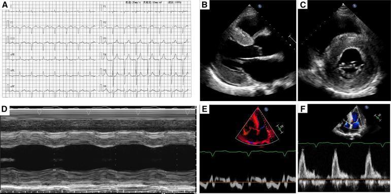
This composite image illustrates diagnostic findings for a patient with restrictive cardiomyopathy, specifically cardiac amyloidosis. Panel A shows a 12-lead electrocardiogram (ECG) with low-voltage QRS complexes and signs of prior myocardial infarction. Panels B and C are two-dimensional transthoracic echocardiograms (parasternal long-axis and short-axis views) showing severe concentric left ventricular hypertrophy and a characteristic 'speckled' or 'ground-glass' myocardial appearance. Panel D is an M-mode echocardiogram demonstrating reduced left ventricular contractility and wall motion abnormalities. Panels E and F provide pulse-waved and color Doppler echocardiograms showing restrictive filling patterns and flow velocity curves consistent with Stage III diastolic dysfunction. The collection highlights key imaging markers for systemic amyloidosis infiltrating the myocardium, including wall thickening, altered echogenicity, and diastolic impairment, serving as a comprehensive clinical example for cardiology education.
Amyloidosis
Structure of Amyloid
- Beta-pleated sheet conformation - the defining structural feature; makes the protein insoluble and protease-resistant
- X-ray diffraction: shows a characteristic cross-beta pattern
- Congo red staining: deposits appear salmon-pink under bright field; under polarized light they show apple-green birefringence - the gold standard diagnostic finding
- Electron microscopy: non-branching fibrils, 7.5-10 nm in diameter
- Also contains a non-fibrillar component - serum amyloid P (SAP) component, a glycoprotein present in all amyloid deposits
Pathogenesis
- Normal proteins with an inherent tendency to misfold - do so when produced in excess or when degradation is impaired (e.g., wild-type transthyretin in senile ATTR)
- Variant (mutant) proteins that are structurally prone to misfolding and aggregation (e.g., variant transthyretin in hereditary amyloidosis)
- Robbins, Cotran & Kumar Pathologic Basis of Disease (Table 6.17)
Classification
| Type | Fibril Protein | Precursor | Associated Condition |
|---|---|---|---|
| AL | Immunoglobulin light chain (esp. lambda) | Free Ig light chains | Multiple myeloma, monoclonal plasma cell proliferation |
| AA | Serum amyloid A (SAA) derived protein | SAA (acute-phase reactant) | Chronic inflammatory diseases (RA, Crohn's, TB, bronchiectasis, FMF) |
| ATTRwt | Wild-type transthyretin | Transthyretin | Senile systemic amyloidosis (males >70 years) |
| ATTRv | Variant transthyretin | Mutant transthyretin (TTR gene) | Familial amyloidotic neuropathy/cardiomyopathy |
| Aβ | Beta-amyloid protein | Amyloid precursor protein (APP) | Alzheimer disease (localized, CNS) |
| Aβ2m | Beta-2 microglobulin | MHC class I component | Long-term hemodialysis (carpal tunnel) |
| AIAPP | Islet amyloid polypeptide | IAPP | Type 2 diabetes mellitus (localized, pancreas) |
| ACal | Calcitonin-derived | Calcitonin | Medullary carcinoma of thyroid |
| AANF | Atrial natriuretic factor | ANF | Isolated atrial amyloidosis |
AL Amyloidosis (Primary Amyloidosis)
- Most common form - ~2,000-3,000 new cases/year in the US
- Caused by clonal plasma cell proliferation; occurs in 5-15% of multiple myeloma cases
- Free lambda light chains (~6x more amyloidogenic than kappa) deposit as AL amyloid
- Most patients do NOT have overt myeloma but have monoclonal immunoglobulins detectable in serum/urine
- Distribution: systemic
AA Amyloidosis (Reactive/Secondary Amyloidosis)
- Previously called "secondary" amyloidosis
- Complicates rheumatoid arthritis (most common today), ankylosing spondylitis, IBD, Familial Mediterranean Fever, and chronic infections
- SAA is an acute-phase protein synthesized in the liver; chronically elevated SAA leads to accumulation of AA fibrils
- Kidneys, liver, and spleen most commonly affected
- Distribution: systemic
ATTR Amyloidosis (Transthyretin Amyloidosis)
- ATTRwt (wild-type, formerly "senile systemic"): affects men >70 years; heart predominantly
- ATTRv (hereditary): point mutations in TTR gene; familial amyloidotic neuropathy (most common: Val30Met mutation); also cardiac involvement
- A 2025 systematic review (PMID 39819351) confirms ATTR amyloidosis is more prevalent than previously appreciated
Morphology / Organ Involvement
Kidneys (most common and serious site)
- Deposits begin in mesangium and glomerular basement membrane
- Glomerular architecture progressively obliterated
- Leads to nephrotic-range proteinuria → nephrotic syndrome → progressive renal failure
- Kidney failure is a common cause of death in systemic amyloidosis
Heart
- Deposits between myocardial fibers (perimyocytic pattern) → restrictive cardiomyopathy
- Echocardiogram: concentric LV hypertrophy with "sparkling/granular" myocardial texture, dilated atria
- Conduction disturbances and arrhythmias (can be fatal)
- Mimics chronic constrictive pericarditis
Liver
- Deposits in space of Disse (along hepatic sinusoids) and portal tracts
- Hepatomegaly; usually minimal liver dysfunction
Spleen
- Two patterns: "lardaceous" (diffuse) or "sago spleen" (discrete follicular deposits)
- Splenomegaly; rarely causes functional problems
Tongue / GI Tract
- Macroglossia - enlargement causing difficulty speaking/swallowing (classic feature of AL amyloidosis)
- GI: malabsorption, diarrhea, disturbances in digestion
Peripheral/Autonomic Nerves
- Peripheral neuropathy (esp. in familial ATTR amyloidosis)
- Carpal tunnel syndrome (esp. in dialysis-associated Aβ2m amyloidosis)
Vasculature
- Amyloid deposits in vessel walls → vascular fragility → spontaneous bleeding
- AL amyloid can bind and inactivate Factor X → life-threatening coagulopathy
Other Sites
- Adrenals, thyroid, pituitary (often clinically silent)
- Respiratory tract (larynx to bronchioles)
- Joints (hemodialysis patients)
Clinical Features
| System | Manifestations |
|---|---|
| Renal | Proteinuria, nephrotic syndrome, progressive CKD/ESRD |
| Cardiac | Congestive heart failure, restrictive cardiomyopathy, arrhythmias, heart block |
| GI | Macroglossia, malabsorption, diarrhea, hepatomegaly |
| Neurological | Peripheral neuropathy, autonomic neuropathy, carpal tunnel syndrome |
| Hematologic | Bleeding (Factor X deficiency), periorbital purpura ("raccoon eyes") |
| Endocrine | Adrenal insufficiency (rare) |
| Constitutional | Weight loss, fatigue |
Diagnosis
-
Tissue biopsy with Congo red staining - confirmatory; shows apple-green birefringence under polarized light
- Sites: kidney (if renal involvement), rectal mucosa, gingival tissue, or abdominal fat pad aspirate
- Abdominal fat aspirate: highly specific but low sensitivity
-
Typing the amyloid - critical for treatment:
- Immunohistochemistry or immunofluorescence
- Mass spectrometry - most definitive method for amyloid protein typing
- Serum/urine protein electrophoresis + immunofixation (for AL)
- Bone marrow biopsy (for AL)
- TTR gene sequencing (for hereditary ATTR)
-
Nuclear medicine: Technetium-labeled bone scan (DPD/PYP/HMDP) - sensitive and specific for ATTR cardiac amyloidosis (non-biopsy diagnosis when serum/urine paraprotein negative)
-
Serum free light chain assay - for AL amyloidosis
-
SAA levels - elevated in AA amyloidosis; monitoring helps guide treatment
Treatment
| Type | Treatment Strategy |
|---|---|
| AL | Eliminate clone producing light chains: high-dose chemotherapy + autologous stem cell transplant (ASCT); or bortezomib-based regimens (daratumumab + bortezomib + dexamethasone) |
| AA | Suppress underlying inflammation (treat underlying disease); colchicine for FMF; IL-1 inhibitors (anakinra, canakinumab) |
| ATTRv | Liver transplantation (removes source of mutant TTR); patisiran/inotersen (RNA-targeted therapies) to silence TTR gene; tafamidis (TTR stabilizer) |
| ATTRwt | Tafamidis (stabilizes TTR tetramer); diflunisal; patisiran/vutrisiran |
Note: Digoxin is potentially dangerous in cardiac amyloidosis - amyloid fibrils bind digoxin, causing unpredictable toxicity.
Key Points Summary
- Amyloid = misfolded protein fibrils in beta-pleated sheet configuration depositing extracellularly
- Diagnosed by Congo red staining + apple-green birefringence under polarized light
- Three main types: AL (plasma cell disorder), AA (chronic inflammation), ATTR (transthyretin - hereditary or senile)
- Most common cause of death: renal failure (from glomerular amyloid) or cardiac arrhythmias/heart failure
- Treatment is directed at the precursor protein source - eliminate the clone or suppress inflammation
- ATTR amyloidosis is increasingly recognized as a cause of heart failure in elderly men; new therapies (tafamidis, RNA silencers) have transformed management