
Development of interatrial septum embryology
Reading File
Reading File
Reading File
Reading File
Finding Sources
Finding Sources
Searching PubMed
"atrial septum" AND embryology
Searching the Web
interatrial septum embryology septum primum secundum foramen ovale authoritative
Reading File
Reading File
Searching Images
embryological formation interatrial septum septum primum septum secundum foramen ovale diagram
Reading Web Page
https://www.ncbi.nlm.nih.gov/books/NBK545203
Reading File
Reading File
Development of the interatrial septum
Atrial septation converts the single primordial atrium into right and left atria. It occurs mainly during the 4th to 6th weeks and creates a fetal right-to-left shunt that later closes after birth.
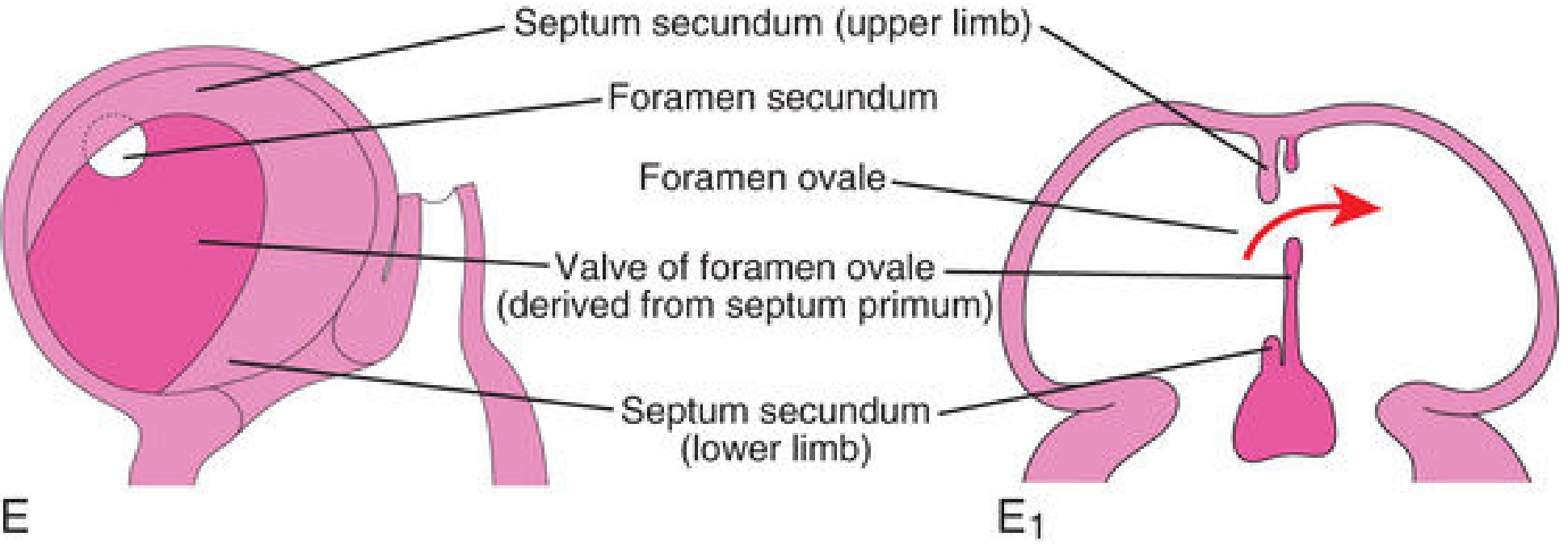
1. Formation of septum primum
- A thin, crescent-shaped septum primum descends from the roof of the common atrium toward the fused atrioventricular endocardial cushions.
- The gap between its free inferior edge and the endocardial cushions is the ostium primum (foramen primum).
- This opening allows blood to pass from the right atrium to the left atrium while the septum is growing.
2. Closure of ostium primum and formation of ostium secundum
- As the septum primum continues to grow, its inferior edge fuses with the endocardial cushions.
- Thus, the ostium primum closes.
- Before it is completely closed, apoptosis produces perforations in the upper part of septum primum.
- These perforations unite to form the ostium secundum (foramen secundum), maintaining right-to-left atrial flow.
3. Formation of septum secundum
- A thicker, crescentic septum secundum develops just to the right of septum primum.
- It grows downward during the 5th and 6th weeks, overlapping the ostium secundum.
- Its inferior free edge does not completely close the interatrial communication. The remaining oblique opening is the foramen ovale.
4. Valve of the foramen ovale
- The remaining inferior portion of the septum primum acts as the thin, flap-like valve of the foramen ovale.
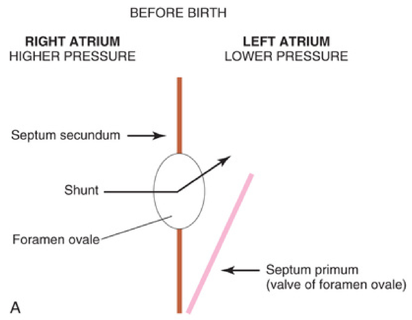
- In fetal life, right atrial pressure exceeds left atrial pressure. Blood can therefore pass:
Right atrium → foramen ovale → left atrium
- This bypasses the non-functioning fetal lungs.
- Reverse flow is prevented because the septum primum is pressed against the more rigid septum secundum, acting as a one-way valve.
5. Closure after birth
At birth:
- Expansion of the lungs decreases pulmonary vascular resistance.
- Pulmonary blood flow and pulmonary venous return increase.
- Left atrial pressure becomes greater than right atrial pressure.
- Septum primum is pushed against septum secundum, producing functional closure of the foramen ovale.
- Usually, the two septa later fuse anatomically, forming the fossa ovalis.
In the adult:
- Floor of fossa ovalis: septum primum
- Limbus/rim of fossa ovalis: free edge of septum secundum
Clinical correlations
- Ostium primum ASD: failure of septum primum to fuse with the endocardial cushions. Often associated with atrioventricular septal defects.
- Ostium secundum ASD: excessive resorption of septum primum or inadequate development of septum secundum.
- Patent foramen ovale (PFO): septum primum and septum secundum fail to fuse after birth. It is not a true tissue-deficiency ASD, because the flap valve is present but incompletely sealed.
- Common atrium: rare failure of both septum primum and septum secundum to develop.
One-line sequence:
Septum primum descends → ostium primum closes → ostium secundum forms → septum secundum overlaps it → foramen ovale remains → septum primum becomes its valve → postnatal fusion forms fossa ovalis.
Septum primum descends → ostium primum closes → ostium secundum forms → septum secundum overlaps it → foramen ovale remains → septum primum becomes its valve → postnatal fusion forms fossa ovalis.
Source: The Developing Human: Clinically Oriented Embryology, pp. 804-808. The mechanism is also summarized in the NCBI embryology review.
This is a shared conversation. Sign in to Orris to start your own chat.