
Hey von hippel landau syndrome
Reading File
Finding Sources
Searching PubMed
"von Hippel-Lindau" syndrome
Reading File
Reading File
Reading File
Reading File
Reading File
Loading Image
I now have comprehensive information from multiple authoritative textbooks. Here is the full overview:
Von Hippel-Lindau (VHL) Syndrome
Overview
VHL syndrome is a rare autosomal dominant multisystem tumor-predisposition disorder. Incidence is approximately 1 in 36,000 live births. The term was coined in 1936 to honor Eugen von Hippel (who described retinal angiomas) and Arvid Lindau (who described spinal hemangioblastomas). About 20% of cases arise from de novo mutations; the rest are inherited.
Genetics & Molecular Basis
- Gene: VHL tumor suppressor gene, mapped to chromosome 3p25
- Encodes two protein isoforms: pVHL30 (213 aa, 30 kD) and pVHL19 (19 kD, lacking 53 amino acids)
- Follows the two-hit hypothesis - biallelic loss is required for tumor development
- Normal function of pVHL: Under normoxic conditions, pVHL binds and ubiquitinates the alpha-subunits of hypoxia-inducible factor (HIF), targeting them for degradation
- Effect of VHL loss: HIF-alpha subunits accumulate, translocate to the nucleus, form heterodimers with HIF-beta, and activate hypoxia-response genes including:
- VEGF (angiogenesis - explains the vascular tumors)
- Erythropoietin (EPO) - causes paraneoplastic polycythemia
- PDGFB, TGF-alpha, GLUT1, Carbonic anhydrase IX
- pVHL also plays roles in microtubule stability and primary cilia integrity (explaining cyst formation)
Classification (Genotype-Phenotype Correlation)
| Type | Features | Mutation type |
|---|---|---|
| Type 1 | No pheochromocytoma | Nonsense, frameshift, deletions |
| Type 2A | PCC + hemangioblastomas, no RCC | Missense |
| Type 2B | PCC + RCC + hemangioblastomas | Missense |
| Type 2C | PCC only | Missense |
Clinical Features (Organ by Organ)
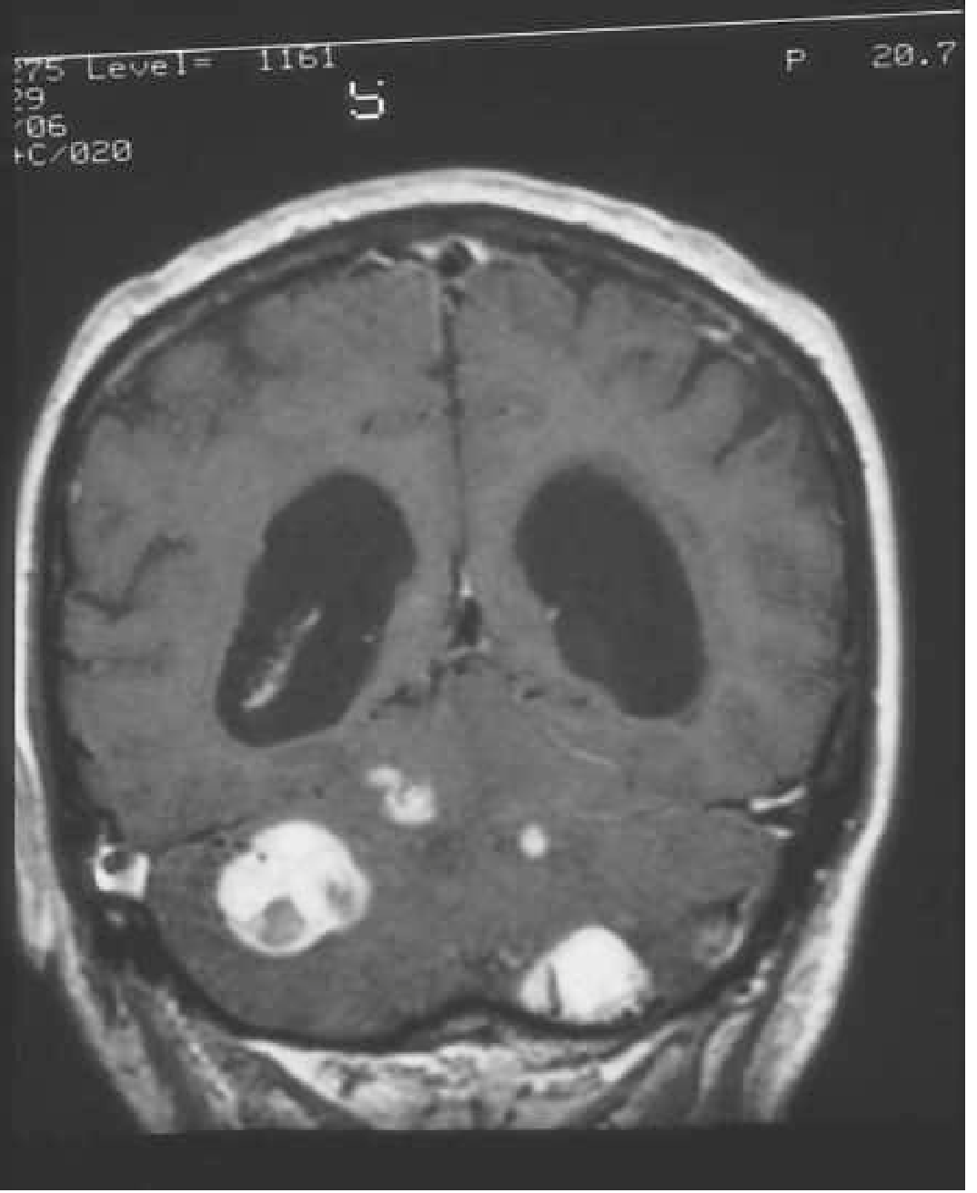
1. CNS Hemangioblastomas (60-80% of patients)
- Most common site: cerebellum (~50%), followed by spinal cord and brainstem; cerebral hemispheres <5%
- Benign but slow-growing; behavior is unpredictable with phases of growth and quiescence
- Histology: thin-walled capillary vessels + stromal cells with vacuolated, lipid-rich cytoplasm (PAS-positive); stain positive for inhibin (diagnostic marker); mast cells present and may produce EPO
- Appear as a mural nodule within a large fluid-filled cyst
- Symptoms: headache (most common), ataxia, nausea/vomiting, nystagmus; spinal lesions cause back/neck pain, sensory loss, weakness, and syringomyelia
- 84% of VHL patients develop at least one cerebellar hemangioblastoma by age 60
- Imaging: contrast-enhanced MRI (pre + post T1-weighted with thin posterior fossa cuts)
2. Retinal Hemangioblastomas / Angiomas
- Most common presenting feature of VHL
- Histopathologically identical to CNS hemangioblastomas
- Typically bilateral; can occur as young as age 1
- Lead to vision loss in 35-55% of cases via arteriovenous shunting, fluid extravasation, hemorrhage, retinal detachment, glaucoma, macular edema
- Treatment: laser photocoagulation for most; VEGF inhibitors (ranibizumab) or tyrosine kinase inhibitors (sunitinib) for severe exudative lesions
3. Renal Cell Carcinoma (RCC)
- Most common malignant tumor in VHL and leading cause of death
- Lifetime risk up to 70%; mean age at presentation ~40 years (younger than sporadic RCC)
- Almost always clear cell type; typically multifocal and bilateral
- Surveillance: abdominal MRI with and without contrast every 2 years from age 16
- Surgery indicated when tumor reaches 3 cm (risk of metastasis increases)
- Kidney-sparing surgery or ablation preferred
4. Pheochromocytoma (PCC)
- Occurs in 7-20% of patients; mean age ~28 years
- Can be bilateral and extraadrenal
- May be the only clinical manifestation in some patients
- Symptoms: episodic/sustained hypertension, severe headache, flushing with profuse sweating; can cause hypertensive crisis, stroke, MI, heart failure
- Diagnosis: excessive catecholamines in serum and urine
- Screening: annually from age 5
- Important: always screen for PCC before any elective surgery
5. Renal Cysts
- Present in >50% of patients; simple cysts from distal tubular epithelium (vs. RCC from proximal tubular epithelium)
- Rarely cause renal failure
6. Pancreatic Lesions
- Pancreatic cysts (most common): usually asymptomatic unless obstructing bile duct
- Pancreatic islet cell tumors: typically non-secretory, occur in 5-10%; surgery when >3 cm
- Serous microcystic adenomas, rarely adenocarcinoma
- Islet cell tumors frequently co-occur with PCC (both from neural crest cells)
7. Endolymphatic Sac Tumors (ELSTs)
- Arise from membranous labyrinth of inner ear
- Bilateral ELSTs are pathognomonic of VHL
- Cause: tinnitus, vertigo, hearing loss
- Occur in ~10% of patients
8. Epididymal Cystadenomas
- May be palpable and cause discomfort; often asymptomatic
Diagnostic Criteria
Clinical diagnosis requires one of the characteristic tumors (CNS/retinal HB, clear cell RCC, PCC, pancreatic islet tumor, ELST) plus a positive family history - OR two characteristic tumors without a family history.
Genetic testing is recommended for:
- At-risk relatives with family history
- Isolated retinal or CNS HB
- PCC or ELST
- Clear cell RCC before age 50, bilateral or multifocal RCC
Mutation detection rate: 95-100% in classic cases; penetrance >90%.
Surveillance Summary
| Organ | Test | Starting Age | Frequency |
|---|---|---|---|
| CNS | Brain + spine MRI | Age 16 | Every 1-2 years |
| Retina | Indirect ophthalmoscopy | Birth | Annually |
| Kidney/Abdomen | Abdominal MRI | Age 16 | Every 2 years |
| PCC | Plasma/urine catecholamines | Age 5 | Annually |
Treatment
- CNS hemangioblastomas: surgical resection when symptomatic; laser ablation for retinal lesions
- RCC: kidney-sparing surgery or ablation at 3 cm threshold
- PCC: surgery (pre-op catecholamine blockade mandatory)
- Targeted therapies: Tyrosine kinase inhibitors (sunitinib) show promise across multiple VHL-associated lesions; ongoing trials for RCC, pancreatic lesions, PCCs, CNS/retinal HBs
- HIF-2alpha inhibitor belzutifan (approved 2021 by FDA) is a newer targeted therapy specifically for VHL-related tumors
Sources: Robbins & Kumar Basic Pathology; National Kidney Foundation Primer on Kidney Diseases (8e); Bradley and Daroff's Neurology in Clinical Practice; Campbell-Walsh Wein Urology
This is a shared conversation. Sign in to Orris to start your own chat.