
DAMAGE CONTROL RESUSCITATION According to latest ATLS guideline,rosens and tintinalli book
"damage control resuscitation" trauma hemorrhage
ATLS 10th edition damage control resuscitation guidelines 2024 2025
damage control resuscitation hemorrhagic shock massive transfusion protocol
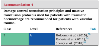
<table> <tr> <td colspan="4" style="color:#b22222;"><b>Recommendation 4</b></td> </tr> <tr> <td colspan="4">Damage control resuscitation principles and massive transfusion protocols used for patients with traumatic haemorrhage are recommended for patients with vascular trauma.</td> </tr> <tr> <th>Class</th> <th>Level</th> <th>References</th> <th>ToE</th> </tr> <tr> <td style="background-color:#7fcdbb;">I</td> <td style="background-color:#225ea8; color:#fff;">A</td> <td>Holcomb <i>et al.</i> (2015),<sup>58</sup><br>Roberts <i>et al.</i> (2011),<sup>64</sup><br>Sperry <i>et al.</i> (2018)<sup>59</sup></td> <td></td> </tr> </table>
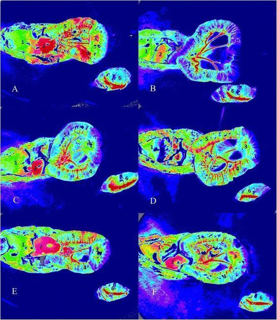
This diagnostic image set showcases Laser Speckle Contrast Imaging (LSCI) used to evaluate microcirculatory blood flow intensity in an animal model of hemorrhagic shock and resuscitation. Six pseudo-colored panels (A-F) represent different experimental groups: sham (a), control (b), 0.9% saline (c), hypertonic saline (d), gelatin (e), and hydroxyethyl starch (f) at time point T2. The color scale indicates blood flow intensity, where red and yellow represent higher perfusion, green intermediate, and blue/purple lower perfusion. Each panel identifies six distinct anatomical regions of interest (ROIs): (1) liver, (2) kidney, (3) intestinal mucosa, (4) serosal muscular layer, (5) Peyer's patch, and (6) gracilis muscle. The comparison highlights significant perfusion deficits in the control and saline groups (cooler tones in kidney and intestine) versus the restoration of microcirculatory flow in groups resuscitated with hypertonic saline or colloids (warmer tones). This visualization is essential for understanding splanchnic organ perfusion and the efficacy of different fluid resuscitation strategies in critical care medicine.
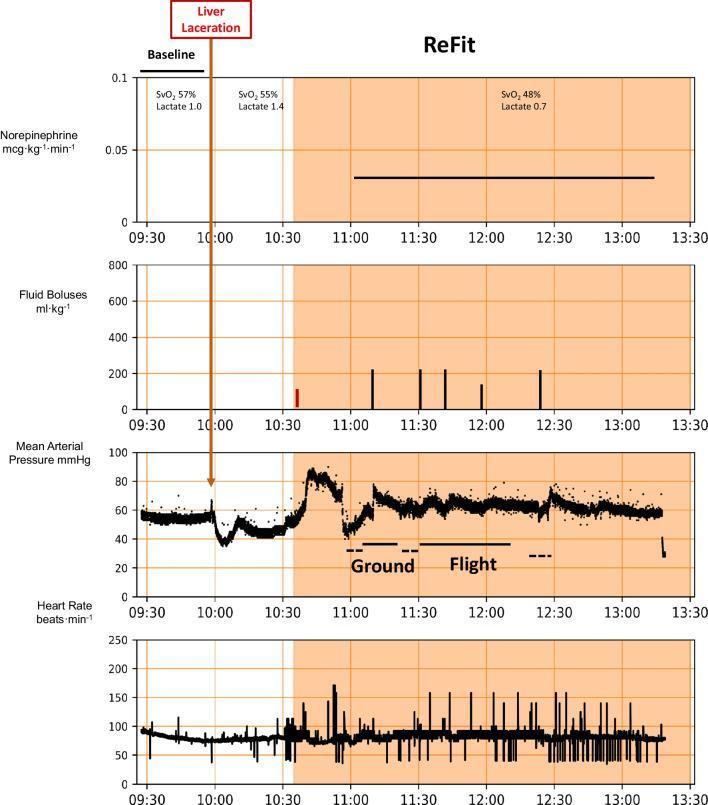
This physiological trend chart displays real-time hemodynamic monitoring data during a 'ReFit' resuscitation protocol following a liver laceration event (indicated at 10:00). The image consists of four vertically stacked graphs sharing a common timeline from 09:30 to 13:30. The parameters tracked include Norepinephrine infusion rate (mcg/kg/min), Fluid Boluses (ml/kg), Mean Arterial Pressure (MAP, mmHg), and Heart Rate (beats/min). Shaded areas indicate the active protocol phase and transportation periods (Ground and Flight). Post-injury, MAP shows a precipitous drop below 40 mmHg, followed by stabilization via the ReFit algorithm through fluid boluses (represented by vertical bars) and a constant norepinephrine infusion of approximately 0.03 mcg/kg/min. Periodic values for SvO2 and Lactate levels are annotated at specific time points. The 'Ground' and 'Flight' transport phases illustrate the stability of MAP (maintained at ~60 mmHg) and heart rate during patient transfer. This educational material demonstrates physiological management of hemorrhagic shock using automated algorithms during multi-modal medical evacuation.
DAMAGE CONTROL RESUSCITATION (DCR)
1. DEFINITION AND CONCEPT
- Worsen trauma-induced coagulopathy (TIC) by diluting clotting factors
- Cause abdominal compartment syndrome
- Exacerbate hypothermia, acidosis, and inflammation
- Can "pop the clot" by raising hydrostatic pressure at sites of non-compressible hemorrhage
2. THE LETHAL TRIAD (Bloody Vicious Cycle)
| Component | Driver |
|---|---|
| Hypothermia | Exposure, cold fluids, decreased cellular respiration |
| Acidosis | Tissue hypoperfusion, lactic acidosis |
| Coagulopathy | Dilutional + consumptive; worsened by hypothermia and acidosis |
3. ATLS 11th EDITION (2025) - KEY UPDATES FOR DCR
x-ABCDE Framework
DCR Principles embedded in ATLS 11e:
- Immediate hemorrhage control as the first priority
- Permissive hypotension targeting SBP ~90 mmHg (MAP ~60 mmHg) until surgical hemostasis
- Restrict crystalloids - use blood products from the start
- Early whole blood or balanced component therapy (1:1:1) - pRBC : FFP : platelets
- Tranexamic acid (TXA) - given as early as possible (ideally within 3 hours of injury)
- Avoid over-resuscitation
4. COMPONENTS OF DCR (Box-by-Box)
A. HEMORRHAGE CONTROL (First Priority)
- External hemorrhage: Tourniquet, wound packing, direct pressure, hemostatic dressings
- Internal hemorrhage: Expedited surgical or interventional radiology (IR) control
- Prehospital DCR has been shown to confer survival benefit (Cannon JW, NEJM 2018)
B. PERMISSIVE HYPOTENSION
| Setting | SBP Target |
|---|---|
| General hemorrhagic shock (penetrating, blunt) | ~90 mmHg (MAP ~60 mmHg) |
| Civilian trauma | ~110 mmHg (Tintinalli) |
| Combat casualties | ~100 mmHg (Tintinalli) |
| Traumatic Brain Injury (TBI) | NOT recommended - maintain normal BP |
| Elderly / hypertensive / CAD patients | NOT recommended |
- Traumatic Brain Injury (TBI)
- Blunt trauma with suspected occult hemorrhage in elderly
- Cardiac disease (myocardial ischemia), cerebrovascular disease
C. RESTRICTIVE CRYSTALLOID USE
- Isotonic crystalloids (NS, LR) are NOT preferred as primary resuscitation fluid in hemorrhagic shock
- Problems with large-volume crystalloid:
- Only ~25-30% stays intravascular (LR/NS distributes across interstitial space); need 3:1 ratio to replace 1 L blood loss
- Causes dilutional coagulopathy, hypothermia, hyperchloremic acidosis (NS), and abdominal compartment syndrome
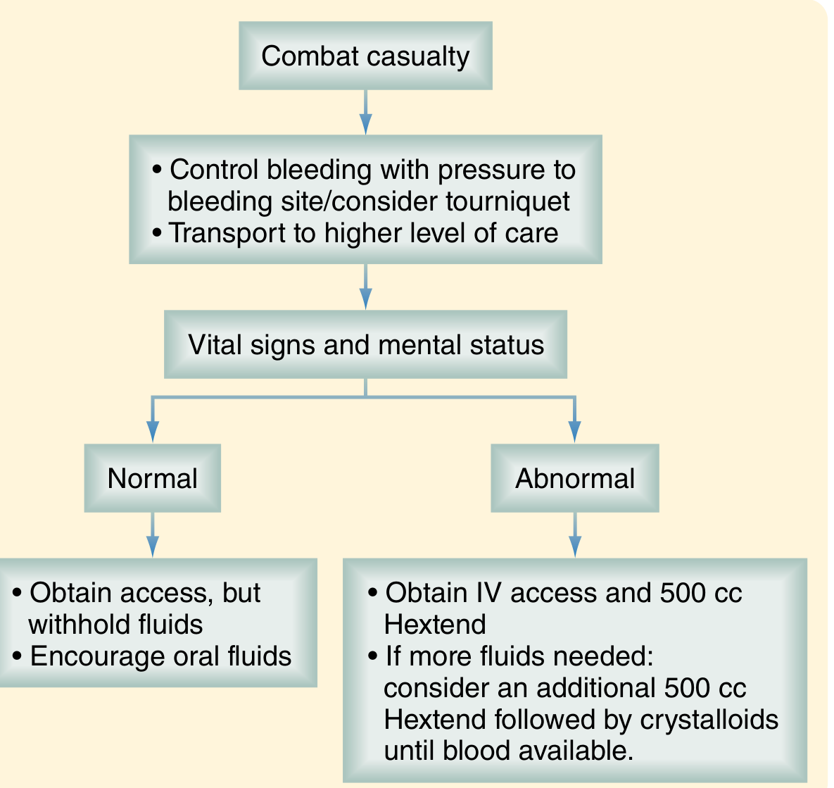
- Current approach: Limit crystalloid; if needed initially, give 250-500 mL aliquots while blood products are prepared
- Transition immediately to a plasma-based Massive Transfusion Protocol (MTP) once ongoing hemorrhage is recognized
D. BLOOD PRODUCT RESUSCITATION (Hemostatic Resuscitation)
Massive Transfusion
- Defined as >10 units pRBCs in 24 hours (or >3 units in 1 hour in some definitions)
- Affects ~3-5% civilian trauma, ~10% military trauma patients
Predicting Massive Transfusion - ABC Score
- Penetrating mechanism
- Positive FAST
- SBP <90 mmHg
- HR >120 bpm
- Score ≥2 = activate MTP (sensitivity 76-90%, specificity 67-87%)
Blood Product Ratios - 1:1:1
| Product | Ratio | Notes |
|---|---|---|
| pRBC | 1 | Restore O2-carrying capacity |
| FFP | 1 | Restores all coagulation factors |
| Platelets | 1 | Support primary hemostasis |
- The PROPPR trial (Holcomb et al., JAMA 2015) confirmed 1:1:1 pRBC:FFP:platelets vs 1:1:2 ratio showed improved 24-hour and 30-day hemostasis and survival benefit
- Improved survival also seen with early high plasma:pRBC ratios (~1:1.4) from combat data
- Use ABO-compatible blood; type O negative pRBC and AB+ FFP for emergencies
- Fresh whole blood is superior (currently mainly military setting); civilian equivalent is 1:1:1 component therapy
E. CORRECTION OF THE LETHAL TRIAD
1. Hypothermia Prevention and Treatment
- Target temperature: >36°C
- Apply active external warming immediately
- Use warmed IV fluids and blood products (all resuscitation fluids should be warmed)
- Keep patient covered; reduce exposure time
2. Acidosis Correction
- Target: normalize base deficit and lactate
- Serial lactate and base deficit measurements guide adequacy of resuscitation
- Central venous O2 saturation (ScvO2) target: 60-70%
- Tissue O2 saturation (StO2) by near-infrared spectroscopy: maintain >50%
- Acidosis resolves with hemorrhage control and adequate perfusion - not with bicarb
3. Coagulopathy Management
- Use balanced blood products (FFP, cryoprecipitate for fibrinogen replacement)
- Calcium replacement: pRBCs and FFP contain citrate that chelates calcium - administer calcium chloride (preferred over gluconate) to maintain ionized calcium ≥0.9 mmol/L
- Viscoelastic testing (see below)
F. TRANEXAMIC ACID (TXA)
- Antifibrinolytic agent - blocks plasminogen activation
- CRASH-2 trial (Roberts et al., Lancet 2011): TXA reduced mortality in bleeding trauma patients by ~1.5% absolute risk reduction
- MILITARY DATA (MATTERs study): TXA with MTP reduced mortality (OR 0.46) in combat casualties
- Dosing: 1 g IV over 10 min, then 1 g IV over 8 hours
- Timing: Must be given within 3 hours of injury - after 3 hours, may be harmful (paradoxically increases mortality)
- The ATLS 11th edition emphasizes early TXA as a standard component of DCR
- Ongoing research to identify which specific patient subgroups benefit most
G. VISCOELASTIC HEMOSTATIC ASSAYS (VHA)
- They are slow (30-45 min turnaround)
- Performed at 37°C (don't reflect hypothermic coagulopathy)
- Don't assess fibrinolysis or platelet function
- Clot formation time (reaction time)
- Clot kinetics (alpha angle)
- Clot strength (maximum amplitude - MA)
- Fibrinolysis
- Prolonged reaction time/clot time - give FFP
- Low MA/amplitude - give platelets or cryoprecipitate
- Fibrinolysis detected - give TXA
H. ADJUNCT HEMOSTATIC AGENTS
| Agent | Indication | Notes |
|---|---|---|
| TXA | Early hemorrhage, <3 hrs | Standard of care |
| Prothrombin Complex Concentrate (PCC) | Anticoagulated patients, rapid reversal | 4-factor PCC preferred |
| Recombinant Factor VIIa (rFVIIa) | Refractory coagulopathy | High cost, thrombotic risk |
| Fibrinogen concentrate / Cryoprecipitate | Fibrinogen <1.5-2 g/L | Especially if hypofibrinogenemia on TEG |
| Desmopressin (DDAVP) | Platelet dysfunction (e.g. aspirin use, uremia) | Releases vWF |
| Calcium chloride | All massive transfusions | Prevents citrate-induced hypocalcemia |
I. HYPERTONIC SALINE
- 7.5% hypertonic saline can pull water into the vascular space (table below)
- Volume effect: 1 L of 7.5% saline expands plasma by ~990 mL (vs 275 mL for NS)
- Useful as a small-volume resuscitator
- May have immunomodulatory effects
- Use limited by sodium load and risk of hypernatremia
5. DCR IN CONTEXT: THE THREE PHASES
| Phase | What Happens |
|---|---|
| Phase 0 (Prehospital) | Tourniquet, wound packing, TXA, restrict fluids, rapid transport |
| Phase 1 (Initial DCR in ED/OR) | Hemorrhage control, permissive hypotension, MTP activation, TXA, blood products 1:1:1 |
| Phase 2 (ICU Resuscitation) | Full resuscitation, correction of lethal triad, optimization before definitive surgery |
| Phase 3 (Definitive Surgery) | Formal repair once physiology corrected |
6. SUMMARY TABLE - DCR COMPONENTS (from Sabiston Box 33.2 & ATLS 11e)
| Component | Goal |
|---|---|
| Rapid hemorrhage control | Stop source of bleeding immediately |
| Permissive hypotension | SBP ~90 mmHg until surgical hemostasis |
| Restrict / avoid crystalloids | Prevent dilutional coagulopathy, ACS |
| Early blood products (1:1:1) | Approximate whole blood |
| Whole blood (if available) | Ideal resuscitation fluid |
| TXA <3 hours | Prevent fibrinolysis |
| Correct hypothermia | Active warming; warm fluids |
| Correct acidosis | Restore perfusion; serial lactate |
| Calcium replacement | Ionized Ca++ ≥0.9 mmol/L |
| TEG/ROTEM-guided therapy | Goal-directed correction of coagulopathy |
| Consider PCC / rFVIIa | Refractory or anticoagulant-related coagulopathy |
7. SPECIAL POPULATIONS
| Population | Modification |
|---|---|
| TBI + hemorrhagic shock | Permissive hypotension is CONTRAINDICATED; maintain SBP ≥90 mmHg (some guidelines >110 mmHg) |
| Elderly patients | Avoid permissive hypotension; more likely to have cardiac/vascular comorbidities |
| Pediatric trauma | DCR principles extend; avoid crystalloids >60 mL/kg/day; evidence still limited vs adults |
| Anticoagulated patients | Reversal agents (PCC, andexanet, idarucizumab) as appropriate |
8. KEY CLINICAL TRIAL REFERENCES
- CRASH-2 (Lancet 2011) - TXA reduces mortality in bleeding trauma within 3 hours (PMID: 20554319)
- PROPPR (Holcomb et al., JAMA 2015) - 1:1:1 vs 1:1:2 pRBC:FFP:Plt; 1:1:1 superior for hemostasis and 24-hr survival
- PAMPer Trial (Sperry et al., NEJM 2018) - Prehospital plasma transfusion improved 30-day survival in blunt trauma hypotensive patients transported by air
- MATTERs - Military TXA with MTP reduced mortality in combat casualties
KEY TAKEAWAYS
- DCR = stop bleeding + permissive hypotension + blood products 1:1:1 + TXA + correct lethal triad
- ATLS 11e (2025): DCR is now core doctrine; x-ABCDE prioritizes exsanguination control before airway
- TXA must be given within 3 hours - after that it may harm
- Crystalloids are restricted - they worsen coagulopathy, dilute clotting factors, cause ACS
- Whole blood is the ideal resuscitation fluid; 1:1:1 component therapy approximates it in civilian practice
- Permissive hypotension is contraindicated in TBI, elderly, and cardiac/cerebrovascular patients
- TEG/ROTEM enables real-time, goal-directed hemostatic resuscitation
Recent evidence alert: A 2025 review of ATLS 11th edition updates (PMID: 41671886) confirms DCR integration as a core principle alongside x-ABCDE; no contradictions to the textbook content above.