
Is dialysis that we insert on neck region is risky while handling a old age women
dialysis catheter internal jugular complications elderly women
internal jugular vein dialysis catheter placement neck complications
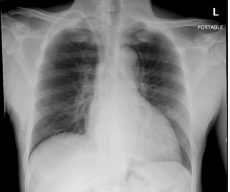
This diagnostic image is a front-on chest radiograph (X-ray), labeled as a portable study, demonstrating the placement of a right internal jugular dialysis catheter. The radiopaque catheter is seen entering via the right neck, coursing inferiorly through the superior vena cava. The distal tip is correctly positioned, projecting at the right cavoatrial junction. The anatomical structures, including the cardiac silhouette, mediastinum, and hila, appear within normal limits. The lung fields are clear bilaterally with no evidence of focal consolidation, pleural effusion, or pneumothorax. Bony structures, including the ribs, clavicles, and scapulae, are unremarkable. This radiograph serves as a clinical evaluation for catheter tip placement and to rule out acute cardiopulmonary complications, such as infectious processes or procedural trauma, in a patient with end-stage renal disease (ESRD).
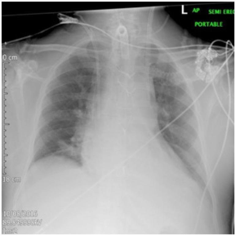
This diagnostic image is an anteroposterior (AP) portable semi-erect chest radiograph demonstrating a malpositioned dialysis catheter. The primary finding is a radiopaque double-lumen dialysis catheter entering via the left internal jugular vein. Rather than following the expected course into the superior vena cava, the catheter tip is positioned anomalously within the mediastinum, directed toward the left hilum. The image displays the cardiothoracic silhouette, ribs, and clavicles, with an endotracheal tube and additional monitoring lines also visible. The lung fields are largely clear, though limited by image quality, with sharp costophrenic angles. This visual serves as a critical educational example of central venous access complications, illustrating the importance of post-procedural radiographic verification to identify improper placement and potential vascular anomalies, such as an anomalous left superior pulmonary vein or persistent left superior vena cava.
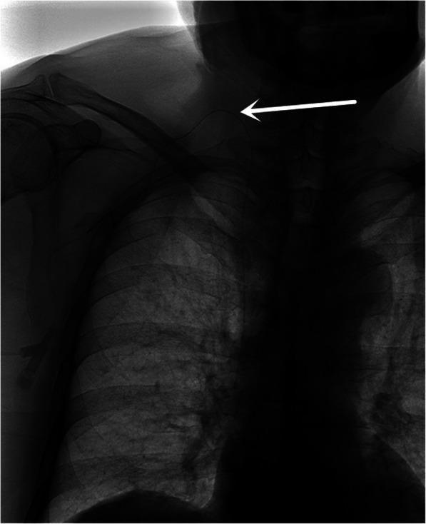
Anteroposterior (AP) chest X-ray radiograph demonstrating the placement of a Totally Implantable Venous Access Device (TIVAD) via the right internal jugular vein (IJV). The radiopaque catheter is visible entering the soft tissues of the right neck, crossing over the medial third of the right clavicle, and descending toward the superior vena cava. A prominent white arrow indicates a sharp acute angle or folding point where the catheter traverses the clavicle, reflecting potential mechanical stress or kinking due to a high puncture site. Anatomical structures including the right clavicle, ribs, and mediastinal shadow are visualized. This image serves as a clinical example of procedural outcomes and potential catheter complications, such as bending or restricted flow, associated with the IJV approach for venous port implantation.
Dialysis Catheter in the Neck Region - Risks in Elderly Women
What Is the Catheter You Are Referring To?
- Non-tunneled (temporary) - placed at the bedside for acute dialysis
- Tunneled-cuffed (long-term) - placed for maintenance hemodialysis when a fistula or graft is not possible
Immediate (Procedural) Risks at the Time of Insertion
| Risk | Description |
|---|---|
| Pneumothorax | Accidental puncture of the lung apex, especially risky with subclavian approach |
| Arterial puncture / perforation | The carotid artery lies right next to the IJV; inadvertent cannulation can cause massive hemorrhage |
| Local hematoma | Can extend into neck soft tissue and cause airway obstruction - especially dangerous |
| Air embolism | If the catheter system is opened to air |
| Cardiac tamponade | From pericardial perforation if the catheter tip is placed too deep |
| Mediastinal/pericardial perforation | Can cause hemothorax |
- Veins are more fragile, smaller, and less compliant
- Osteoporosis and kyphosis can distort the anatomy
- Reduced subcutaneous fat over the neck makes hemostasis harder to achieve
- Anticoagulants or antiplatelet drugs (commonly used in the elderly) increase bleeding risk
- Poor cardiovascular reserve means they tolerate complications like tamponade or hemothorax less well
Catheter Images (X-ray)
Long-Term Complications (After the Catheter Is In Place)
-
Infection / Bacteremia (most common serious complication)
- Catheter-related bacteremia is the leading infectious complication
- Risk is proportional to duration of catheter use
- There is a ~25% frequency of symptomatic ipsilateral deep vein thrombosis with internal jugular dialysis catheters (Brenner and Rector's The Kidney)
- Elderly patients with impaired immunity are at higher infection risk
-
Central Vein Stenosis / Thrombosis
- Repeated catheter use causes scarring and narrowing of the vein
- This can cause arm swelling, or in severe cases, superior vena cava (SVC) syndrome
- Once central vein stenosis occurs, it prevents use of the same-side arm for a future fistula or graft - a major long-term loss
-
Fibrin Sheath Formation
- A fibrin sheath builds around the catheter over time, reducing flow rates and making the catheter dysfunctional
-
Catheter Malposition
- Tip placed in the carotid artery, pericardial sac, or wrong vessel has been documented (see textbook figures)
Why Is the Right IJV Preferred?
"Most tunneled catheters are placed in the internal jugular veins... Nephrologists, interventional radiologists, and vascular surgeons generally prefer to avoid placement of catheters into the subclavian veins; while flow rates are usually excellent, subclavian stenosis is a frequent complication and, if present, will likely prohibit future access creation."
Special Considerations in Elderly Women
| Factor | Why It Matters |
|---|---|
| Fragile, small veins | Harder to cannulate; higher perforation risk |
| Osteoporosis/kyphosis | Distorted anatomy, harder landmark identification |
| Multiple comorbidities | Diabetes, hypertension, heart disease reduce tolerance for complications |
| Anticoagulant/antiplatelet use | Higher bleeding risk at insertion |
| Lower immunity | Higher risk of catheter-related bloodstream infection |
| Thin neck/minimal tissue | Hematoma more likely to compress airway |
| Skin fragility | Exit site care and dressing adherence can be challenging |
Bottom Line
- Use ultrasound guidance during insertion
- Confirm placement with a chest X-ray before starting dialysis
- Use strict sterile technique to minimize infection
- Limit catheter dwell time as much as possible
- Transition to a fistula or graft as soon as feasible ("fistula first" principle)