
Liver chir
Reading File
It looks like your message got cut off - "Liver chir" seems incomplete. Could you clarify what you're looking for?
Asking for Preferences
What aspect of liver surgery (chirurgie) are you asking about?
Fibroendenoma of breast
Reading File
Now let me search the medical library for fibroadenoma of the breast.
Finding Sources
Searching PubMed
fibroadenoma breast
Finding Sources
Finding Sources
Excellent - multiple rich sources found. Let me read the key sections in parallel.
Reading File
Reading File
Reading File
Reading File
Reading File
Loading Image
Here is a comprehensive overview of Fibroadenoma of the Breast drawn from multiple authoritative textbooks.
Fibroadenoma of the Breast
Definition & Epidemiology
Fibroadenoma is the most common benign solid tumor of the female breast and the most common breast lump in women aged 15-25 years. It is the second most common breast tumor overall (after carcinoma). New fibroadenomas are rarely seen after age 40-45.
- Robbins Pathologic Basis of Disease, p. 984
- Sabiston Textbook of Surgery, p. 1385
Pathogenesis / Molecular Basis
Fibroadenomas arise from hyperplasia of a breast lobule (intralobular stroma + epithelium). At the molecular level:
-
~2/3 harbor driver mutations in MED12 - a subunit of the mediator complex that regulates RNA polymerase II-dependent gene transcription. This is the same mutation found in uterine leiomyomas, both arising from stromal cells in hormone-responsive organs.
-
~1/3 also harbor mutations in RARA (retinoic acid receptor alpha), an estrogen target gene that may cooperate with estrogen receptor (ER) in regulating transcription.
-
They are hormonally responsive: grow during pregnancy, regress after menopause.
-
Robbins Pathologic Basis of Disease, p. 984
Histological Types
| Type | Histology | Size / Consistency |
|---|---|---|
| Pericanalicular | Fibrous tissue surrounds small tubular glands | Smaller, harder |
| Intracanalicular | Fibrous tissue indents and compresses glands into elongated/cleft-like spaces | Larger, softer |
-
In older females, stroma becomes densely hyalinized and epithelium may be atrophic.
-
In younger females, stroma is delicate and often myxoid.
-
S Das Manual on Clinical Surgery, p. 434
-
Robbins Pathologic Basis of Disease, p. 984
Pathology Image
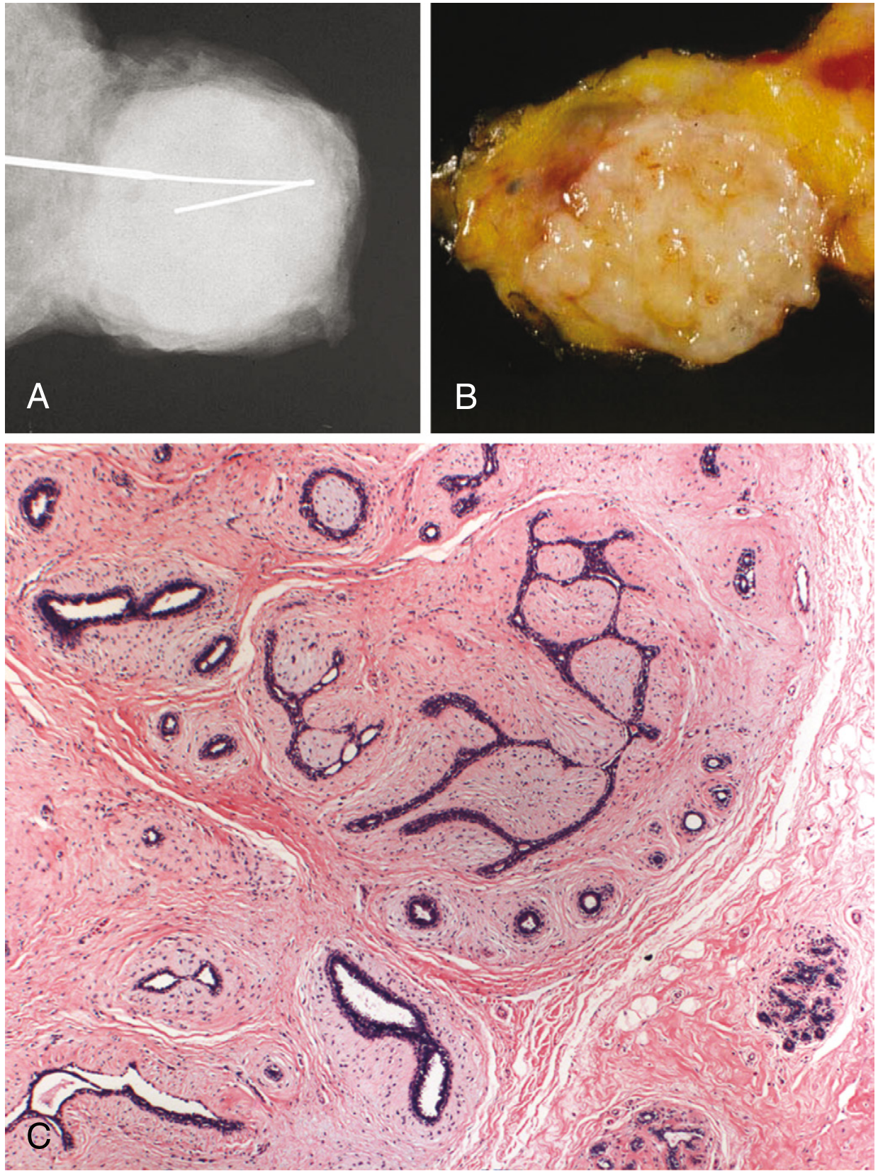
Fig. 23.24 Fibroadenoma. (A) Mammogram: well-circumscribed mass. (B) Gross: rubbery, white, well-circumscribed nodule. (C) Histology: intralobular stroma surrounds and distorts the epithelium; sharp border from surrounding tissue.
- Robbins Pathologic Basis of Disease, p. 984
Clinical Features
-
Presentation: Painless, smooth, firm, well-defined, highly mobile lump - classically described as a "breast mouse" or "floating tumour" because it slides freely under examining fingers.
-
No skin fixation, no tethering, no axillary lymphadenopathy.
-
Usually 2-3 cm in size; often multiple and bilateral.
-
May wax and wane with the menstrual cycle.
-
Rapid growth (including infarction) during pregnancy can mimic carcinoma.
-
Pericanalicular type: ages 15-30 years (young girls).
-
Intracanalicular type: ages 35-50 years (middle-aged women).
-
Bailey & Love's Surgery 28th Ed., p. 944
-
S Das Manual on Clinical Surgery, p. 434
Subtypes
| Subtype | Description | Management |
|---|---|---|
| Simple fibroadenoma | Standard form, RR for cancer 1.5-1.7 | Observe if typical |
| Complex fibroadenoma | Contains cysts >0.3 cm, sclerosing adenosis, epithelial calcifications, or papillary apocrine change | RR for cancer 3.4-3.7 (with hyperplasia); excise |
| Giant fibroadenoma | >5 cm; occurs at puberty or young adulthood | Excision recommended |
| Juvenile fibroadenoma | Large, more cellular, in adolescents | Observe if <5 cm; excise if >5 cm, growing, or persisting to adulthood |
| Myxoid fibroadenoma | Association with Carney complex (PRKAR1A mutation) | Consider genetic work-up |
- Robbins Pathologic Basis of Disease, p. 984
- Sabiston Textbook of Surgery, p. 1385
Investigations
-
Ultrasound: First-line - readily distinguishes fibroadenoma from cysts; shows well-defined, hypoechoic, oval mass. Mammography is less useful for differentiation.
-
Biopsy: Recommended if:
- Patient is over 25 years old
- Atypical features on ultrasound
- Rapidly growing or >3 cm (concern for phyllodes tumor)
-
Triple assessment (clinical + imaging + FNAC/core biopsy) is standard for any breast lump.
-
Bailey & Love's Surgery 28th Ed., p. 944
-
Sabiston Textbook of Surgery, p. 1385
Cancer Risk
| Condition | Relative Risk (RR) |
|---|---|
| Simple fibroadenoma | 1.5-1.7 |
| Complex fibroadenoma + epithelial hyperplasia | 3.4-3.7 |
| Complex fibroadenoma + family history | 3.0-4.0 (especially lobular carcinoma) |
| Cancer within a fibroadenoma (newly discovered) | 0.2% (exceedingly rare) |
- Bailey & Love's Surgery 28th Ed., p. 944
- Sabiston Textbook of Surgery, p. 1385
Management
Conservative (Observation)
- Clinically typical fibroadenoma confirmed on ultrasound in a patient under 25 - can be observed without biopsy.
- Confirmed fibroadenoma on tissue diagnosis - surgical excision is not needed unless symptomatic or patient prefers removal.
- Medical: Some regression observed with tamoxifen and ormeloxifene (antioestrogens).
Indications for Surgical Excision
- Age over 30 years
- Suspicious features on imaging (e.g., microlobulation)
- Atypia on histology
- Size >5 cm
- Family history of breast cancer
- Patient preference
- Lesion >3 cm or rapidly growing (to exclude phyllodes tumor)
- In elderly patients - excise with a rim of normal tissue (may harbor malignancy or phyllodes tumor)
Surgical Technique
- Open excisional biopsy or percutaneous approach for smaller masses.
- Giant fibroadenoma - can be enucleated through a submammary (Gaillard Thomas) incision.
Minimally Invasive (Emerging)
Recent systematic reviews (2025-2026) support cryoablation and high-intensity focused ultrasound (HIFU) as effective alternatives to surgery for appropriate candidates - both show good efficacy and safety profiles (PMID: 41061430, PMID: 42147239).
- Bailey & Love's Surgery 28th Ed., p. 944
- Sabiston Textbook of Surgery, p. 1385
Key Differentials
| Condition | Distinguishing Feature |
|---|---|
| Phyllodes tumor | Occurs 10-20 years later (peak age ~50s); larger, bosselated, more stromal cellularity, MED12 + TERT/TP53/RB mutations |
| Breast cyst | Fluctuant; ultrasound shows anechoic fluid-filled lesion |
| Breast carcinoma | Hard, irregular, fixed; skin/nipple changes; lymphadenopathy |
| Breast abscess | Tender, warm, erythematous; fever |
Summary Points for Exam
- Most common breast lump in 15-25 year olds
- Arises from intralobular stromal + epithelial hyperplasia
- MED12 mutation is the main driver (same as uterine fibroid)
- Clinically: smooth, firm, mobile - "breast mouse"
- Two histological types: pericanalicular (hard, young) and intracanalicular (soft, older)
- Hormonally responsive (grows in pregnancy, regresses post-menopause)
- Cancer risk is low but not zero - complex type with family history carries higher risk
- Management is largely conservative; surgical excision for specific indications
- Giant (>5 cm): excise; Juvenile: observe if small, excise if >5 cm or persisting
This is a shared conversation. Sign in to Orris to start your own chat.