
Cervical mass management
"neck mass" OR "cervical mass" management diagnosis
neck mass management algorithm differential diagnosis
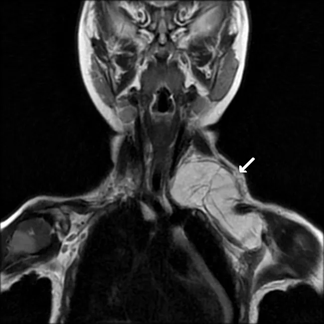
Imaging modality and technique: Magnetic Resonance Imaging (MRI) with T2-weighted coronal sequence. Clinical context: a 2-year-old female with a rapidly enlarging neck mass. Anatomic distribution: a large, well-defined lipomatous soft tissue mass encompasses both the supra- and infraclavicular neck regions. Morphology: internal septations are evident within the lesion. Signal characteristics: lesion is fatty in nature, showing high signal on T1-weighted and T2-weighted images with near-complete suppression on fat-saturated sequences, consistent with adipocytic tissue. Anatomic relations: the mass displaces adjacent soft tissues without definite invasion; margins are well circumscribed, suggesting benign behavior. Pathology and diagnosis: histologic examination after excision confirmed lipoblastoma, a pediatric adipocytic tumor displaying lobulated architecture with vacuolated adipocytes and immature lipoblasts amid a myxoid stromal background. Clinical significance: lipoblastoma should be included in the differential diagnosis of pediatric neck masses with fatty signal; MRI features—multiloculated fat-containing mass with internal septations and fat suppression behavior—help distinguish from lipoma and other lipomatous lesions. Differential considerations: lipoma, fibrolipoma, and rare pediatric liposarcoma; correlation with symptoms and growth rate is essential. Management and prognosis: complete surgical excision is standard; prognosis is favorable though recurrence can occur. Educational use: case emphasizes pediatric soft-tissue neoplasms and imaging-pathology correlation.
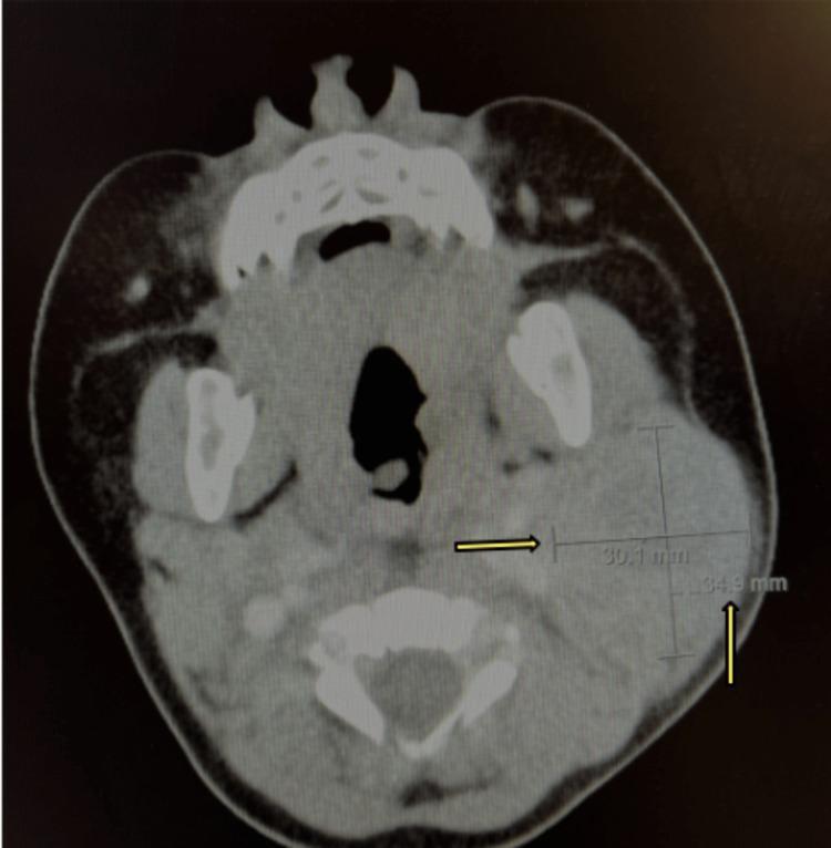
This diagnostic image is an axial contrast-enhanced computerized tomography (CT) scan of the neck at the level of the mandible and cervical vertebrae. The image demonstrates a large, ill-defined, heterogeneous mass located in the left anterior cervical region. Yellow arrows and electronic calipers highlight the lesion, providing cross-sectional measurements of 30.1 mm and 34.9 mm. The mass exhibits non-uniform internal density with areas of heterogeneous enhancement, suggesting internal necrosis, inflammation, or complex tissue composition. Anatomically, the lesion causes significant effacement of the surrounding soft tissue planes and exerts a mass effect on adjacent structures. Educational focus includes the radiologic evaluation of neck masses and the differential diagnosis of cervical lymphadenopathy, which may include neoplasia, infectious lymphadenitis with central necrosis, or inflammatory processes. This material is suitable for radiology and otolaryngology medical education for students and residents learning to interpret cervical CT pathology.
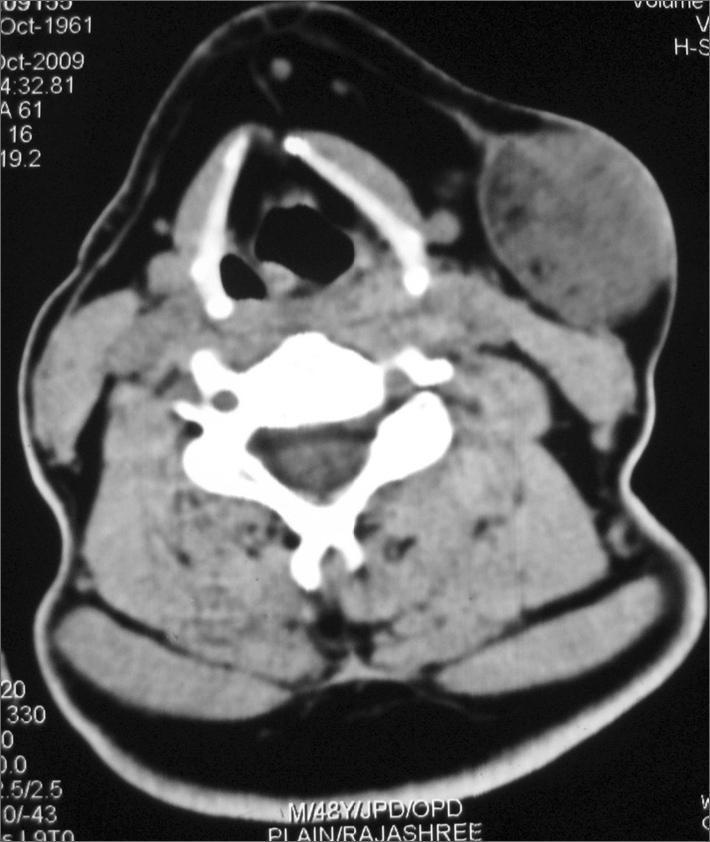
This diagnostic image is an axial computed tomography (CT) scan of the neck at the level of the cervical spine and larynx. The scan demonstrates a prominent, well-circumscribed soft tissue mass measuring approximately 3.2 x 4.2 cm in the left submandibular/parotid region. The lesion is primarily composed of soft tissue density with scattered internal hypodensities consistent with fat attenuation, particularly visible at the periphery. Anatomically, the mass is located lateral to the superficial lobe of the left parotid gland and extends anteriorly to the sternocleidomastoid muscle. The lesion exerts a significant mass effect, resulting in the outward displacement of the overlying platysma muscle and subcutaneous tissues. The internal architecture appears heterogeneous, which is a key clinical finding for differential diagnosis of parotid gland or soft tissue tumors such as pleomorphic adenoma or liposarcoma. This educational material is suitable for radiology and head and neck surgery students to study the spatial relationships and CT attenuation characteristics of cervical soft tissue lesions.
Cervical Mass Management
Anatomy and Localization
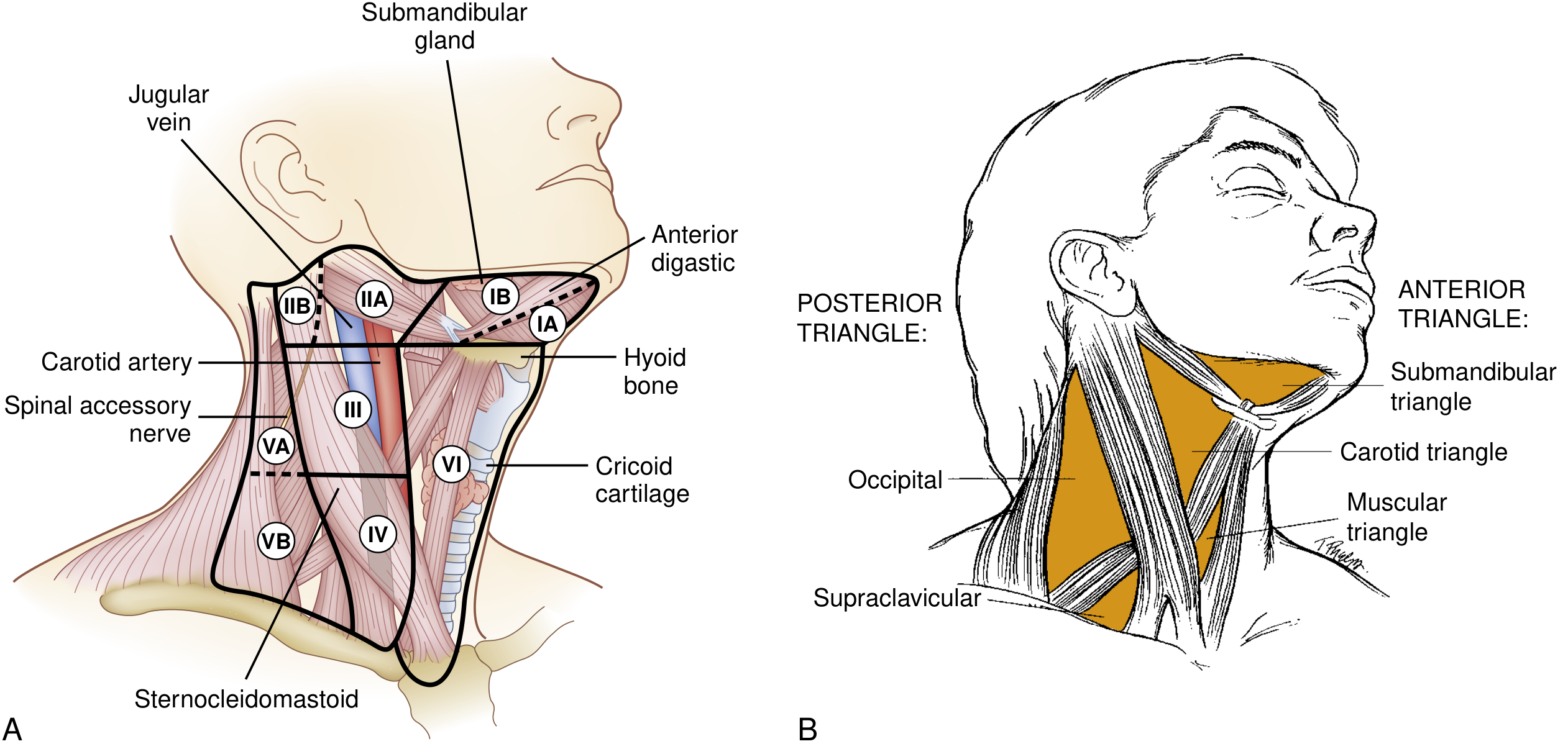
| Level | Node Group | Primary Sites |
|---|---|---|
| IA/IB | Submental / Submandibular | Lip, oral cavity, skin, submandibular gland |
| IIA/IIB | Upper jugular | Parotid, oral cavity, oropharynx, nasopharynx, larynx |
| III | Middle jugular | Oral cavity, oropharynx, hypopharynx, larynx, thyroid |
| IV | Lower jugular | Hypopharynx, larynx, thyroid, esophagus; Virchow's node (lung/GI) |
| VA/VB | Posterior triangle | Nasopharynx, skin/scalp, thyroid |
| VI | Central compartment | Thyroid, larynx, hypopharynx, cervical esophagus |
| VII | Substernal | Extension of Level VI |
Differential Diagnosis
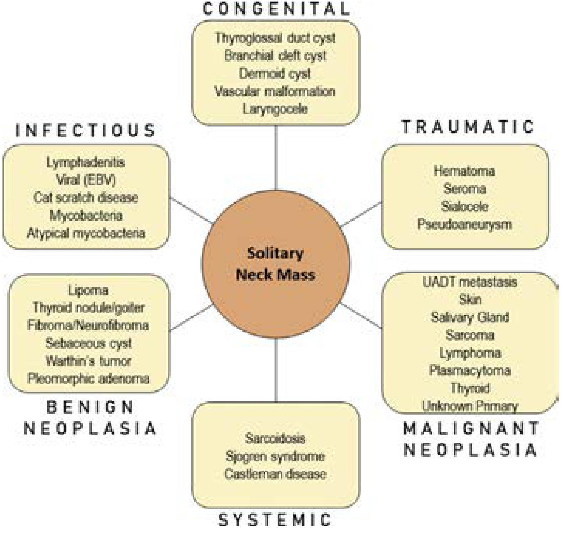
By Etiology
| Category | Entities |
|---|---|
| Congenital | Thyroglossal duct cyst (most common congenital neck mass in children), Branchial cleft cyst, Dermoid cyst, Vascular malformation, Laryngocele |
| Infectious | Cervical lymphadenitis (most common cause in children), EBV (infectious mononucleosis), Cat scratch disease, Mycobacteria / atypical mycobacteria |
| Benign Neoplasia | Lipoma, Thyroid nodule/goiter, Fibroma/Neurofibroma, Sebaceous cyst, Warthin's tumor, Pleomorphic adenoma |
| Malignant Neoplasia | Metastatic SCC (most common adult neck malignancy), Lymphoma (most common pediatric primary malignancy), Thyroid carcinoma, Salivary gland carcinoma, Sarcoma, Unknown primary |
| Systemic | Sarcoidosis, Sjogren syndrome, Castleman disease |
| Traumatic | Hematoma, Seroma, Sialocele, Pseudoaneurysm |
Age-Based Risk Stratification
| Age Group | Likely Etiology |
|---|---|
| Children (0-18 yrs) | Most likely benign - lymphadenitis, congenital (branchial/thyroglossal) |
| Young adults (<35 yrs) | Higher benign probability but malignancy not excluded |
| Adults (>35 yrs) | Strong presumption of malignancy; 80% probability if thyroid excluded |
History: Key Points
- Timing and evolution: Fluctuating/long-standing mass suggests congenital or inflammatory; persistently enlarging mass over a shorter time suggests malignancy
- Associated symptoms: Dysphagia, odynophagia, hoarseness, epistaxis, otalgia, hemoptysis, nasal obstruction, hearing loss → raise suspicion for head and neck malignancy
- Constitutional symptoms: Fever, night sweats, weight loss → lymphoma
- Risk factors: Tobacco and alcohol use, HPV exposure history (multiple sexual partners), prior radiation to head and neck, immunosuppression
- Prior treatment: A mass persisting through antibiotics may indicate an underlying anatomic abnormality or malignancy
- Travel/occupational history: Asbestos exposure, endemic mycobacterial regions
Physical Examination
- Size - masses >2 cm in adults carry >80% probability of malignancy
- Site - anterior vs. posterior triangle; level I-VII
- Shape and surface
- Consistency - firm/hard suggests malignancy; fluctuant suggests cyst or abscess
- Fixation - fixed to deep structures suggests malignancy
- Pulsatility - pulsatile with bruit = vascular (carotid body tumor, pseudoaneurysm)
- Compressibility and transillumination - cystic lesions (branchial cyst, hygroma)
- Overlying skin changes - erythema (infection), tethering (malignancy)
Diagnostic Workup
Step 1 - Imaging
- Borders and consistency (cystic vs. solid)
- Location and level
- Enhancement pattern (necrotic nodes suggest metastatic SCC)
- Relationship to adjacent structures
Step 2 - Fine-Needle Aspiration (FNA)
Step 3 - Core Biopsy
Step 4 - Open Biopsy (use with caution)
Critical principle: Never perform open/excisional biopsy of a cervical mass without a plan to convert to neck dissection if frozen section confirms carcinoma. An unplanned open biopsy can upstage disease and compromise future surgical management.
Specific Entities and Their Management
Thyroglossal Duct Cyst
- Most common congenital neck mass in children; second most common cervical mass in childhood overall
- Midline, moves with tongue protrusion and swallowing
- Treatment: Sistrunk procedure - excision of the cyst, tract, and central hyoid body (to reduce recurrence)
Branchial Cleft Cyst (2nd branchial cleft)
- Fluctuant, anterior border of SCM, upper third-middle third junction
- In patients >35 years: high index of suspicion for cystic metastatic node (especially from HPV+ oropharyngeal SCC or papillary thyroid cancer) - malignancy must be excluded before excision
- Treatment: complete excision when quiescent; passes superficial to hypoglossal and glossopharyngeal nerves
Cervical Lymphadenitis
- Most common cause of neck mass in children
- Rapidly growing, tender mass in a child: first-line treatment is antibiotics (Staph/Strep coverage)
- If no response to antibiotics: consider imaging, FNA, and broader differential (atypical mycobacteria, lymphoma)
Metastatic SCC (Unknown Primary)
- Most common malignancy in the adult neck
- Workup includes PET-CT, panendoscopy with directed biopsies (base of tongue, tonsils, piriform sinus), and HPV testing
- Management: surgery (neck dissection ± primary site treatment) + radiation/chemoradiation
Lymphoma
- Most common primary malignancy in pediatric neck
- Constitutional B-symptoms (fever, night sweats, weight loss), bilateral lymphadenopathy, mediastinal involvement
- Core or excisional biopsy for diagnosis (FNA often insufficient for subtyping); do not perform neck dissection
- Management: chemotherapy ± radiation (depends on Hodgkin vs. Non-Hodgkin subtype)
Management Summary Algorithm
Neck Mass
    ↓
History + Physical Exam (age, risk factors, mass characteristics)
    ↓
Head and Neck Exam (fiberoptic laryngoscopy, palpation of all mucosa)
    ↓
CT Neck with Contrast ± Ultrasound
    ↓
FNA Biopsy (ultrasound/CT-guided if needed)
    ↓
┌─────────────────────────────────────┐
│ Diagnosis established?              │
│ YES → Directed treatment            │
│ NO  → Core biopsy (if lymphoma      │
│        suspected)                   │
│       Open biopsy (last resort;     │
│       plan for neck dissection)     │
└─────────────────────────────────────┘
Red Flags Requiring Urgent Workup
- Adult age >35 with neck mass >2 cm
- Painless, firm, fixed mass
- Associated hoarseness, dysphagia, hemoptysis
- Unilateral serous otitis media in an adult
- Weight loss, night sweats
- Rapid progressive enlargement without infection signs
- Cummings Otolaryngology Head and Neck Surgery - Key Points and Neck Mass chapter
- Current Surgical Therapy 14e - Differential Diagnosis and Diagnostic Evaluation
- Schwartz's Principles of Surgery 11e - Neck mass evaluation
- Bailey and Love's Short Practice of Surgery 28th Ed - Lump in the neck, Branchial cyst
Cervix mass management
"cervical cancer" management treatment
cervical cancer FIGO staging colposcopy CIN management
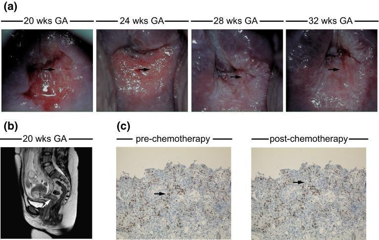
This composite educational image illustrates the longitudinal management of FIGO stage IB1 cervical cancer during pregnancy. (a) A series of colposcopy images at 20, 24, 28, and 32 weeks gestational age (GA) demonstrates the cervical tumor's evolution during neoadjuvant chemotherapy. Initial images show atypical vessels and ulceration (indicated by arrows), with subsequent images following the clinical response. (b) A sagittal T2-weighted MRI at 20 weeks GA shows a gravid uterus and the cervical tumor, characterized by increased size and high signal intensity (arrow), used for staging and excluding lymph node involvement. (c) Immunohistochemistry (IHC) micrographs (100x magnification) compare squamous cell carcinoma (SCC) tissue pre- and post-chemotherapy. Brown staining (arrows) indicates Ki-67 expression, a marker of cellular proliferation, while hematoxylin (blue) provides nuclear counterstaining. These panels demonstrate the integration of clinical colposcopy, diagnostic imaging, and histopathology in monitoring gestational malignancy and treatment response.
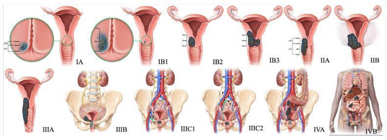
Anatomical diagrams illustrating the 2018 FIGO staging system for cervical cancer, progressing from Stage IA to IVB. The top row depicts cervical and vaginal spread: Stage I is confined to the cervix with subdivisions based on depth (IA) and diameter (IB1-IB3); Stage II shows extension beyond the uterus into the upper two-thirds of the vagina (IIA) and parametrial invasion (IIB). The bottom row illustrates advanced regional and distant spread: Stage IIIA involves the lower one-third of the vagina, while IIIB shows extension to the pelvic sidewall and associated hydroureter/hydronephrosis. Stage IIIC indicates lymphatic involvement, further categorized into pelvic (IIIC1) and para-aortic (IIIC2) lymph nodes. Stage IV demonstrates local invasion of the bladder or rectum (IVA) and distant metastasis beyond the pelvis (IVB). Key anatomical landmarks included are the uterus, cervix, vagina, ureters, kidneys, pelvic bones, and major blood vessels. The diagrams use color-coded masses to represent tumor growth and invasion depth, serving as a comprehensive educational guide for gynecologic oncology staging.
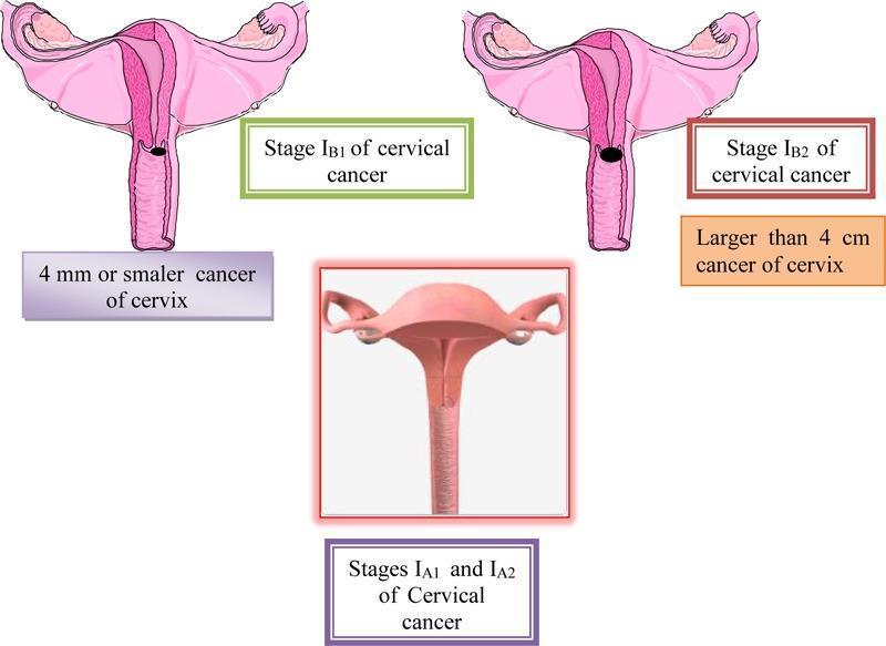
A multi-panel medical illustration and anatomical diagram depicting the FIGO staging of cervical cancer (Stages IA and IB). The central bottom panel displays a 3D-style anatomical diagram representing Stages IA1 and IA2, where the cervix appears macroscopically normal, indicating microinvasive disease that is not visible to the naked eye. The top left panel illustrates Stage IB1, showing a cross-section of the uterus and cervix with a small, localized dark lesion on the cervical canal; it is annotated as a cancer of 4 mm or smaller. The top right panel illustrates Stage IB2, showing a significantly larger, darker, and more prominent exophytic/ulcerative lesion on the cervix, annotated as being larger than 4 cm. All panels show relevant reproductive anatomy including the uterus, fallopian tubes, and ovaries. This educational graphic serves to distinguish clinical stages of cervical carcinoma based on tumor size and macroscopic visibility, relevant for gynecologic oncology and diagnostic pathology training.
Cervix Mass Management
Background and Pathology
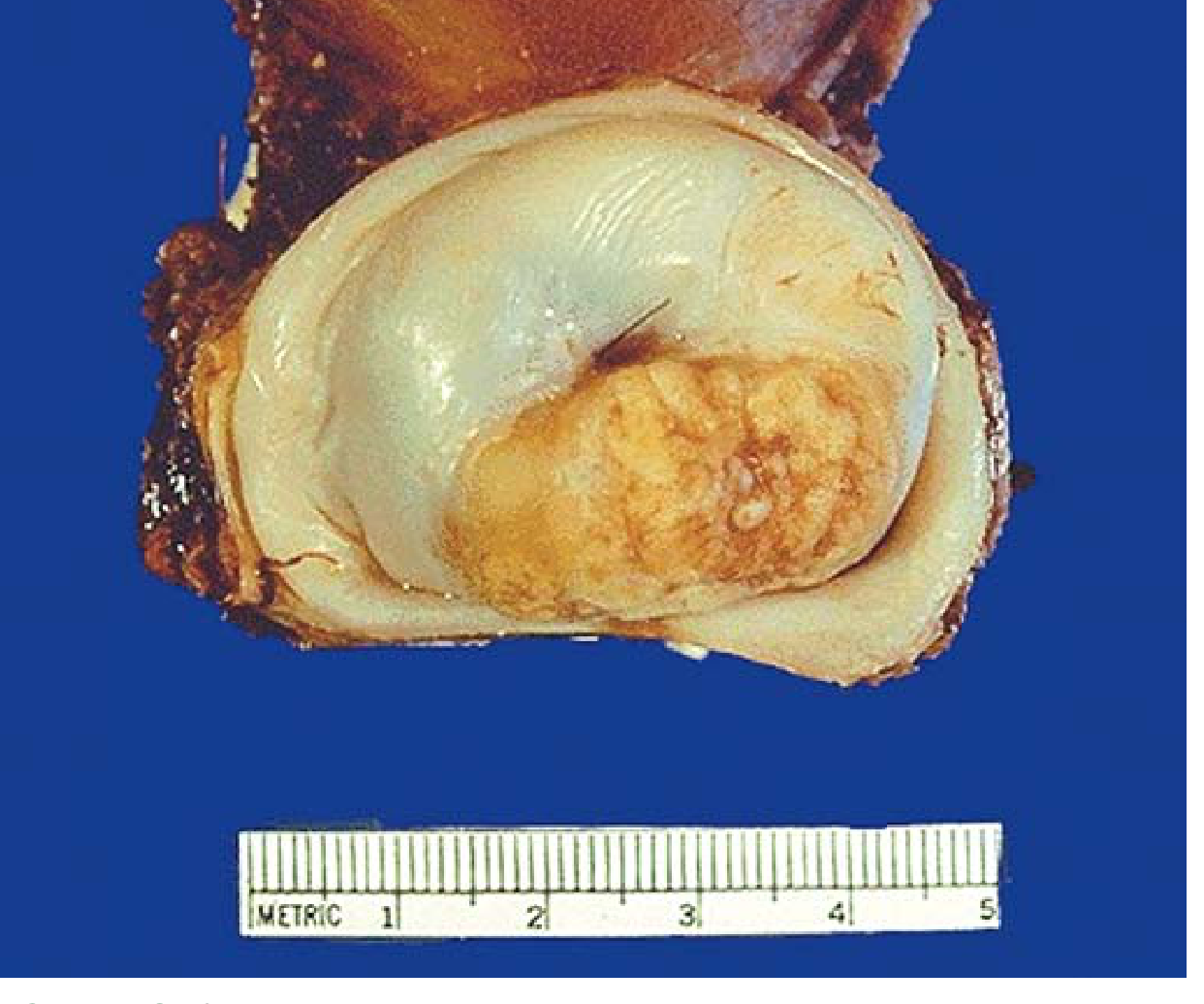
- Squamous cell carcinoma: ~80% of cases; peaks at age ~45 years
- Adenocarcinoma / adenosquamous carcinoma: ~15%; incidence increasing due to limitations of Pap smear in detecting glandular precursors
- Small cell neuroendocrine carcinoma: <5%; very poor prognosis
- Robbins & Kumar Basic Pathology; Berek & Novak's Gynecology
Differential Diagnosis of a Cervical Mass
| Category | Entities |
|---|---|
| Benign | Nabothian cysts, cervical polyp, leiomyoma (fibroid), condyloma acuminatum, Gartner's duct cyst, cervicitis |
| Premalignant | CIN 1 / LSIL, CIN 2, CIN 3 / HSIL, Adenocarcinoma in situ (AIS) |
| Malignant | Squamous cell carcinoma, Adenocarcinoma, Adenosquamous carcinoma, Small cell neuroendocrine carcinoma, Sarcoma (rare) |
Cervical Cancer Screening
| Age Group | Recommendation |
|---|---|
| <21 years | No screening regardless of sexual history |
| 21-29 years | Cytology (Pap smear) alone every 3 years |
| 30-65 years | Co-testing (cytology + hrHPV) every 5 years OR cytology alone every 3 years |
| >65 years | Discontinue if adequate negative history (3 negative cytologies or 2 negative co-tests in past 10 years) |
Premalignant Disease (CIN) Management
CIN Classification
- CIN 1 (LSIL): Mitoses/immature cells confined to lower third of epithelium; a manifestation of HPV infection, not a true cancer precursor
- CIN 2 (HSIL): Middle third involvement; intermediate risk
- CIN 3 (HSIL/CIS): Upper third involvement; significant malignant potential
CIN 1 Management
- Spontaneous regression rate: 60-85% within 2 years
- Adequate colposcopy: Surveillance with co-testing (cytology + hrHPV) at 12 months
- If negative at 12 months: return to age-appropriate screening
- If preceded by ASC-H or HSIL cytology: co-testing at 12 and 24 months or LEEP
- CIN 1 persisting ≥24 months: Patient may choose continued surveillance OR ablation/excision (only if colposcopy adequate)
- Immunosuppressed patients: Expectant management may be inappropriate; treat more aggressively
CIN 2 and CIN 3 Management
- Women ≥25 years: Require destruction or excision of the transformation zone
- CIN 2 progression rates: ~20% progress to carcinoma in situ, ~5% to invasion if untreated
- Preferred approach: Excisional procedure (LEEP or cold-knife conization) over ablation - allows pathologic assessment of margins
- Ablation (cryotherapy/laser) is contraindicated when:
- Colposcopy is inadequate
- CIN 2/3 is recurrent
- Endocervical sampling is positive
- Post-treatment: co-testing at 12 and 24 months before returning to routine screening
Adenocarcinoma In Situ (AIS)
- Must be treated with an excisional procedure (not ablation)
- Hysterectomy recommended for women who have completed childbearing (high recurrence/skip lesion risk)
Treatment Modalities for CIN
| Procedure | Setting | Notes |
|---|---|---|
| Cryotherapy | Office | Ablative; only for adequate colposcopy, small ectocervical CIN |
| LEEP (Loop Electrosurgical Excision) | Office/outpatient | Excisional; allows margin assessment; first-line for HSIL |
| Cold-knife conization (CKC) | OR | Excisional; preferred when endocervical involvement, AIS, or recurrent disease |
| Hysterectomy | OR | Reserved for recurrent high-grade CIN, AIS (fertility completed), or concurrent uterine disease |
- Berek & Novak's Gynecology; Sabiston Textbook of Surgery
Invasive Cervical Cancer: FIGO Staging
| FIGO Stage | Description | Key MRI Finding |
|---|---|---|
| IA1 | Stromal invasion <3 mm depth, <7 mm width | Microscopic only; MRI not usually indicated |
| IA2 | Invasion 3-5 mm depth, ≤7 mm width | As above |
| IB1 | Visible lesion ≤2 cm | Intermediate signal mass; MRI accurately measures size |
| IB2 | Visible lesion 2-4 cm | |
| IB3 | Visible lesion >4 cm | |
| IIA | Extends beyond uterus; upper 2/3 vagina, no parametria | Disruption of vaginal wall low-signal ring |
| IIB | Obvious parametrial invasion | Disruption of cervical stromal ring; spiculated interface |
| IIIA | Involves lower 1/3 vagina | |
| IIIB | Extension to pelvic wall or hydronephrosis | Tumor within 3 mm of pelvic side wall |
| IIIC1 | Pelvic lymph node metastasis | |
| IIIC2 | Para-aortic lymph node metastasis | |
| IVA | Invasion of bladder or rectum | Loss of fat planes; disruption of bladder/rectal mucosa |
| IVB | Distant metastases |
Imaging in Cervical Cancer
- Tumor location (exophytic vs. endocervical)
- Tumor size
- Depth of stromal invasion
- Parametrial invasion (disruption of the low-signal cervical stromal ring)
- Vaginal and lower uterine segment extension
- Pelvic lymph node involvement
Invasive Cervical Cancer Treatment by Stage
Stage IA1 (Microinvasive, <3 mm)
- Cone biopsy (LEEP or CKC) with clear margins - acceptable for fertility preservation
- Simple hysterectomy for women who have completed childbearing
- Lymph node dissection generally not required (risk of nodal metastasis <1%)
Stage IA2 (3-5 mm invasion)
- Radical hysterectomy with pelvic lymph node dissection OR
- Radical trachelectomy (fertility-sparing) with pelvic lymph node dissection
- Risk of nodal metastasis increases to >1%, justifying lymphadenectomy
Stage IB1-IB2 / IIA (Early Locally Confined)
- Radical hysterectomy (Wertheim/type III) with sentinel lymph node biopsy or pelvic ± para-aortic lymphadenectomy = surgical standard of care
- Surgery and primary radiation are equally effective; surgery preferred in younger patients (avoids radiation-induced ovarian failure and vaginal stenosis)
- Important: Open abdominal approach is the standard of care - minimally invasive radical hysterectomy carries higher recurrence risk (especially when tumor >2 cm)
- Radical trachelectomy (fertility-sparing): option for tumors ≤2 cm in young women wanting future pregnancy
Stage IB3 / IIB and Beyond (Locally Advanced)
- Concurrent chemoradiation (external beam radiation + cisplatin-based chemotherapy) is the standard of care
- Surgery alone is NOT recommended for stages IIB and above
- Brachytherapy (intracavitary) is added after external beam for radical intent
Stage III / IVA
- Concurrent chemoradiation + brachytherapy
- IVA: may require pelvic exenteration in select cases
Stage IVB / Recurrent Disease
- Systemic chemotherapy: Carboplatin/paclitaxel ± bevacizumab is first-line
- Pembrolizumab (PD-1 inhibitor): added to chemotherapy for PD-L1 positive tumors (KEYNOTE-826 regimen)
- Pelvic exenteration for central recurrence after radiation (in selected patients)
Surgical Procedures Summary
| Disease | Procedure |
|---|---|
| CIN (adequate colposcopy) | LEEP or cryotherapy |
| CIN / AIS (inadequate colposcopy or recurrent) | Cold-knife conization |
| Microinvasive (IA1) - fertility desired | Cone biopsy |
| Microinvasive (IA1/IA2) - no fertility | Simple hysterectomy |
| IB1 ≤2 cm - fertility desired | Radical trachelectomy + LND |
| IB1-IIA - standard | Radical hysterectomy (open) + pelvic LND |
| IIB and above | Chemoradiation (NOT surgery) |
Adjuvant Therapy after Surgery
- Positive surgical margins
- Positive lymph nodes
- Parametrial involvement
- Large tumor size
- Lymphovascular space invasion
- Deep stromal invasion
Prognosis
| Stage | Approximate 5-Year Survival |
|---|---|
| IA | >95% |
| IB | 80-90% |
| II | 65-75% |
| III | 35-50% |
| IV | <15% |
HPV Vaccination (Primary Prevention)
- Berek & Novak's Gynecology - CIN classification, screening guidelines, CIN management
- Sabiston Textbook of Surgery - Cervical cancer treatment, surgical techniques
- Robbins & Kumar Basic Pathology - Histopathology, morphology, clinical features
- Grainger & Allison's Diagnostic Radiology - FIGO staging table, imaging modalities
- ESGO/ESHRE/ESGE Fertility-sparing treatment guidelines 2024 (PMID 39216500)
How to perform cervical mass biopsy in a older lady
colposcopy cervical biopsy procedure punch biopsy technique
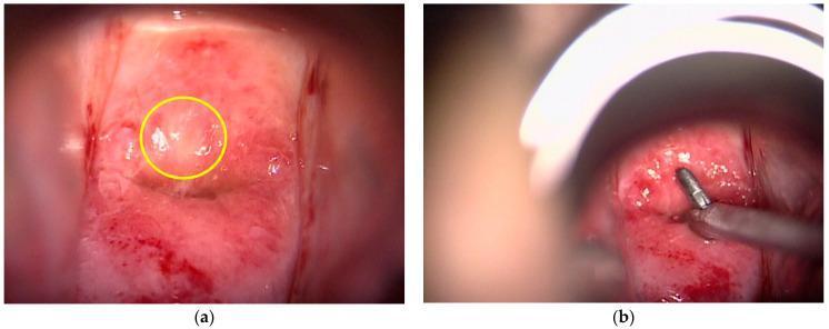
This clinical photograph displays colposcopic findings of the uterine cervix in two panels, focusing on Cervical Intraepithelial Neoplasia (CIN) and diagnostic procedures. Panel (a) shows the cervix after the application of acetic acid, revealing a predominantly pinkish-red mucosal surface. A yellow circle highlights a localized area of thin acetowhite epithelium, a key colposcopic marker for neoplastic change where the tissue appears paler and more opaque than the surrounding mucosa. Panel (b) captures a punch biopsy (P-Bx) procedure performed under colposcopic guidance. Metallic, silver-colored punch biopsy forceps are seen grasping a targeted area of the suspicious cervical tissue. The surrounding tissue in this panel appears hyperemic, glistening, and moist, with minor surface bleeding visible, which is common during the biopsy process. These images demonstrate the transition from visual screening using the Rio 2011 Colposcopy Nomenclature to tissue acquisition for histological confirmation of high-grade CIN lesions.
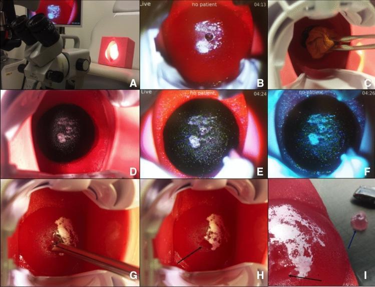
A composite clinical educational image (Panels A–I) demonstrating the use of a cervical colposcopy and biopsy simulator for gynecological training. (A) Setup showing a ZEISS colposcope and a 3D-printed vaginal/cervical phantom. (B-F) Sequential colposcopic views of the artificial cervix: (B) pre-treatment cervix with red surface and simulated white dysplastic areas; (C-E) application of Lugol’s iodine illustrating the Schiller test, where normal glycogen-rich tissue turns dark brown/black (iodine-positive) and abnormal dysplastic tissue remains white/pale (iodine-negative); (F) colposcopy using a green filter to enhance vascular contrast and lesion margins. (G-I) Biopsy procedure: (G) insertion and positioning of biopsy forceps at the simulated lesion site; (H) post-procedural view showing a visible punch biopsy defect (black arrow); (I) macroscopic view of the extracted simulator tissue (blue arrow) alongside the donor site on the cervical model. The material simulates the appearance and texture of the human cervix to facilitate training in identifying squamous intraepithelial lesions and performing diagnostic biopsies.
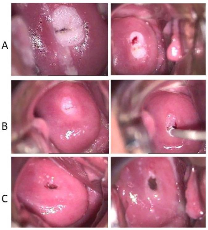
A series of colposcopy images illustrating the therapeutic progression of cervical condyloma acuminata after the application of 3% acetic acid. The clinical timeline is divided into three stages: (A) Baseline visit showing a prominent, raised acetowhite lesion on the ectocervix, consistent with a flat condyloma (low-grade squamous intraepithelial lesion). The right panel of (A) demonstrates the site of a histological punch biopsy. (B) Treatment phase images showing significant regression of the acetowhite area, with the lesion becoming smaller and less defined as pharmacological intervention proceeds. (C) Long-term follow-up at ten months demonstrating restitutio ad integrum, characterized by the complete resolution of the exophytic lesion and return to normal cervical mucosal appearance with a smooth, pink squamous epithelium and no residual acetowhite changes. This timeline serves as a clinical educational tool for monitoring Human Papillomavirus (HPV)-related cervical pathologies and their response to antiviral or topical therapy.
Cervical Mass Biopsy in an Older Woman
Why Older Patients Are Different
- SCJ recession into the endocervical canal - the transformation zone (TZ) is no longer visible on the ectocervix, making colposcopy unsatisfactory in most cases
- Atrophic epithelium - pale, thin, fragile; bleeds easily; mimics acetowhite change
- Vaginal stenosis - the vaginal walls are less distensible, making speculum insertion difficult and sometimes painful
- Lugol's staining unreliable - both normal atrophic epithelium and dysplasia fail to take up iodine due to low glycogen, making Schiller's test non-discriminatory
- Dense acetowhitening from atrophic change can be confused with dysplasia
Key principle: In a postmenopausal woman without HRT, colposcopy is almost always unsatisfactory (SCJ not visible) → colposcopy-directed biopsy alone is insufficient → endocervical curettage (ECC) and/or excisional procedure (cone biopsy/LEEP) are usually required.
- Pfenninger and Fowler's Procedures for Primary Care
Pre-Procedure Assessment
History
- Last Pap smear result and HPV test result
- Prior cervical procedures (LEEP, cone, cryotherapy) - prior excision further recesses the TZ
- Postmenopausal bleeding or vaginal discharge (postmenopausal bleeding = malignancy until proven otherwise; ~10% have endometrial or cervical malignancy)
- Anticoagulants (warfarin, aspirin, clopidogrel) - may need to be held
- Vaginal estrogen use (if on local estrogen, atrophy is less severe)
- Comorbidities: diabetes, immunosuppression, cardiovascular disease
Pre-Treatment with Topical Estrogen
- Restores glycogen to squamous cells, making Lugol's test more reliable
- Reduces atrophic fragility and bleeding
- May slightly evert the SCJ, improving visualization
Pre-Procedure Medications
- Ibuprofen or naproxen 500 mg orally, 1 hour before the procedure (reduces discomfort)
- Do not schedule during heavy vaginal bleeding
- Obtain informed consent - explain the procedure, risks (bleeding, infection, discomfort, rare perforation), and planned biopsy type
Equipment Required
| Equipment | Purpose |
|---|---|
| Appropriate-size vaginal speculum (narrow/small for atrophic vagina) | Exposure |
| Colposcope (2x-15x magnification) | Visualization |
| 3-5% acetic acid (white vinegar) | Acetowhite lesion identification |
| Lugol's iodine solution | Schiller's test (limited utility in atrophic cervix) |
| Green filter | Vascular pattern assessment |
| Ring/sponge forceps | Cleaning/application of solutions |
| Punch biopsy forceps (Kevorkian, Schubert, Eppendorfer) | Directed punch biopsy |
| Endocervical curette (Kevorkian) | ECC - essential in postmenopausal women |
| Endocervical speculum (Kogan's) | Opens os to visualize endocervix |
| Tenaculum (single-tooth) | Stabilizes/straightens the cervix if needed |
| Silver nitrate sticks or Monsel's solution (ferric subsulfate paste) | Hemostasis |
| Pathology containers (formalin) labeled per site | Histology |
| Topical lidocaine gel or injectable 1% lidocaine with epinephrine | Local anesthesia |
Step-by-Step Biopsy Procedure
Step 1 - Patient Positioning and Speculum Insertion
- Position in dorsal lithotomy with adequate lighting
- In older women with vaginal atrophy/stenosis, use a smaller, narrow-blade speculum (e.g., pediatric or small Graves speculum) - a standard bivalve speculum may cause significant discomfort or trauma
- Insert gently with warm lubricant on the outer blades (not on the cervix itself, as lubricant interferes with cytology/biopsy)
- Once the cervix is visualized, adjust speculum blades to optimize exposure
Step 2 - Initial Visualization Without Staining
- Use the colposcope to inspect the cervix at low magnification first
- Note: the os in an older woman may appear stenotic, patulous (if previously parous), or obscured
- Identify the SCJ location - in postmenopausal women it is usually within or beyond the canal (type 3 TZ)
- Note any gross lesion: exophytic mass, ulceration, friability, abnormal vascularity → if gross tumor is present, a simple directed punch biopsy of the grossest area is sufficient; colposcopy is unnecessary
If obvious tumor growth is present, a cervical biopsy is usually sufficient for diagnosis. If gross disease is not present, colposcopic examination with cervical biopsies and endocervical curettage is the next step.
- Berek & Novak's Gynecology
Step 3 - Application of 3-5% Acetic Acid
- Remove excess cervical mucus with a cotton swab or ring forceps
- Apply acetic acid liberally to the entire ectocervix and lower endocervix using a cotton swab or gauze-soaked sponge
- Wait 60 seconds - acetic acid dehydrates cells; dysplastic cells with dense nuclei turn white (acetowhite epithelium, AWE)
- In atrophic older women: atrophic epithelium itself may appear acetowhite - this is a source of false positives; more densely white, raised, thickened areas with sharp borders and abnormal vessels are more suspicious
- Dense, rapidly appearing, uniform acetowhitening in the transformation zone, especially with coarse punctation or mosaicism = high-grade CIN or cancer
Step 4 - Green Filter and Vascular Assessment
- Apply the green filter to enhance blood vessel visualization (vascular patterns appear as darker lines against green background)
- Punctation (dots) and mosaicism (tile pattern) suggest dysplasia; coarse patterns indicate higher-grade lesions
- Atypical vessels (irregular, corkscrew, branching vessels that do not taper) = suspect invasive carcinoma
Step 5 - Assess Adequacy of Colposcopy
| Satisfactory Colposcopy | Unsatisfactory Colposcopy |
|---|---|
| Entire SCJ visible | SCJ not fully visible (typical of postmenopausal) |
| All lesion borders fully visible | Lesion extends into canal beyond view |
| Absence of active infection/bleeding | Active inflammation or atrophy obscuring view |
| → Directed biopsy is adequate | → ECC mandatory; consider cone biopsy/LEEP |
Step 6 - Optional Lugol's Solution (Schiller's Test)
- Apply Lugol's iodine to ectocervix after acetic acid has faded
- Normal estrogenized epithelium: dark brown/mahogany (iodine-positive; glycogen-rich)
- Dysplastic/CIN tissue: remains pale yellow/white (iodine-negative = Lugol's-negative = Schiller's positive)
- Atrophic postmenopausal epithelium: also fails to stain → limited utility in older women unless pre-treated with estrogen
- Can be helpful to delineate SCJ boundaries for the colposcopist
Step 7 - Directed Punch Biopsy (Ectocervical)
- Identify the most abnormal appearing area (densest acetowhitening, coarsest vessels, most raised/thickened lesion)
- Apply topical lidocaine gel to the biopsy site, or inject 1% lidocaine with epinephrine into the cervical stroma at the biopsy site (especially helpful in older women who tolerate pain less well)
- Place the jaw of the punch biopsy forceps (Kevorkian, Schubert, or Tischler) directly over the targeted acetowhite area at the SCJ margin or the most abnormal area
- Close the jaws firmly and rotate slightly to detach the specimen; avoid crushing
- Sample multiple sites if multiple abnormal areas are present - label each separately and note the clock position (e.g., "12 o'clock", "6 o'clock")
- Biopsy the most advanced area at each site - the histopathologist makes the diagnosis, not the colposcopist
- Biopsy samples should be 2-3 mm in diameter and 2-3 mm deep to include stroma
- Place immediately in formalin-labeled containers per site
Step 8 - Endocervical Curettage (ECC) - Essential in Older Women
- Insert the Kevorkian endocervical curette (a narrow curette with a basket tip) through the cervical os into the endocervical canal
- In older women with a stenotic os: gently dilate with cervical dilators (Hegar #3 → #4 → #5) if needed before curettage; be gentle to avoid perforation
- Apply systematic circumferential scraping from the internal os down to the external os, using firm but controlled pressure in all four quadrants
- Collect the curetted material and blood with the curette basket or by swabbing the canal with a cytobrush
- Place in a separate labeled container - ECC result is interpreted separately from punch biopsy
- A negative ECC shows orderly benign columnar epithelium; a positive result changes management toward excisional procedure
Step 9 - Hemostasis
- Monsel's solution (ferric subsulfate paste): apply with a cotton swab directly to the biopsy site; causes chemical coagulation; highly effective
- Silver nitrate sticks: gentle pressure to the biopsy site; useful for smaller bleeding points
- Direct pressure with a cotton swab for 1-2 minutes
- Rarely: if brisk bleeding occurs from a vascular lesion, a vaginal pack or electrocautery may be needed
- Avoid: blind suturing into an atrophic, friable cervix
Special Considerations in the Elderly Patient
| Challenge | Solution |
|---|---|
| Atrophic, stenotic vagina | Use smaller speculum; warm speculum; topical estrogen pretreatment |
| Stenotic cervical os | Use Kogan's endocervical speculum; gentle dilation before ECC |
| SCJ not visible (Type 3 TZ) | Always perform ECC; proceed to cone/LEEP if ECC positive or colposcopy unsatisfactory with high-grade cytology |
| Atrophic acetowhitening mimicking CIN | Pre-treat with vaginal estrogen; interpret acetowhite changes cautiously |
| Increased pain sensitivity / vasovagal risk | Pre-medicate with NSAIDs; use local anesthetic (lidocaine injection); have patient supine; monitor vitals |
| Anticoagulation | Hold anticoagulants per protocol; have Monsel's solution ready |
| Friable tissue / easy bleeding | Handle gently; have hemostatic agents prepared |
| Discordant cytology and biopsy | Lower threshold to proceed to cold-knife conization |
When to Proceed Directly to Cone Biopsy / LEEP (Not Just Punch Biopsy)
- Unsatisfactory colposcopy + high-grade cytology (HSIL) → cone biopsy or LEEP
- Positive ECC (CIN 2+ in curettings) → excisional procedure
- Suspected AIS or endocervical adenocarcinoma
- Gross lesion extending into canal
- Significant discordance between cytology and biopsy (e.g., cytology = CIS, biopsy = mild dysplasia) → re-biopsy or conization
- Recurrent CIN 2/3 after prior treatment
"An unsatisfactory colposcopic examination coupled with cytologic evidence of significant dysplasia may necessitate a cervical conization for evaluation."
- Pfenninger and Fowler's Procedures for Primary Care
Post-Procedure Instructions
- Normal: dark, coffee-ground-like vaginal discharge for 24-48 hours (Monsel's solution + blood)
- Normal: mild spotting for up to 48 hours; may resume intercourse after 24-48 hours
- Seek evaluation for: passage of clots, fresh profuse bleeding, foul vaginal odor, fever, or pelvic pain
Complications
| Complication | Management |
|---|---|
| Bleeding | Monsel's solution, silver nitrate, pressure; rarely vaginal pack or electrocautery |
| Infection/cervicitis | Antibiotics directed at culture results |
| Vasovagal reaction | Trendelenburg, atropine if needed; more common in older/anxious patients |
| Cervical stenosis post-procedure | More common in postmenopausal; monitor |
| Inadvertent perforation (ECC) | Rare; usually self-limiting; monitor |
Interpretation Pitfalls in the Elderly
- A biopsy result one degree less advanced than cytology is acceptable
- If biopsy is two or more degrees less advanced than cytology → suspect sampling error → repeat colposcopy with biopsy or proceed to conization
- Never ablate (cryotherapy, laser) a cervix with discordant cytology/histology
- Never ablate if invasive cancer is suspected - freezing invasive cancer is absolutely contraindicated
- Pfenninger and Fowler's Procedures for Primary Care, 3rd Ed - Full colposcopy technique, equipment, complications, postprocedure care
- Schwartz's Principles of Surgery 11e - Colposcopy and cervical biopsy procedure
- Berek & Novak's Gynecology - CIN management, cone biopsy indications, unsatisfactory colposcopy
- Sabiston Textbook of Surgery - Cervical surgical techniques
Patient concieved on cabergolin should be continued in pregnancy also
cabergoline pregnancy prolactinoma safety
PMID: 40629810
PMID: 40116053
Cabergoline in Pregnancy: Should It Be Continued?
The Standard Recommendation
Discontinue cabergoline at the time pregnancy is confirmed in most cases - this is the current standard practice.
- Microprolactinomas (tumor <1 cm, no extrasellar extension)
- Macroprolactinomas that have already shrunk significantly on therapy and do not abut the optic chiasm
- Creasy & Resnik's Maternal-Fetal Medicine; Scott-Brown's Otorhinolaryngology
When Cabergoline SHOULD Be Continued (or Restarted)
| Situation | Action |
|---|---|
| Macroadenoma (>1 cm) with invasion into cavernous sinus | Continue cabergoline throughout pregnancy |
| Macroadenoma abutting or compressing the optic chiasm | Continue cabergoline |
| Macroadenoma that did NOT shrink adequately (<50% reduction) before pregnancy | Higher risk of expansion; continue or restart promptly at signs of growth |
| Symptomatic tumor expansion during pregnancy (headache, visual field defects) | Restart cabergoline (0.25-0.5 mg twice weekly) or bromocriptine |
- Creasy & Resnik's Maternal-Fetal Medicine, p. 1531
Safety Data for Cabergoline in Pregnancy
What the evidence shows:
- No increased risk of spontaneous abortion
- No increased risk of multiple pregnancy
- No increased risk of congenital malformations
- Long-term follow-up of >200 children exposed to cabergoline for up to 12 years showed no adverse developmental outcomes
-
Otis et al. (2025) - Systematic review of 1,662 pregnancies exposed to cabergoline: no increased risk of major congenital malformations or spontaneous abortions compared to other treatments or no treatment. (PMID 40116053)
-
Chakraborty & Rastogi (2025) - Systematic review and meta-analysis of 1,387 pregnancies: no increased risk of congenital malformations (RR 0.99), preterm birth (RR 1.00), or low birth weight (RR 1.02). However, live birth rates were slightly lower among those who continued cabergoline during pregnancy vs. those who discontinued at diagnosis (RR 0.81, 95% CI 0.67-0.98, p=0.03). The authors recommend careful consideration rather than routine continuation. (PMID 40629810)
Interpretation of the live birth finding: This likely reflects indication bias - women continuing cabergoline are those with larger, higher-risk tumors, not that the drug itself causes pregnancy loss. Congenital malformations, preterm birth, and birth weight were identical between groups.
Risk of NOT Continuing (Tumor Expansion During Pregnancy)
- Prolactin levels rise 10-fold physiologically
- Estrogen stimulates lactotroph cell hypertrophy
- Microprolactinoma: ~2.5% risk of symptomatic expansion - low risk, stop is safe
- Macroprolactinoma (medically treated only): ~18% symptomatic expansion - higher risk
- Macroprolactinoma (post-surgery/radiation): ~4.7% symptomatic expansion
Monitoring Protocol During Pregnancy
| Situation | Monitoring |
|---|---|
| Microprolactinoma | Clinical review each trimester; prolactin levels NOT useful (rise 10-fold normally); no routine MRI needed |
| Macroprolactinoma | Review every 1-3 months; formal visual field testing each trimester; MRI (without gadolinium) if symptoms develop |
| Symptoms develop (headache, visual changes) | Urgent visual field + MRI without gadolinium → restart cabergoline or bromocriptine |
Prolactin levels during pregnancy are not a reliable marker of tumor activity because they rise 10-fold physiologically - do not use them to monitor disease.
Management Summary Table (from Creasy & Resnik's MFM)
| Timing | Action |
|---|---|
| Prior to pregnancy | Ensure macroprolactinoma has shrunk ≥50%; if no shrinkage or drug intolerance → consider surgical resection before conception |
| At confirmation of pregnancy | Discontinue cabergoline (standard); continue if macroadenoma with cavernous sinus invasion or abutting optic chiasm |
| If symptomatic during pregnancy | Visual fields + MRI → restart bromocriptine or cabergoline; transsphenoidal surgery only if vision threatened and unresponsive to medical therapy |
| Postpartum | MRI and visual fields at 2-3 months postpartum; hold cabergoline during lactation unless tumor growth documented; restart when breastfeeding stops |
Regarding the Valvular Heart Disease Concern
- Scott-Brown's Otorhinolaryngology Head & Neck Surgery
Bottom Line
| Tumor Type | Cabergoline in Pregnancy |
|---|---|
| Microprolactinoma | Stop at pregnancy confirmation - safe; tumor unlikely to grow |
| Macroprolactinoma (well-controlled, not abutting chiasm) | Stop at confirmation, monitor closely; restart if symptoms |
| Macroprolactinoma with chiasm/cavernous sinus involvement | Continue throughout pregnancy |
| Symptomatic tumor expansion during pregnancy | Restart cabergoline (0.25-0.5 mg twice weekly) or bromocriptine |
- Creasy & Resnik's Maternal-Fetal Medicine - Prolactin-Producing Pituitary Adenomas, p. 1530-1531
- Scott-Brown's Otorhinolaryngology Head & Neck Surgery Vol 1 - Prolactinoma
- Otis et al. 2025 - Systematic review, 1,662 pregnancies (PMID 40116053)
- Chakraborty & Rastogi 2025 - Meta-analysis, 1,387 pregnancies (PMID 40629810)