
Memory
Reading File
"Memory" is quite broad. Let me check what you're looking for before diving in.
Asking for Preferences
What aspect of memory are you interested in?
Quick answer: Memory is encoded, stored, and retrieved through a distributed network of brain structures. The hippocampus is central to forming new declarative (explicit) memories, while the amygdala modulates emotional memories, the cerebellum handles procedural memory, and the prefrontal cortex manages working memory. At the cellular level, memory formation relies on synaptic plasticity - particularly long-term potentiation (LTP), where repeated synaptic firing strengthens connections via NMDA receptor activation and structural synaptic changes.
Searching sources...
Finding Sources
Finding Sources
Searching the Web
neuroscience of memory formation hippocampus synaptic plasticity LTP 2025
Good results. Now let me read from the best textbook sources - the Neuroanatomy clinical cases book and Bradley's Neurology, which are most relevant to the neuroscience/physiology of memory.
Reading File
Reading File
Finding Sources
Excellent - Kandel's Principles of Neural Science is in the library. Let me read those key sections on memory from the definitive neuroscience textbook.
Reading File
Reading File
Reading File
I now have rich, textbook-level content. Let me read a bit more from Kandel's Principles of Neural Science to get the explicit/implicit memory taxonomy and LTP molecular mechanism sections.
Reading File
Finding Sources
Reading File
Reading File
There's a brain anatomy image in Kandel. Let me fetch it to display alongside the answer.
Loading Image
I now have everything I need for a thorough, well-illustrated answer. Let me compile it.
The Neuroscience and Physiology of Memory
What Is Memory?
Memory is "the process by which knowledge is encoded, stored, and later retrieved." It is not a single function located in one brain region - different memory types depend on distinct neural circuits. Three major insights from decades of research:
- There are several forms of memory, each with different cognitive properties and brain substrates.
- Memory involves four operations: encoding, storage, consolidation, and retrieval.
- Imperfections in memory (amnesia, confabulation) reveal how normal memory works.
- Eric Kandel, Principles of Neural Science, 6th ed.
Taxonomy of Memory
Memory is classified along two main axes: time course and type of content.
By Time Course
| Stage | Clinical Term | Neuroscience Term | Duration | Anatomy |
|---|---|---|---|---|
| 1 | Immediate memory | Working memory | Seconds to ~20 sec without rehearsal | Prefrontal cortex, striatum |
| 2 | Short-term / recent memory | Episodic (recent) | Minutes to hours | Hippocampus + medial temporal lobe |
| 3 | Long-term / remote memory | Episodic (remote) + semantic | Weeks to lifetime | Neocortex (less hippocampus over time) |
Working memory holds roughly 5-9 meaningful items in conscious awareness. Without rehearsal, it fades in approximately 18-20 seconds. It relies primarily on prefrontal cortex (dorsolateral for spatial, ventrolateral for objects), with dopaminergic input from the midbrain and striatal modulation.
- Bradley and Daroff's Neurology in Clinical Practice
By Content: Explicit vs. Implicit
Explicit (declarative) memory - Conscious, can be verbalized. Damaged by medial temporal lobe lesions.
- Episodic memory: personal events ("what I did last Tuesday") - requires "mental time travel"
- Semantic memory: factual world knowledge ("Paris is the capital of France") - distributed across lateral temporal and association cortices
Implicit (non-declarative) memory - Unconscious, not verbalized. Survives medial temporal lobe damage.
- Procedural memory (skills and habits): processed in the striatum
- Classical/operant conditioning: amygdala (emotional responses), cerebellum (motor responses)
- Priming: improved recall from prior exposure; survives even in profound amnesia
- Habituation/sensitization: depends on basic reflex pathways
The landmark case of patient H.M. (bilateral hippocampal resection) showed that he could learn new motor skills (mirror drawing) and improve with priming tasks, but had no conscious recollection of having done them - directly proving the explicit/implicit dissociation.
- Kandel, Principles of Neural Science, pp. 1341-1342
Brain Structures: The Medial Temporal Lobe System
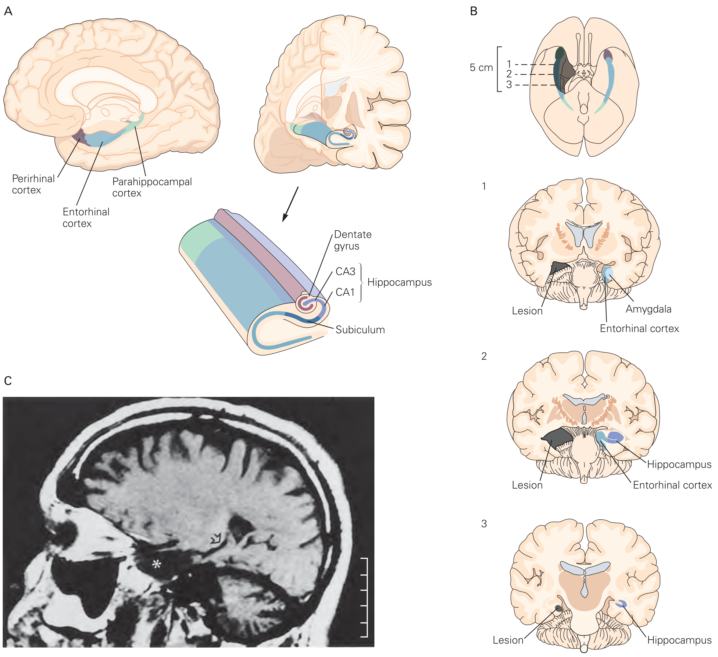
Figure: The medial temporal lobe (A), showing the hippocampus (CA1, CA3, dentate gyrus, subiculum), entorhinal cortex, perirhinal cortex, and parahippocampal cortex. Panel B shows H.M.'s bilateral lesion extent across 3 coronal levels. Panel C is H.M.'s actual MRI scan.
The hippocampal formation is the core structure for forming new episodic memories. It builds relational associations - linking objects, places, times, and people into a coherent episode. The hippocampus is NOT the final storage site; it acts as a temporary binding hub while memories are being consolidated to the neocortex.
A useful anatomical analogy (from Bradley's Neurology):
- Frontal lobes = the "filing clerk" that decides what to retrieve and from where
- Medial temporal lobes = the "recent memory filing cabinet" where new memories are stored
- Subcortical white matter = the pathway the filing clerk must travel to reach the cabinet
| Lesion site | Effect on memory |
|---|---|
| Medial temporal lobe (e.g., Alzheimer disease) | Damaged file cabinet - memories cannot be stored |
| Frontal lobe (e.g., stroke, tumor) | Disorganized filing clerk - poor organization and retrieval |
| Subcortical white matter (e.g., MS, ischemia) | Blocked pathway - slow/impaired access, intact on recognition testing |
Consolidation: From Short-Term to Long-Term
Consolidation occurs at two levels:
Synaptic Consolidation (Hours)
Driven by long-term potentiation (LTP) - a durable increase in synaptic transmission efficiency after repeated stimulation. LTP proceeds in two phases:
- Early LTP (E-LTP): protein synthesis-independent; lasts minutes to hours. The synapse becomes "tagged" via a protein synthesis-independent mechanism.
- Late LTP (L-LTP): requires intracellular signaling cascades and new protein synthesis in soma and dendrites; lasts days to years. The tagged synapse must capture plasticity-related proteins (PRPs) to stabilize.
The molecular trigger for most LTP:
- High-frequency presynaptic activity (or theta-burst stimulation, 4-8 Hz) depolarizes the postsynaptic membrane
- NMDA receptors (glutamate-gated, voltage-dependent) become unblocked - Na⁺ and Ca²⁺ flow in
- Calcium rise activates CaMKII (calcium-calmodulin-dependent kinase II), which autophosphorylates
- Cytoskeletal reconfiguration and AMPA receptor insertion strengthen the synapse
- Gene activation and new protein synthesis produce structural synaptic changes (dendritic spine growth)
"The key to memory is alteration in the strength of selected synaptic connections... the alteration involves the synthesis of proteins and the activation of genes. This occurs during the change from short-term working memory to long-term memory."
- Ganong's Review of Medical Physiology, 26th ed.
Systems Consolidation (Weeks to Years)
Over time, repeated reactivation of hippocampal-neocortical circuits gradually transfers memory representations to the neocortex. Once fully consolidated there, retrieval no longer requires the hippocampus - which is why patients with hippocampal damage (H.M., early Alzheimer disease) can recall childhood events (stored in neocortex) but not recent ones (not yet transferred).
Reconsolidation: Memory Is Not Fixed
A retrieved memory becomes transiently plastic again - a window called the reconsolidation window. After retrieval, the memory must re-stabilize through protein synthesis-dependent mechanisms similar to initial consolidation. This means:
- Existing memories can be strengthened or updated during reconsolidation
- Fear memories can be weakened or erased by pharmacologic or behavioral interventions during this window
This has generated significant clinical interest for PTSD treatment.
- Miller's Anesthesia, 10th ed.
The Amygdala and Emotional Memory
The amygdala (specifically the basolateral nucleus, BLA) is not essential for standard episodic memory, but it modulates the encoding and consolidation of emotionally significant events in the hippocampus via noradrenergic mechanisms. This explains why emotionally charged events are remembered more vividly and durably - a process that can become maladaptive in conditions like PTSD.
Summary Table: Brain Regions and Memory Functions
| Brain Region | Memory Role |
|---|---|
| Hippocampus (CA1, CA3, dentate gyrus) | New episodic memory encoding; relational binding; consolidation hub |
| Entorhinal/perirhinal/parahippocampal cortex | Gateway to hippocampus; object/context representation |
| Prefrontal cortex (dorsolateral/ventrolateral) | Working memory; retrieval strategy; executive control |
| Amygdala (basolateral nucleus) | Emotional memory modulation; fear conditioning |
| Striatum (basal ganglia) | Procedural memory; habit learning |
| Cerebellum | Motor conditioning (e.g., eyeblink conditioning) |
| Neocortex (association areas) | Long-term semantic and remote episodic storage |
Sources: Eric Kandel, Principles of Neural Science (6th ed.); Bradley and Daroff's Neurology in Clinical Practice; Ganong's Review of Medical Physiology (26th ed.); Miller's Anesthesia (10th ed.); Synaptic plasticity and memory hypothesis (PMC)
For a visual walkthrough of LTP and memory formation:
Synaptic Plasticity
Synaptic plasticity is the ability of synapses to strengthen or weaken over time in response to activity. The main forms are long-term potentiation (LTP) and long-term depression (LTD), both driven by NMDA receptor activation and calcium signaling. At a molecular level, LTP involves AMPA receptor insertion and CaMKII activation, while LTD involves receptor removal via phosphatase activity. Structural changes - dendritic spine growth and pruning - underlie the persistence of these changes.
Searching sources...
Finding Sources
Finding Sources
Searching the Web
synaptic plasticity LTP LTD molecular mechanisms neuroscience 2025
Excellent. Now let me read the core sections in parallel - Kandel (best molecular detail), Neuroscience Exploring the Brain (timing/STDP), and Ganong's (physiology overview).
Reading File
Reading File
Reading File
Good content. Now let me get the LTP/LTD molecular detail from Kandel block 10 and the Kaplan & Sadock section on synaptic plasticity (which covers neurotrophins and structural changes).
Reading File
Reading File
Excellent - there's a detailed LTP diagram. Let me fetch it, and also read a bit more of the Kaplan text on late LTP and LTD.
Loading Image
Reading File
Loading Image
I now have rich content from multiple authoritative textbooks and two excellent diagrams. Let me also quickly check what Kaplan says about late LTP (the continuation of what was cut off) and check for LTD content and homeostatic plasticity.
Finding Sources
Finding Sources
Finding Sources
Reading File
Reading File
I have comprehensive content. Now I'll compile the full answer.
Synaptic Plasticity
What It Is
Synaptic plasticity is the activity-dependent ability of synapses to change their strength - either strengthening or weakening - in response to patterns of use. It is the primary neurochemical foundation of learning and memory, and it also maintains network stability and governs how the brain adapts to experience.
"Changes in the strength and efficiency of synaptic signaling, termed synaptic plasticity, underlie one of the most important neurochemical foundations of learning and memory."
- Kaplan & Sadock's Comprehensive Textbook of Psychiatry
Overview: Three Major Categories
| Category | Direction | Duration | Function |
|---|---|---|---|
| Short-term plasticity | Up (facilitation) or down (depression) | Seconds to minutes | Filters and shapes signal transmission |
| Long-term potentiation (LTP) | Strengthening | Hours to years | Memory encoding and storage |
| Long-term depression (LTD) | Weakening | Hours to years | Memory refinement, forgetting, motor learning |
Plus two regulatory forms: homeostatic plasticity (network-wide gain control) and metaplasticity (plasticity of plasticity itself).
Short-Term Synaptic Plasticity
Short-term plasticity depends on the release probability (P) of a synapse:
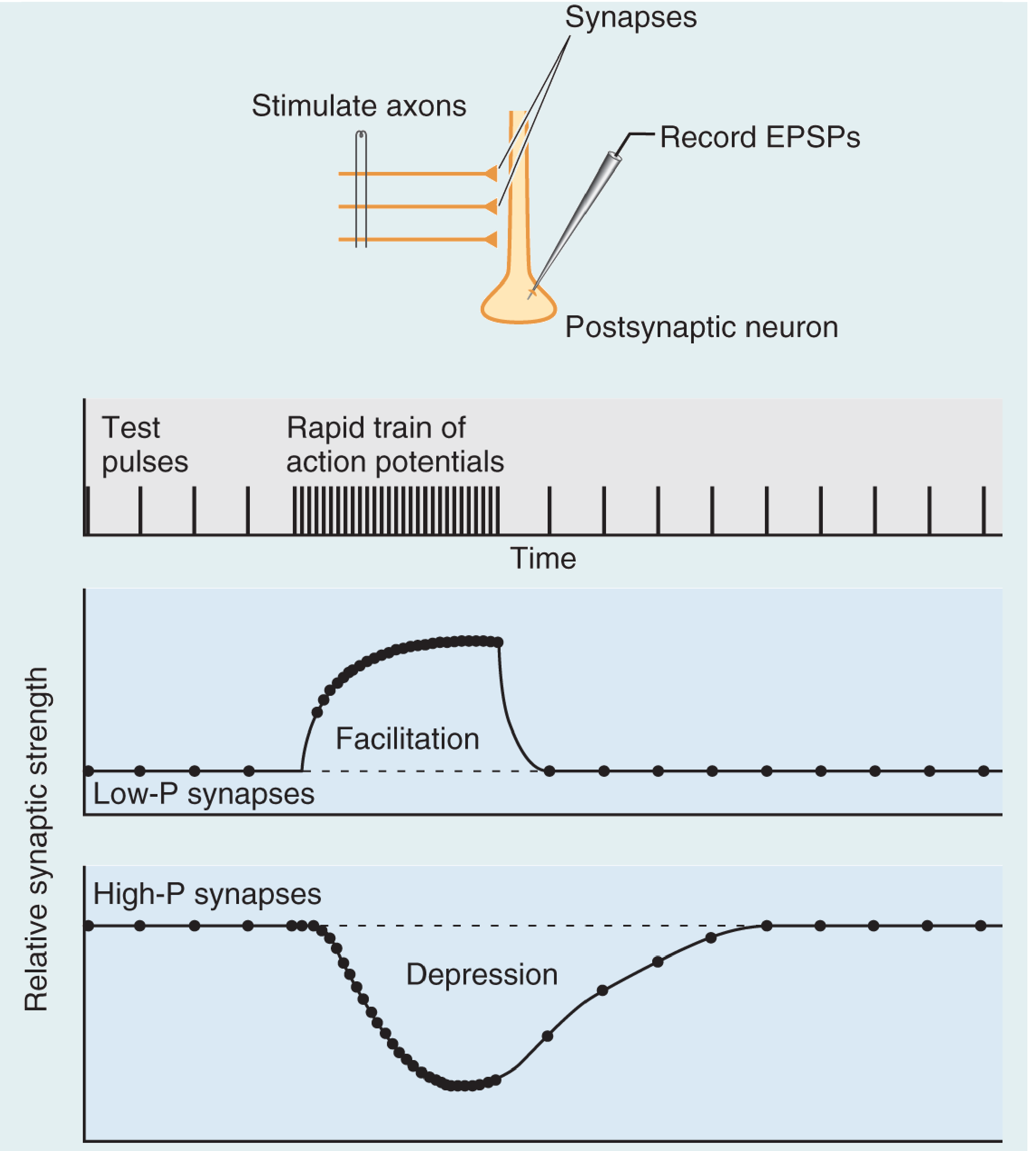
Figure: Repetitive stimulation of a presynaptic axon produces facilitation at low-P synapses (Ca²⁺ builds up, raising release probability) and depression at high-P synapses (vesicle pool depleted). Both reset to baseline after stimulation ends. From Neuroscience: Exploring the Brain, 5th ed.
Facilitation (low-P synapses): Infrequent spikes are unreliable, but rapid bursts cause Ca²⁺ to accumulate in the axon terminal before it can be cleared - making release virtually assured. These synapses are specialized to filter low-frequency signals while faithfully transmitting high-frequency bursts.
Depression (high-P synapses): Because they release reliably on each spike, the vesicle pool depletes during rapid trains. Transmission recovers only when vesicles are replenished from the reserve pool.
- Neuroscience: Exploring the Brain, 5th ed.
Long-Term Potentiation (LTP)
LTP is "a durable increase in synaptic transmission efficiency following a stimulation protocol" that persists from hours to years and is the dominant cellular model for memory storage.
The Hebbian Principle
Donald Hebb proposed that a synapse strengthens when it successfully participates in firing the postsynaptic neuron - "neurons that fire together, wire together." LTP is the biophysical implementation of this idea.
Three Key Properties (Kandel)
LTP at NMDA-receptor-dependent synapses (e.g. hippocampal CA1) has three properties that make it ideal for information storage:
-
Cooperativity - A single weak input cannot induce LTP (can't expel Mg²⁺ from NMDA channel). Only convergent activation of many inputs simultaneously achieves the strong depolarization required. This ensures only significant events trigger memory formation.
-
Associativity - A weak input paired with a strong one achieves LTP in both, because the strong input provides the depolarization. This is the cellular analog of Pavlovian conditioning - a neutral stimulus gains meaning when paired with a meaningful one.
-
Synapse specificity - Only activated synapses undergo LTP, even when neighboring synapses on the same cell receive strong stimulation. This allows a single neuron to store vast amounts of independent information across its thousands of synapses.
- Kandel, Principles of Neural Science, 6th ed., p. 1397
Molecular Mechanism of LTP
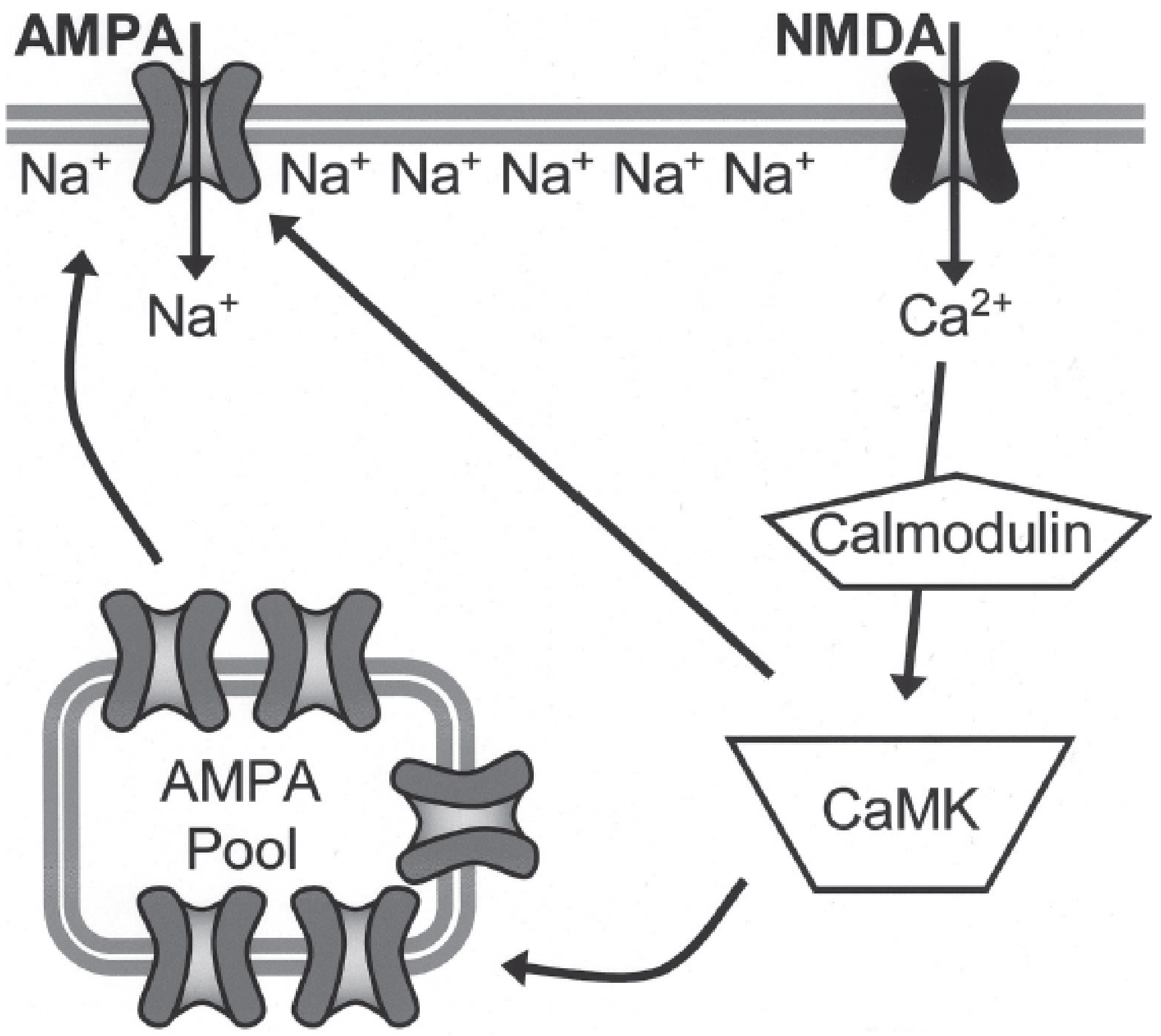
Figure: Postsynaptic mechanism of LTP. Glutamate activates AMPA receptors (Na⁺ influx, depolarization) → NMDA receptor Mg²⁺ block relieved → Ca²⁺ entry → calmodulin → CaMKII activation → AMPA receptor phosphorylation + trafficking of new AMPA receptors to the membrane. From Kaplan & Sadock's Comprehensive Textbook of Psychiatry.
Step-by-step:
- Glutamate binds to AMPA receptors → Na⁺ influx → membrane depolarization
- Sufficient depolarization expels Mg²⁺ from NMDA receptor channel
- NMDA receptor opens → large Ca²⁺ influx (the critical trigger)
- Ca²⁺ binds calmodulin → activates CaMKII (and PKC)
- CaMKII phosphorylates existing AMPA receptors → increased Na⁺ conductance
- CaMKII drives insertion of additional AMPA receptors from an intracellular pool into the postsynaptic membrane
- More AMPA receptors = larger future EPSPs = stronger synapse
Spike Timing-Dependent Plasticity (STDP)
Researchers found that the exact timing of the postsynaptic action potential matters. If a back-propagating action potential (generated in the soma, propagating back into dendrites) arrives within ~50 ms after the EPSP, NMDA receptors - which still have glutamate bound - are depolarized and open. Ca²⁺ floods in and LTP is triggered. If the spike arrives before the EPSP, LTD results instead. This is STDP - the synapse acts as a coincidence detector with millisecond-level precision.
In 2017, a distinct form called behavioral time-scale plasticity (BTSP) was discovered in hippocampal CA1, where "plateau potentials" (abrupt depolarizations with burst firing) can trigger LTP even seconds after a prior synaptic event - relevant to how place fields form during spatial navigation.
- Neuroscience: Exploring the Brain, 5th ed.
Two Phases: E-LTP and L-LTP
| Phase | Duration | Mechanism |
|---|---|---|
| Early LTP (E-LTP) | Minutes to hours | Protein synthesis-independent; CaMKII phosphorylation, AMPA receptor trafficking |
| Late LTP (L-LTP) | Days to years | Requires gene activation and new protein synthesis; structural synapse remodeling |
In L-LTP, the activated synapse receives a molecular "tag" (protein synthesis-independent). This tag allows it to capture plasticity-related proteins (PRPs) synthesized in the soma and dendrites. The tag-and-capture mechanism explains how thousands of synapses on a single neuron can be in varying states of stabilization simultaneously - the synaptic tagging hypothesis.
Key late-stage molecular players include:
- CaMKIV (nuclear) and PKA (cAMP-dependent) → phosphorylate CREB
- CREB recruits RNA polymerase II → transcription of plasticity genes (Arc, Homer, ΔFosB)
- New proteins cause dendritic spine enlargement (thin → mushroom-shaped spines) and structural synapse growth
Long-Term Depression (LTD)
LTD is the weakening of synaptic strength and is equally important as LTP - it refines neural circuits, enables forgetting of irrelevant information, and is critical for cerebellar motor learning.
The key to LTP vs. LTD is Ca²⁺ magnitude:
- High Ca²⁺ (from strong, high-frequency stimulation) → activates CaMKII → LTP
- Low Ca²⁺ (from weak, low-frequency stimulation, partial Mg²⁺ relief) → activates calcineurin (a Ca²⁺-dependent phosphatase with a higher affinity for Ca²⁺ than CaMKII) → dephosphorylates AMPA receptors → receptor endocytosis (removal from membrane) → LTD
In summary: the same NMDA receptor that drives LTP also drives LTD, with the concentration of Ca²⁺ acting as a molecular switch between kinase activation (LTP) and phosphatase activation (LTD).
- Kandel, Principles of Neural Science, 6th ed.
Homeostatic Plasticity
While Hebbian plasticity (LTP/LTD) is synapse-specific and driven by coincident activity, homeostatic plasticity operates at the whole-neuron or network level to maintain stability.
Synaptic scaling: When a neuron's overall activity is persistently too high or too low, it globally scales all its synaptic strengths up or down proportionally - preserving the relative weights while normalizing the total input. This involves adjusting the total number of AMPA receptors across all synapses.
Firing rate homeostasis: Networks regulate average firing rates through intrinsic excitability changes (ion channel expression).
Metaplasticity ("plasticity of plasticity"): Prior activity shifts the threshold for inducing future LTP or LTD. High ongoing activity raises the LTP induction threshold (making further potentiation harder), protecting against runaway excitation. This is regulated by NR2 subunit composition of NMDA receptors and other mechanisms.
"Unlike Hebbian plasticity, homeostatic plasticity is unlikely to drive information storage but rather helps maintain longer-term function in response to local changes in excitation and inhibition."
- Kaplan & Sadock's Comprehensive Textbook of Psychiatry
Clinical relevance: Ketamine (antidepressant) rapidly upregulates excitatory synaptic responses in hippocampus and cortex via non-Hebbian homeostatic mechanisms. Lithium dampens synaptic function in a similar homeostatic fashion. ECT and other brain stimulation methods are also believed to act partly through these homeostatic pathways.
Clinical and Disease Relevance
| Condition | Plasticity Abnormality |
|---|---|
| Alzheimer disease | Amyloid-β impairs NMDA receptor function and disrupts LTP |
| PTSD | Maladaptive amygdala LTP of fear memories; reconsolidation window targeted by behavioral therapy |
| Drug addiction | Psychostimulants and opiates induce LTD-like changes in nucleus accumbens during early withdrawal; LTP-like changes and mature dendritic spines after prolonged withdrawal (silent synapse mechanism) |
| Schizophrenia | NMDA receptor hypofunction impairs LTP-dependent cognition |
| Intellectual disability (e.g. fragile X) | Excessive LTD due to mGluR5 overactivation (the "mGluR theory" of fragile X) |
Summary
SYNAPTIC ACTIVITY
|
Ca²⁺ concentration in postsynaptic cell
|
HIGH Ca²⁺ LOW Ca²⁺
| |
CaMKII active Calcineurin active
| |
AMPA ↑ (LTP) AMPA ↓ (LTD)
Spine enlargement Spine shrinkage
Protein synthesis Receptor endocytosis
(if L-LTP)
Sources: Kandel, Principles of Neural Science (6th ed.); Kaplan & Sadock's Comprehensive Textbook of Psychiatry; Neuroscience: Exploring the Brain (5th ed.); Miller's Anesthesia (10th ed.); Frontiers - Hebbian and homeostatic plasticity 2026
Lipoprotic factors
Lipoproteins are macromolecular complexes that transport lipids through the aqueous bloodstream. Their key structural components include a hydrophobic core (triglycerides, cholesterol esters), a hydrophilic surface (phospholipids, free cholesterol), and apolipoproteins that determine metabolic fate. The major classes are chylomicrons, VLDL, IDL, LDL, and HDL - each with distinct densities, compositions, and roles in lipid transport.
Searching sources...
Finding Sources
Finding Sources
Searching the Web
lipoprotein classes structure apolipoproteins lipid transport metabolism
Good start. The Tietz and Henry's books look most relevant for lipoprotein structure/metabolism. Let me search more specifically and read those sections.
Finding Sources
Finding Sources
Excellent - Harrison's has a dedicated "LIPOPROTEIN STRUCTURE AND METABOLISM" section, and Harper's Biochemistry has formation details. Let me read both in parallel along with the Tietz apolipoproteins section.
Reading File
Reading File
Reading File
Excellent content. Let me now fetch the key pathway diagrams from Harper's and read more of the Harrison's section on VLDL/LDL/HDL metabolism.
Loading Image
Loading Image
Reading File
I now have comprehensive content from Harrison's, Harper's, and Tietz. Let me now read the VLDL/LDL and HDL sections from Harrison's more closely, and also get the key lipoprotein classification table from Tietz.
Reading File
Reading File
I now have rich content from Harrison's, Harper's, and Tietz. Let me fetch the key Harrison's exogenous/endogenous pathway diagram.
Loading Image
I have all the content and diagrams needed for a comprehensive answer. Let me now compose it.
Lipoprotein Structure, Classification, and Metabolism
What Are Lipoproteins?
Lipoproteins are spherical macromolecular complexes that solve a fundamental biological problem: transporting water-insoluble lipids (fats) through the aqueous bloodstream. Each particle has:
- A hydrophobic core packed with triglycerides (TG) and cholesterol esters (CE)
- A hydrophilic surface shell of phospholipids and unesterified (free) cholesterol
- Apolipoproteins embedded in the surface, which determine particle identity, dictate enzyme interactions, and serve as ligands for receptors
Classification of Lipoproteins
Lipoproteins are classified by density (measured by ultracentrifugation) - the higher the lipid-to-protein ratio, the lower the density. The major classes from largest/least dense to smallest/most dense:
| Class | Density (g/mL) | Size (nm) | Major Lipid | Key Apo | Origin | Function |
|---|---|---|---|---|---|---|
| Chylomicrons | <0.930 | 100-1000 | TG (85-90%) | ApoB-48, ApoE, ApoCII | Intestine | Transport dietary (exogenous) lipids |
| VLDL | 0.930-1.006 | 30-80 | TG (55-65%) | ApoB-100, ApoE, ApoCII | Liver | Transport hepatic (endogenous) TG to periphery |
| IDL | 1.006-1.019 | 25-35 | TG + CE (equal) | ApoB-100, ApoE | VLDL catabolism | Transitional particle; cleared by liver or converted to LDL |
| LDL | 1.019-1.063 | 18-25 | CE (~45%) | ApoB-100 only | IDL catabolism | Deliver cholesterol to peripheral tissues |
| HDL | 1.063-1.210 | 5-12 | CE + PL | ApoA-I, ApoA-II | Liver, intestine | Reverse cholesterol transport |
| Lp(a) | ~1.05 | 25-30 | CE | ApoB-100 + Apo(a) | Liver | Independent ASCVD risk factor |
Apolipoproteins: The Key Regulatory Proteins
Apolipoproteins are not merely structural - they are the functional "address labels" of lipoproteins. Most (except ApoB and Apo(a)) can transfer freely between lipoprotein particles in the blood.
| Apolipoprotein | MW (Da) | Chromosome | Carrier | Key Function |
|---|---|---|---|---|
| ApoA-I | 29,016 | 11 | HDL, chylomicrons | Core structural protein of HDL; ligand for ABCA1 (cholesterol efflux); cofactor for LCAT |
| ApoA-II | 17,414 | 1 | HDL | Structural protein of HDL (~2/3 of HDL particles) |
| ApoA-V | - | - | VLDL, chylomicrons | Promotes LPL-mediated TG lipolysis |
| ApoB-100 | 512,723 | 2 | VLDL, IDL, LDL, Lp(a) | One of the largest proteins in humans; ligand for LDL receptor; required for VLDL assembly/secretion |
| ApoB-48 | 240,800 | 2 | Chylomicrons | Intestinal form (48% of ApoB-100 sequence, from same gene via mRNA editing); required for chylomicron assembly; lacks LDL receptor-binding domain |
| ApoC-I | 6,630 | 19 | CM, VLDL, HDL | Activates LCAT; inhibits chylomicron clearance |
| ApoC-II | 8,900 | 19 | CM, VLDL, HDL | Essential cofactor for LPL (lipoprotein lipase) - without it, TG-rich lipoproteins cannot be hydrolyzed |
| ApoC-III | 8,800 | 11 | CM, VLDL, HDL | Inhibits LPL and inhibits remnant receptor binding - raises plasma TG |
| ApoE | 34,145 | 19 | CM remnants, IDL, HDL | Ligand for LDL receptor and LRP - essential for hepatic clearance of remnants |
Structural motif: Most apolipoproteins (except ApoB) contain amphipathic helices - one hydrophobic face inserts into the lipid core, the other polar face faces outward. This weak, reversible binding allows exchange between particles. ApoB, by contrast, is irreversibly embedded and cannot transfer.
- Tietz Textbook of Laboratory Medicine, 7th ed.
The Three Major Metabolic Pathways
Pathway 1: Exogenous (Dietary) Pathway - Chylomicrons
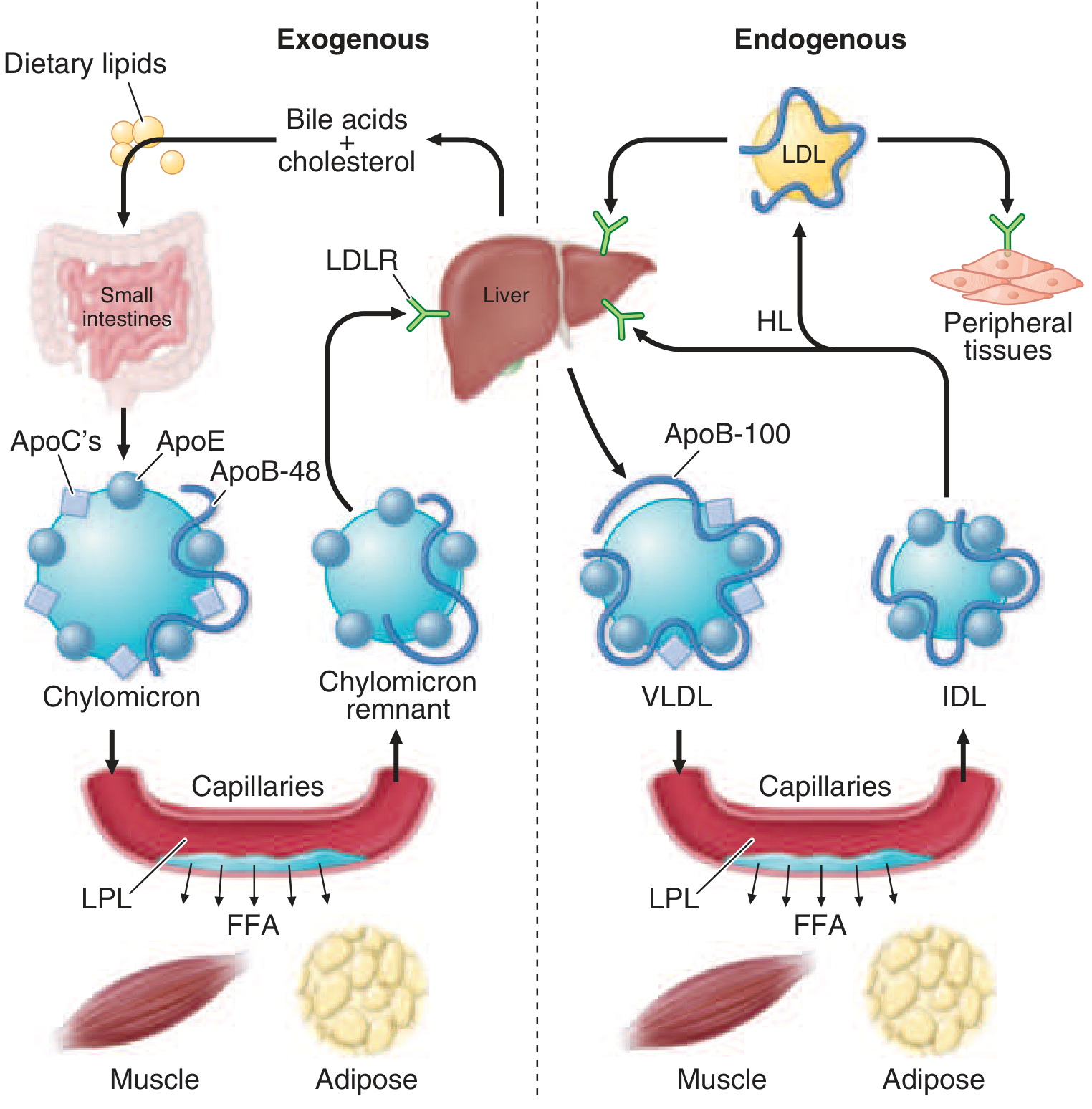
Figure: Left (exogenous): dietary lipids → chylomicrons → LPL in capillaries → FFA to muscle/adipose → chylomicron remnants → liver. Right (endogenous): liver → VLDL → LPL → IDL → LDL → peripheral tissues via LDLR. From Harrison's Principles of Internal Medicine, 22nd ed.
- Dietary fats are digested in the intestinal lumen and absorbed in the proximal small intestine
- Cholesterol and fatty acids are esterified in enterocytes; longer-chain FAs (>12C) are incorporated into TGs
- Microsomal TG transfer protein (MTP) packages TGs with ApoB-48, phospholipids, cholesteryl esters, retinyl esters, and vitamin E to form nascent chylomicrons
- Secreted into intestinal lymph → thoracic duct → systemic circulation
- In blood, chylomicrons acquire ApoC-II and ApoE from HDL
- ApoC-II activates LPL (anchored to endothelium of capillaries in adipose, heart, skeletal muscle by GPHBP1 protein) → TGs hydrolyzed → free fatty acids released to muscle (oxidation) and adipose (storage)
- Excess surface phospholipids, cholesterol, and apolipoproteins transfer to HDL
- Chylomicron remnants (enriched in cholesterol, carrying ApoB-48 + ApoE) are rapidly cleared by the liver via LRP and LDL receptor-related receptors (ApoE is the critical ligand)
- Half-life of chylomicrons < 1 hour; absent after 12-h fast
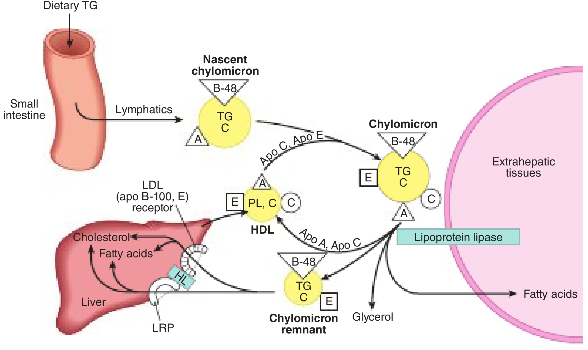
Figure: Nascent chylomicron leaves small intestine via lymphatics, acquires ApoC/E from HDL, undergoes LPL-mediated lipolysis at extrahepatic tissues, returns as chylomicron remnant to liver via LRP. From Harper's Illustrated Biochemistry, 32nd ed.
Pathway 2: Endogenous (Hepatic) Pathway - VLDL → IDL → LDL
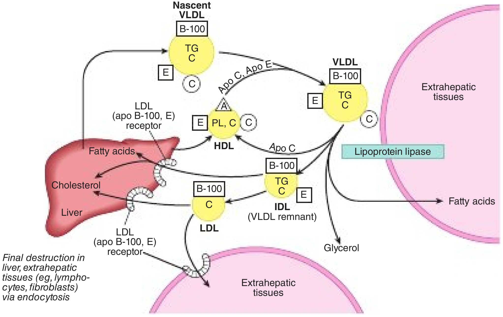
Figure: Nascent VLDL (with ApoB-100) leaves liver, acquires ApoC/E from HDL, undergoes LPL lipolysis → IDL (VLDL remnant) → either cleared by liver (via ApoE/ApoB-100 binding to LDLR) or further processed by hepatic lipase → LDL. LDL taken up in liver and extrahepatic tissues via LDLR. From Harper's Illustrated Biochemistry, 32nd ed.
- Liver assembles nascent VLDL: packages TGs (from de novo lipogenesis or fatty acids) with ApoB-100, cholesteryl esters, phospholipids via MTP → secreted into plasma
- Nascent VLDL acquires ApoC-II, ApoC-III, ApoE from circulating HDL
- LPL hydrolyzes VLDL TGs at capillary surfaces → FFAs released to tissues → VLDL shrinks to IDL (VLDL remnant)
- IDL contains ApoB-100 + ApoE → approximately half is taken up directly by the liver (via LDL receptor and LRP, using ApoE as ligand)
- Remaining IDL is further hydrolyzed by hepatic lipase (HL) → most apolipoproteins (including ApoE) transferred to HDL, leaving only ApoB-100 → LDL is formed
- LDL circulates with a half-life of ~2-3 days, delivering cholesterol to:
- Liver (~70%) via LDL receptor (LDLR) - ApoB-100 is the ligand
- Peripheral tissues (lymphocytes, fibroblasts, adrenal glands, etc.) via endocytosis
LDL Receptor (LDLR) Regulation (Brown & Goldstein pathway):
- When cell cholesterol is adequate → LDLR synthesis is suppressed (SREBP pathway inactive)
- When cell cholesterol is low → LDLR expression increases → more LDL uptake
- This is the target of statins (inhibit HMG-CoA reductase → lower intracellular cholesterol → upregulate LDLR → lower plasma LDL)
- Familial hypercholesterolemia = loss-of-function mutations in LDLR → markedly elevated LDL
Pathway 3: Reverse Cholesterol Transport - HDL
HDL is responsible for transporting excess cholesterol from peripheral tissues (including arterial wall macrophages) back to the liver for excretion - the anti-atherogenic "reverse cholesterol transport."
- Nascent HDL (disc-shaped, lipid-poor) is secreted by the liver and intestine, carrying ApoA-I
- ABCA1 transporter on macrophages and other peripheral cells effluxes free cholesterol and phospholipids onto ApoA-I → forming nascent HDL discs (defective in Tangier disease)
- LCAT (lecithin-cholesterol acyltransferase), activated by ApoA-I, esterifies free cholesterol → cholesteryl esters sink into the core → HDL matures into spherical HDL3 → HDL2
- CETP (cholesterol ester transfer protein) transfers CE from HDL to VLDL/chylomicrons in exchange for TGs (allowing indirect return of cholesterol to liver)
- SR-BI (scavenger receptor class B) on hepatocytes takes up CE selectively from HDL without degrading the particle ("selective uptake")
- Phospholipid transfer protein (PLTP) transfers phospholipids between lipoproteins; hepatic lipase and endothelial lipase (EL) remodel HDL, generating smaller particles
Lipoprotein(a) - Lp(a)
Lp(a) is a special atherogenic lipoprotein:
-
Structure: LDL-like particle with ApoB-100 covalently linked (via single disulfide bond) to Apo(a) - a large protein with kringle domains homologous to plasminogen
-
Synthesized in the liver; plasma levels are >90% genetically determined (by LPA gene kringle copy number)
-
Pathogenic mechanisms:
- Atherogenic (like LDL, deposits cholesterol in arterial wall)
- Thrombogenic: Apo(a) competitively inhibits fibrinolysis by interfering with plasminogen activation
-
Levels >125 nmol/L (or >50 mg/dL) are an independent ASCVD and aortic stenosis risk factor
-
Not reduced by statins (may modestly increase); niacin reduces it ~20-30%; PCSK9 inhibitors reduce ~25%; antisense oligonucleotides (pelacarsen) and small interfering RNA (olpasiran) reduce Lp(a) by >80% - now in phase 3 trials
-
Harrison's Principles of Internal Medicine, 22nd ed.; Lp(a) in clinical practice - CCJM 2026
Friedewald Formula and Clinical Measurement
From Harrison's:
LDL-C = Total Cholesterol - (TG/5) - HDL-C
The TG/5 term estimates VLDL-C (based on the 5:1 TG-to-cholesterol ratio in VLDL). This formula is invalid when TG > 400 mg/dL (e.g. in severe hypertriglyceridemia or type III hyperlipoproteinemia).
Non-HDL-C = Total Cholesterol - HDL-C (captures all atherogenic apoB-containing particles: VLDL + IDL + LDL + Lp(a)).
There is growing evidence that ApoB measurement directly (one molecule per atherogenic particle) may better predict ASCVD risk than LDL-C alone.
Summary: The Flow of Lipid Traffic
DIETARY LIPIDS HEPATIC LIPIDS
| |
Chylomicrons Nascent VLDL
(ApoB-48) (ApoB-100)
| |
LPL (ApoC-II) LPL (ApoC-II)
| |
Chylomicron IDL (VLDL remnant)
remnants → Liver Liver (50%) + HL
(ApoE → LRP) |
LDL
(ApoB-100 → LDLR)
|
Peripheral tissues
|
HDL (reverse transport)
ApoA-I → ABCA1 → LCAT
|
Liver (SR-BI, CETP)
Sources: Harrison's Principles of Internal Medicine (22nd ed.); Harper's Illustrated Biochemistry (32nd ed.); Tietz Textbook of Laboratory Medicine (7th ed.); Henry's Clinical Diagnosis and Management by Laboratory Methods
Free Radicles
Free radicals are highly reactive molecules with one or more unpaired electrons. In biology, the most important are reactive oxygen species (ROS) like superoxide (O₂•⁻), hydroxyl radical (•OH), and reactive nitrogen species like nitric oxide (NO•). They are generated during normal metabolism (especially mitochondrial electron transport) but cause damage to lipids, proteins, and DNA when antioxidant defenses are overwhelmed - a state called oxidative stress.
Searching sources...
Finding Sources
Finding Sources
Searching the Web
free radicals reactive oxygen species ROS oxidative stress biology mechanisms 2025
The FTS heading search didn't find exact matches. Let me try broader paragraph searches.
Finding Sources
Finding Sources
Good finds. The Tietz textbook has a dedicated "FREE RADICALS AND ANTIOXIDANTS" section, and Medical Physiology has the ROS/aging content. Let me read both in parallel, plus the surgical physiology section.
Reading File
Reading File
Reading File
Excellent content. Now let me fetch the key ROS diagrams from Medical Physiology and get more content on lipid peroxidation and DNA damage.
Loading Image
Loading Image
Finding Sources
Finding Sources
I have the two key ROS diagrams. Now let me get more content on lipid peroxidation, DNA damage, and disease associations, plus read more from the Tietz FREE RADICALS section.
Reading File
Reading File
Finding Sources
Excellent. Now let me read the NADPH oxidase section and fetch the antioxidant enzyme diagram.
Reading File
Loading Image
I now have all the diagrams and content needed for a comprehensive answer. Let me compile it.
Free Radicals (Reactive Oxygen and Nitrogen Species)
Definition
A free radical is any molecule or atom with one or more unpaired electrons in its outer orbital. This unpaired electron makes the species chemically unstable and highly reactive - it rapidly attacks neighboring molecules to capture an electron, turning the victim molecule into a new radical, and initiating a chain reaction that continues until two radicals meet and form a stable covalent bond.
"Free radicals are highly reactive chemical entities containing unpaired outer orbital electrons... these highly reactive entities damage biologically important molecules."
- Medical Physiology (Boron & Boulpaep), N62-5
The major biologically relevant free radicals are reactive oxygen species (ROS) and reactive nitrogen species (RNS).
Types of ROS and RNS
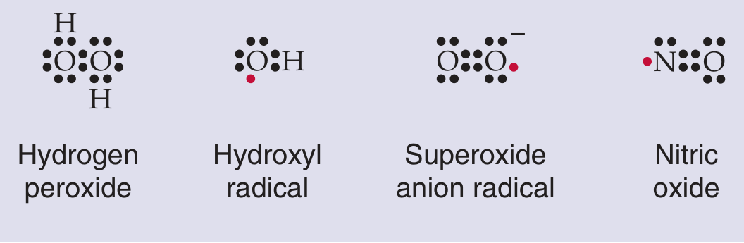
Figure: The four major reactive oxygen/nitrogen species. Red dots indicate unpaired electrons. H₂O₂ is not itself a radical but is a key precursor to •OH. From Medical Physiology.
| Species | Symbol | Radical? | Reactivity | Notes |
|---|---|---|---|---|
| Superoxide anion | O₂•⁻ | Yes | Moderate | Primary product of mitochondrial/NADPH oxidase leakage |
| Hydroxyl radical | •OH | Yes | Extremely high | Most reactive ROS; attacks DNA, proteins, lipids indiscriminately |
| Hydrogen peroxide | H₂O₂ | No | Moderate | Not a radical but generates •OH via Fenton reaction; crosses membranes |
| Nitric oxide | NO• | Yes | Low-moderate | Signaling molecule; reacts with O₂•⁻ to form peroxynitrite |
| Peroxynitrite | ONOO⁻ | No (but RNS) | Very high | Formed from NO• + O₂•⁻; nitrates tyrosine residues in proteins |
| Hypochlorous acid | HOCl | No | High | Generated by myeloperoxidase in neutrophils; kills bacteria |
| Singlet oxygen | ¹O₂ | No | High | Excited-state O₂; generated by photosensitizers and during immune killing |
Sources of Free Radical Generation
1. Mitochondrial Electron Transport Chain (Endogenous - Primary)
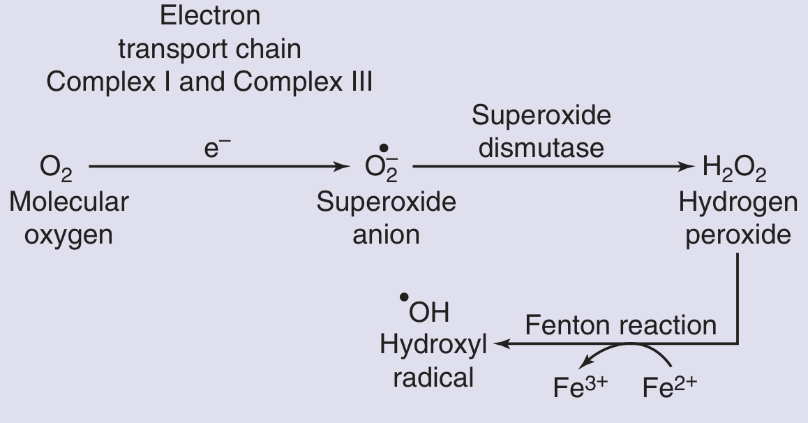
Figure: The main ROS-generating pathway. Complex I and III of the electron transport chain leak electrons onto O₂ to form superoxide. SOD converts this to H₂O₂. The Fenton reaction (Fe²⁺ + H₂O₂) produces the highly reactive •OH. From Medical Physiology.
Complex I and Complex III of the mitochondrial ETC are the dominant sources of O₂•⁻ in aerobic cells. During normal respiration, a small fraction of electrons "leak" onto molecular oxygen instead of being passed along the chain to the final acceptor. This generates superoxide.
"Only a small fraction of the oxygen used in aerobic metabolism (<1%) generates ROS. However, even that amount would be lethal in the absence of protective mechanisms."
- Medical Physiology, p. 1816
Key reactions:
- O₂ + e⁻ → O₂•⁻ (at Complex I/III)
- O₂•⁻ + O₂•⁻ + 2H⁺ → H₂O₂ + O₂ (via superoxide dismutase)
- H₂O₂ + Fe²⁺ → •OH + OH⁻ + Fe³⁺ (Fenton reaction - the most dangerous step)
- O₂•⁻ + Fe³⁺ → O₂ + Fe²⁺ (Haber-Weiss reaction - regenerates Fe²⁺ for Fenton)
This is why iron and copper are so tightly controlled in biology - free transition metals catalytically amplify hydroxyl radical formation.
2. NADPH Oxidase - Phagocytes (Immune/Deliberate)
Neutrophils and macrophages deliberately generate massive bursts of ROS to kill pathogens - the respiratory burst:
- NADPH oxidase (phox system) is assembled on the phagosome membrane
- NADPH + 2O₂ → NADP⁺ + H⁺ + 2O₂•⁻ (superoxide released into phagolysosome)
- SOD converts O₂•⁻ → H₂O₂
- Myeloperoxidase (neutrophil-specific) converts H₂O₂ + Cl⁻ → HOCl (hypochlorous acid, the active ingredient of bleach) - the most potent bactericidal agent
- H₂O₂ also generates •OH via the Fenton reaction
- iNOS activation generates NO•, which combines with O₂•⁻ → peroxynitrite (ONOO⁻)
The entire burst lasts 30-60 minutes and results in bacterial lysis.
Clinical consequence - Chronic Granulomatous Disease (CGD): Loss-of-function mutations in NADPH oxidase components → no respiratory burst → deficient killing of catalase-positive organisms (S. aureus, Aspergillus) → recurrent life-threatening infections. Diagnosed by DHR or NBT test (no superoxide production).
- Basic Medical Biochemistry, 6th ed.; Harrison's Principles of Internal Medicine, 22nd ed.
3. Ischemia-Reperfusion Injury
During ischemia, electron transport chain components accumulate electrons (become "saturated"). When oxygen is reintroduced during reperfusion, the saturated CoQ and ETC components rapidly donate electrons to O₂, causing a sudden massive burst of O₂•⁻ and •OH - often causing more tissue damage than the ischemia itself. This is the basis for myocardial reperfusion injury.
4. Exogenous Sources
| Source | Mechanism |
|---|---|
| Ionizing radiation (X-rays, gamma) | Radiolysis of water → •OH |
| UV radiation | Excites chromophores → singlet oxygen, lipid peroxidation |
| Cigarette smoke | Contains pre-formed radicals; activates inflammatory NADPH oxidase |
| Air pollutants (O₃, NO₂) | Direct radical generation in lung |
| Xenobiotics/drugs (e.g. paraquat, CCl₄) | Metabolized to radical intermediates |
Mechanisms of Molecular Damage
Lipid Peroxidation
The most destructive chain reaction. Polyunsaturated fatty acids (PUFAs) in membrane phospholipids are especially vulnerable to •OH attack:
- Initiation: •OH abstracts a hydrogen from a PUFA → carbon-centered lipid radical (L•)
- Propagation: L• + O₂ → lipid peroxyl radical (LOO•) → reacts with adjacent PUFA → new L• + lipid hydroperoxide (LOOH)
- Termination: Two radicals combine → stable products
End-products: malondialdehyde (MDA) and 4-hydroxynonenal (4-HNE) (both are biomarkers of oxidative stress), and F₂-isoprostanes (the gold-standard clinical marker).
Consequences: Membrane fluidity loss, disrupted ion gradients, cell lysis; oxidation of LDL cholesterol (ox-LDL is taken up by macrophages to form foam cells → atherosclerotic plaques).
Protein Oxidation
- Carbonylation of lysine, arginine, proline residues (measured as protein carbonyls - a clinical biomarker)
- Methionine and cysteine oxidation → loss of enzyme active sites
- Cross-linking and aggregation → proteasome dysfunction
- Nitration of tyrosine residues by peroxynitrite (3-nitrotyrosine biomarker)
DNA Damage
- 8-hydroxy-2'-deoxyguanosine (8-OHdG) - most common base modification; a well-validated biomarker of oxidative DNA damage (measured in urine)
- Single- and double-strand breaks
- Base modifications → mutagenesis if unrepaired → cancer initiation
- Cross-links between DNA strands or between DNA and proteins
Antioxidant Defenses
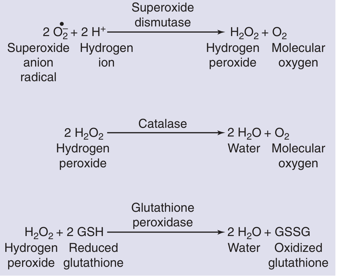
Figure: The three key enzymatic antioxidants. SOD eliminates superoxide but generates H₂O₂ (which can still make •OH). Catalase and glutathione peroxidase then neutralize H₂O₂. From Medical Physiology.
Enzymatic Antioxidants
| Enzyme | Reaction | Location | Cofactor |
|---|---|---|---|
| Superoxide dismutase (SOD) | 2O₂•⁻ + 2H⁺ → H₂O₂ + O₂ | Mitochondria (Mn-SOD), cytosol (Cu/Zn-SOD) | Mn, Cu, Zn |
| Catalase | 2H₂O₂ → 2H₂O + O₂ | Peroxisomes | Heme (Fe) |
| Glutathione peroxidase (GPx) | H₂O₂ + 2GSH → 2H₂O + GSSG | Cytosol, mitochondria | Selenium |
| Glutathione reductase | GSSG + NADPH → 2GSH + NADP⁺ | Cytosol | FAD |
| Thioredoxin reductase | Oxidized Trx + NADPH → reduced Trx | Cytosol, mitochondria | Selenium, FAD |
| Myeloperoxidase | H₂O₂ + Cl⁻ → HOCl | Neutrophil granules | Heme |
Non-Enzymatic Antioxidants
| Antioxidant | Mechanism | Notes |
|---|---|---|
| Vitamin E (α-tocopherol) | Chain-breaking antioxidant in membranes; scavenges LOO• radicals | Fat-soluble; regenerated by vitamin C |
| Vitamin C (ascorbate) | Scavenges •OH, O₂•⁻, HOCl; regenerates vitamin E | Water-soluble; provides ~24% of plasma antioxidant capacity |
| Glutathione (GSH) | Donates electrons to GPx; key intracellular thiol buffer | Rate-limiting substrate for GPx; restored by N-acetylcysteine (NAC) |
| Uric acid | Scavenges •OH, HOCl, singlet oxygen; chelates Cu/Fe | Most abundant water-soluble antioxidant in human plasma |
| Carotenoids (β-carotene) | Long conjugated double bonds quench singlet oxygen and LOO• | Fat-soluble; found in membranes |
| Albumin | Binds Cu²⁺/Fe²⁺, preventing Fenton reaction; directly scavenges radicals | Major plasma protein antioxidant |
| Transferrin, ceruloplasmin | Sequester Fe³⁺ and Cu²⁺ | Prevent metal-catalyzed •OH generation |
| Bilirubin | Scavenges peroxyl radicals | Product of heme oxygenase-1 (HO-1) |
Important caveat: Antioxidants can sometimes become pro-oxidants. For example, vitamin C can reduce Fe³⁺ to Fe²⁺, promoting the Fenton reaction. Selenium in excess generates methyl-selenite → superoxide. This is why antioxidant supplementation trials have largely failed to show clinical benefit and some have even shown harm.
- Tietz Textbook of Laboratory Medicine, 7th ed.
Physiological Roles (Not Just Harmful)
Free radicals are not purely destructive. At low concentrations they are essential:
| Function | Mechanism |
|---|---|
| Immune defense | NADPH oxidase/myeloperoxidase → bactericidal HOCl, •OH |
| Cell signaling | H₂O₂ and NO• activate NF-κB, AP-1, MAPK pathways → inflammation, cell growth |
| Thyroid hormone synthesis | Thyroid peroxidase uses H₂O₂ to oxidize iodide → iodination of thyroglobulin |
| Vasodilation | NO• from eNOS → activates guanylyl cyclase → cGMP → smooth muscle relaxation |
| Redox-regulated gene expression | ROS modulate Nrf2 pathway → upregulation of antioxidant genes (SOD, catalase, GPx) |
Disease Associations
| Disease | Mechanism of ROS involvement |
|---|---|
| Atherosclerosis | LDL oxidation by •OH/HOCl → ox-LDL → macrophage foam cells → plaques |
| Cancer | DNA base modifications → mutations; ROS also used in some cancer therapies |
| Diabetes mellitus | Hyperglycemia → mitochondrial ROS → endothelial dysfunction; glycation + glyoxidation → AGEs |
| Alzheimer disease | Amyloid-β generates H₂O₂; mitochondrial dysfunction → neuronal ROS → tau modification |
| Parkinson disease | Dopamine auto-oxidation → quinones + O₂•⁻; mitochondrial Complex I inhibition |
| COPD/Emphysema | Cigarette smoke radicals → neutrophil ROS → protease-antiprotease imbalance |
| Ischemia-reperfusion injury | Burst of •OH at reperfusion → membrane damage, arrhythmia, infarct extension |
| Aging | Harman's free radical theory (1954): cumulative ROS damage → mitochondrial DNA mutations → cellular senescence |
Clinical Measurement of Oxidative Stress
Because O₂•⁻ and •OH are too short-lived to measure directly, clinicians measure downstream products:
| Biomarker | Target | Sample |
|---|---|---|
| F₂-isoprostanes | Lipid peroxidation of arachidonic acid | Urine, plasma (gold standard) |
| Malondialdehyde (MDA) | Lipid peroxidation end-product | Plasma (TBARS assay) |
| 8-OHdG | Oxidized guanine in DNA | Urine |
| Protein carbonyls | Oxidized lysine/arginine/proline | Plasma |
| 3-Nitrotyrosine | Peroxynitrite protein oxidation | Plasma |
| Oxidized glutathione (GSSG/GSH ratio) | Intracellular redox state | Red blood cells |
Summary
ENDOGENOUS SOURCES EXOGENOUS SOURCES
Mitochondria (Complex I/III) Radiation, smoke,
NADPH oxidase (immune cells) pollutants, drugs
Xanthine oxidase |
P450 enzymes ↓
↓ O₂•⁻ → H₂O₂ → •OH
| |
PHYSIOLOGICAL PATHOLOGICAL (excess)
- Immunity - Lipid peroxidation
- Signaling (NF-κB, - Protein carbonylation
NO•, Nrf2) - DNA mutation (8-OHdG)
- Thyroid synthesis - Disease (atherosclerosis,
cancer, neurodegeneration)
↑
ANTIOXIDANT DEFENSES
SOD → Catalase / GPx
Vitamins E & C, GSH,
Uric acid, Albumin
Sources: Medical Physiology (Boron & Boulpaep); Tietz Textbook of Laboratory Medicine (7th ed.); Basic Medical Biochemistry (6th ed.); Mulholland & Greenfield's Surgery (7th ed.); Free Radicals in Health and Disease - Wiley 2025
Free Radicals Biochemistry
Free radical biochemistry covers the chemical reactions, molecular targets, and enzymatic pathways involved in radical generation and neutralization. Key reactions include the Fenton reaction (Fe²⁺ + H₂O₂ → •OH), radical chain reactions in lipid peroxidation, glutathione redox cycling, and the superoxide dismutase/catalase cascade. These reactions underpin oxidative stress in disease.
Searching sources...
Finding Sources
Finding Sources
Searching the Web
free radical biochemistry reactions lipid peroxidation glutathione antioxidant enzymes mechanisms
Let me search the library with more targeted biochemistry terms.
Finding Sources
Finding Sources
Good. Robbins has a good section. Let me also search for Harper's Biochemistry which should have the best biochemical detail on free radicals.
Finding Sources
Finding Sources
Good - Harper's has the pentose phosphate/glutathione section. Let me now read the key sections in parallel: Harper's glutathione/PPP, Robbins on free radical injury, Basic Medical Biochemistry on NADPH oxidase/ROS, and Murray's respiratory medicine oxidant injury section.
Reading File
Reading File
Reading File
Good content. Now let me get the Harper's pentose phosphate/glutathione diagram and read Robbins Chapter 2 on free radical injury (which is the most biochemistry-focused pathology source).
Loading Image
Finding Sources
Finding Sources
Finding Sources
Reading File
Reading File
Excellent - I have the compartmentalization diagram reference. Let me fetch it and compile the full answer.
Loading Image
I now have all the diagrams and biochemical content needed. Let me compile the comprehensive biochemistry-focused answer.
Free Radical Biochemistry
This builds on the overview from our previous session and goes deeper into the chemistry, reaction mechanisms, enzymatic pathways, and molecular targets of free radical biology.
1. Chemistry of Free Radicals
A free radical is defined by having one or more unpaired electrons in an outer orbital. This gives it three key chemical properties:
- Paramagnetism - detectable by electron spin resonance (ESR/EPR spectroscopy)
- High electrophilicity - the radical abstracts electrons from neighboring molecules
- Chain reaction propagation - each radical generates a new radical in the target molecule
The Radical Chain Reaction (General)
INITIATION: RH + X• → R• + XH
PROPAGATION: R• + O₂ → ROO• (peroxyl radical)
ROO• + R'H → ROOH + R'• (new radical formed)
TERMINATION: R• + R• → R-R (stable product)
R• + ROO• → ROOR (stable product)
The chain continues until two radicals collide to form a stable covalent bond, or an antioxidant donates an electron to quench the chain without producing a new reactive species.
2. Key Reactive Species: Chemistry and Reactions
Superoxide Anion (O₂•⁻)
Formed by one-electron reduction of O₂:
O₂ + e⁻ → O₂•⁻
Generated primarily by:
- Mitochondrial Complex I and Complex III (electron leakage)
- NADPH oxidase (deliberate; immune cells)
- Xanthine oxidase (during ischemia-reperfusion, purine catabolism)
- Cytochrome P450 reactions (liver, during xenobiotic metabolism)
O₂•⁻ is a moderate oxidant in aqueous solution but is much more reactive in lipid environments. It cannot cross membranes except via anion channels. Its most important biochemical role is as the precursor to H₂O₂ and •OH.
Hydrogen Peroxide (H₂O₂)
Not a radical itself (all electrons paired), but the most important ROS intermediate because:
- Crosses membranes freely via aquaporins
- Relatively stable (half-life minutes vs. nanoseconds for •OH)
- Acts as a second messenger at low concentrations (activates NF-κB, Nrf2, MAPK)
- Substrate for the Fenton reaction (generates •OH)
- Substrate for myeloperoxidase (generates HOCl)
Generated from O₂•⁻ by superoxide dismutase (SOD):
2 O₂•⁻ + 2H⁺ → H₂O₂ + O₂ (SOD - spontaneous or enzymatic)
Also directly produced by several oxidases: xanthine oxidase, glucose oxidase, amino acid oxidases, and monoamine oxidase (MAO).
The Fenton and Haber-Weiss Reactions
These are the most biochemically significant radical-generating reactions:
Fenton reaction (Fe²⁺ catalyzed):
Fe²⁺ + H₂O₂ → Fe³⁺ + •OH + OH⁻
Haber-Weiss reaction (net, with regeneration of Fe²⁺):
O₂•⁻ + Fe³⁺ → O₂ + Fe²⁺ (Haber-Weiss, step 1)
Fe²⁺ + H₂O₂ → Fe³⁺ + •OH + OH⁻ (Fenton, step 2)
─────────────────────────────────
Net: O₂•⁻ + H₂O₂ → O₂ + •OH + OH⁻ (Haber-Weiss overall)
Why this matters: Fe²⁺ acts as a catalyst - it is regenerated and can cycle continuously. This is why iron sequestration (by transferrin, ferritin, ceruloplasmin, lactoferrin) is a major antioxidant defense. Free ionic iron is vanishingly rare in healthy cells precisely because of this danger.
Hydroxyl Radical (•OH)
The most reactive biological oxidant. Half-life ~10⁻⁹ seconds - reacts at the site of generation, cannot diffuse far.
Attacks virtually all biomolecules:
- Abstracts H from C-H bonds (→ lipid peroxidation)
- Adds across C=C double bonds
- Oxidizes DNA bases (especially guanine → 8-OHdG)
- Oxidizes amino acid side chains (especially Cys, Met, Trp, Tyr, His)
No enzyme scavenges •OH directly - the cell's only defense is to prevent its formation (by removing H₂O₂ before the Fenton reaction, and by sequestering iron/copper).
Nitric Oxide (NO•) and Peroxynitrite (ONOO⁻)
NO• is synthesized from L-arginine + O₂ by nitric oxide synthases (NOS):
L-arginine + NADPH + O₂ → NO• + L-citrulline + NADP⁺
Three isoforms:
- eNOS (endothelial): vascular tone, anti-platelet
- nNOS (neuronal): synaptic signaling
- iNOS (inducible): macrophages, immune killing (requires IFN-γ induction)
NO• + O₂•⁻ → ONOO⁻ (peroxynitrite) - far more reactive than either precursor
Peroxynitrite causes:
- Nitration of tyrosine residues → 3-nitrotyrosine (a biomarker of nitrosative stress)
- DNA strand breaks and base modifications
- Oxidation of thiols
- Inactivation of Mn-SOD (removing a key mitochondrial antioxidant defense)
ONOO⁻ + H⁺ → ONOOH → [•OH + NO₂•] (homolytic cleavage)
3. Lipid Peroxidation - The Chain Reaction in Membranes
Polyunsaturated fatty acids (PUFAs - especially arachidonic acid, linoleic acid, DHA) are the primary lipid targets because their bis-allylic C-H bonds have particularly low bond dissociation energies.
Three Stages
Initiation (requires a radical, usually •OH):
PUFA-H + •OH → PUFA• + H₂O
(lipid radical)
Propagation (autocatalytic - the fast, damaging stage):
PUFA• + O₂ → PUFA-OO• (peroxyl radical)
PUFA-OO• + PUFA'-H → PUFA-OOH + PUFA'• (new lipid radical)
↑
(lipid hydroperoxide - LOOH)
This repeats hundreds to thousands of times per initiation event.
Termination (two radicals combine):
PUFA• + PUFA-OO• → stable products
2 PUFA-OO• → stable products + O₂
Vitamin E• + PUFA-OO• → stable products (chain-breaking)
Key End-Products (Biomarkers)
| Product | Origin | Use |
|---|---|---|
| Malondialdehyde (MDA) | β-cleavage of lipid hydroperoxides | TBARS assay; cross-links proteins/DNA |
| 4-Hydroxynonenal (4-HNE) | Linoleic acid peroxidation | Highly electrophilic; forms adducts with Lys, Cys, His; activates Nrf2 at low levels |
| F₂-isoprostanes | Non-enzymatic oxidation of arachidonic acid | Gold-standard in vivo oxidative stress biomarker |
| Acrolein | Lipid peroxidation of ω-3 PUFAs | Reacts with DNA (forms ring adducts); potent carbonyl stress agent |
| Lipid hydroperoxides (LOOH) | Primary propagation products | Substrate for glutathione peroxidase |
Why This Is Clinically Important
- Membrane phospholipid peroxidation disrupts membrane fluidity, ion gradients, and receptor function
- Oxidized LDL (ox-LDL) - from lipid peroxidation in LDL particles - is taken up by macrophage scavenger receptors (SR-A, CD36) → foam cells → atherosclerotic plaques
- MDA and 4-HNE cross-link proteins → inhibits proteasome degradation → protein aggregates (as in Parkinson's, Alzheimer's)
4. Protein Oxidation - Biochemistry
Types of Oxidative Protein Modification
| Modification | Amino Acid Target | Consequence |
|---|---|---|
| Carbonylation | Lys, Arg, Pro, Thr (by •OH or metal-catalyzed) | Loss of function; proteasome substrate |
| Disulfide formation | Cys-SH + Cys-SH → Cys-S-S-Cys | Structural change; may be regulatory |
| Sulfenylation | Cys-SH + H₂O₂ → Cys-SOH | Reversible; redox signaling |
| Sulfinylation | Cys-SOH + H₂O₂ → Cys-SO₂H | Mostly irreversible |
| Methionine oxidation | Met → Met sulfoxide | Repaired by methionine sulfoxide reductase |
| Tyrosine nitration | Tyr + ONOO⁻ → 3-nitrotyrosine | Inhibits phosphorylation (mimics Tyr-P but prevents kinase action) |
| Tryptophan oxidation | Trp → kynurenine, hydroxytryptophan | Loss of structural integrity |
Protein carbonyls are the major clinical biomarker of oxidative protein damage, measured by reaction with 2,4-dinitrophenylhydrazine (DNPH) - the DNPH assay.
5. DNA Oxidation - Biochemistry
•OH and ¹O₂ attack all DNA components, but guanine (lowest ionization potential of all bases) is the most susceptible.
Key Oxidative DNA Lesions
| Lesion | Origin | Consequence |
|---|---|---|
| 8-hydroxy-2'-deoxyguanosine (8-OHdG) | •OH + guanine | G→T transversion mutations; blocks replication |
| Thymine glycol | •OH + thymine | Blocks DNA polymerase |
| 5-hydroxycytosine | •OH + cytosine | C→T transitions |
| Strand breaks (SSB, DSB) | •OH backbone attack; abasic site cleavage | Cell death if unrepaired; oncogenesis |
| DNA-protein cross-links | MDA, 4-HNE, acrolein | Blocks transcription and replication |
Repair Systems Activated
- Base excision repair (BER): removes 8-OHdG via OGG1 glycosylase
- Nucleotide excision repair (NER): removes bulky adducts
- Non-homologous end joining (NHEJ): repairs DSBs (error-prone)
- Homologous recombination (HR): accurate DSB repair in S/G2 phase
Persistent unrepaired oxidative DNA damage → mutagenesis → cancer initiation.
6. The Glutathione System - Central Redox Buffer
Glutathione (γ-L-glutamyl-L-cysteinyl-glycine, GSH) is the most abundant intracellular antioxidant (1-10 mM in most cells; especially high in liver and red blood cells).
The thiol group (-SH) of the cysteine residue is the reactive center.
The Glutathione Redox Cycle
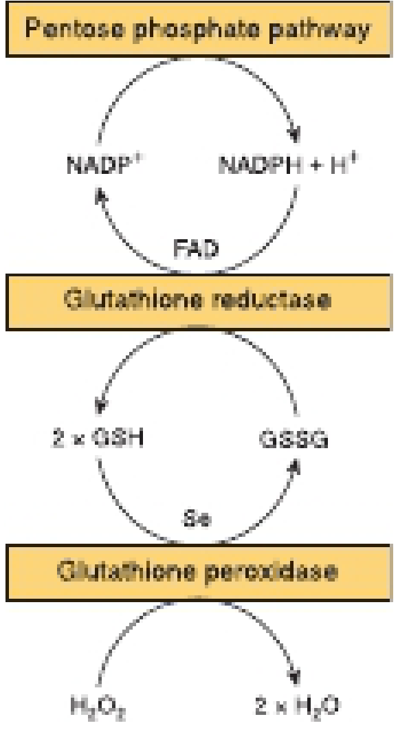
Figure: The coupled pentose phosphate pathway - glutathione reductase - glutathione peroxidase cycle. NADPH from the PPP drives regeneration of GSH from GSSG, enabling continuous H₂O₂ removal. From Harper's Illustrated Biochemistry, 32nd ed.
Key reactions:
H₂O₂ + 2GSH →[GPx, Se]→ 2H₂O + GSSG (H₂O₂ removal)
LOOH + 2GSH →[GPx]→ LOH + H₂O + GSSG (lipid hydroperoxide removal)
GSSG + NADPH + H⁺ →[GR, FAD]→ 2GSH + NADP⁺ (GSH regeneration)
NADP⁺ + G6P →[G6PD]→ NADPH + 6PGL (PPP generates NADPH)
Critical dependency chain:
PPP (G6P → NADPH) → Glutathione Reductase → GSH → Glutathione Peroxidase → H₂O
G6PD deficiency (X-linked; commonest enzyme deficiency worldwide) breaks this chain in red blood cells → H₂O₂ accumulates → hemolytic anemia triggered by oxidant drugs (primaquine, dapsone), infection, or fava beans. RBCs are especially vulnerable because the pentose phosphate pathway is their sole source of NADPH.
Why the Liver Is the Master Antioxidant Organ
The liver:
- Performs cytochrome P450 metabolism (generates ROS as byproduct)
- Synthesizes glutathione (exported to plasma)
- Has G6PD activity up to 30% of glycolytic flux
- Contains the highest SOD, catalase, and GPx activities
"The liver uses more glutathione and NADPH to maintain glutathione reductase and catalase activity than any other tissue."
- Basic Medical Biochemistry, 6th ed.
7. Enzymatic Antioxidants - Biochemical Detail
Superoxide Dismutase (SOD)
Three isoforms, each with a different metal cofactor:
| Isoform | Metal | Location | Gene |
|---|---|---|---|
| Cu/Zn-SOD (SOD1) | Copper + Zinc | Cytosol, nucleus, lysosomes | SOD1 (mutated in familial ALS) |
| Mn-SOD (SOD2) | Manganese | Mitochondrial matrix | SOD2 |
| EC-SOD (SOD3) | Copper + Zinc | Extracellular, around vessels/airways | SOD3 |
Reaction:
2 O₂•⁻ + 2H⁺ → H₂O₂ + O₂
Note: SOD removes superoxide but generates H₂O₂, which must then be cleared by catalase or GPx.
Catalase
- Located in peroxisomes (where H₂O₂-generating oxidases are concentrated)
- Heme iron at active site (4 heme groups per molecule)
- Extremely fast (kcat ~10⁷/s - one of the fastest enzymes known)
2 H₂O₂ → 2 H₂O + O₂ (dismutation)
Glutathione Peroxidase (GPx)
- Selenocysteine at active site (selenium - the unique cofactor)
- Higher affinity for H₂O₂ than catalase at low H₂O₂ concentrations
- Also removes lipid hydroperoxides (LOOHs) from membranes - a function catalase cannot perform
H₂O₂ + 2GSH → 2H₂O + GSSG
LOOH + 2GSH → LOH + H₂O + GSSG
GPx4 (phospholipid hydroperoxide GPx) specifically reduces phospholipid hydroperoxides within membranes - when GPx4 is inactivated, uncontrolled lipid peroxidation leads to ferroptosis (a form of iron-dependent cell death).
Thioredoxin System
A parallel redox system to glutathione:
- Thioredoxin (Trx): small dithiol protein, reduces disulfide bonds on target proteins
- Thioredoxin reductase (TrxR): FAD + selenium enzyme; regenerates reduced Trx using NADPH
- Targets: ribonucleotide reductase, peroxiredoxins, redox-regulated transcription factors
Peroxiredoxins (Prx)
Family of thiol-dependent peroxidases that are emerging as the dominant H₂O₂ scavengers in many cells (faster than catalase at physiological H₂O₂ concentrations). Reduced by thioredoxin.
Heme Oxygenase-1 (HO-1)
- Induced by oxidative stress, heat shock, heavy metals
- Cleaves heme → biliverdin → bilirubin (a potent lipid-soluble antioxidant) + CO (anti-inflammatory) + free Fe²⁺ (exported by ferritin)
- Part of the Nrf2-regulated cytoprotective response
8. Cellular Compartmentalization of Defenses
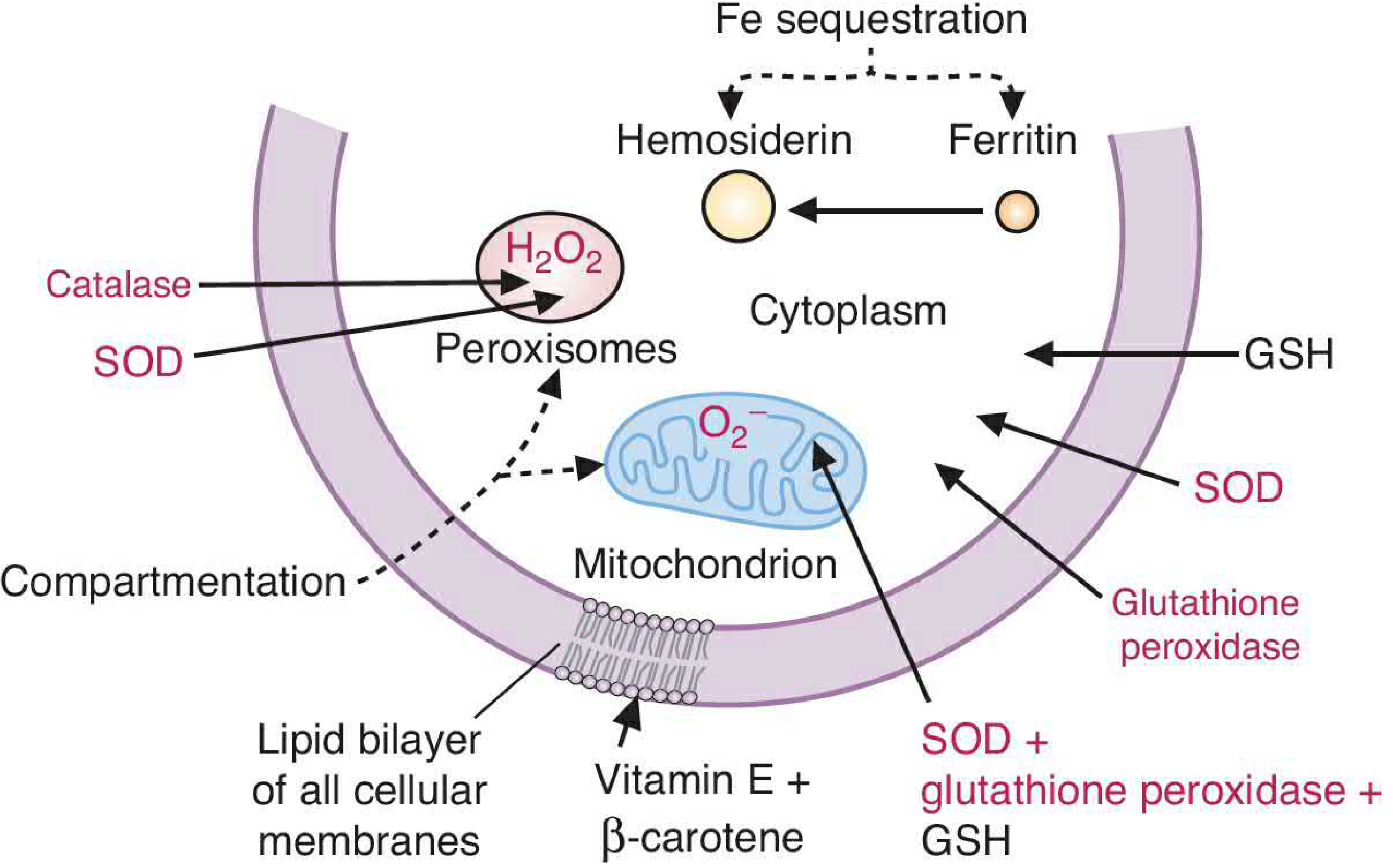
Figure: Location-matched antioxidant defenses. The type and amount of ROS in each subcellular compartment is matched to the antioxidant enzyme present. Peroxisomes contain both H₂O₂-generating enzymes and the catalase that destroys it. Mitochondria have Mn-SOD. Iron is sequestered by ferritin to prevent Fenton chemistry. From Basic Medical Biochemistry, 6th ed.
The principle: defense enzymes are located where ROS are generated.
| Compartment | Main ROS Source | Primary Defense |
|---|---|---|
| Mitochondria | Complex I/III → O₂•⁻ | Mn-SOD, mitochondrial GPx, GSH |
| Peroxisomes | Fatty acid oxidases → H₂O₂ | Catalase (richly concentrated) |
| Cytosol | Multiple | Cu/Zn-SOD, GPx, GSH |
| Plasma membrane | NADPH oxidase, lipid peroxidation | Vitamin E (α-tocopherol), β-carotene |
| Extracellular | Released O₂•⁻, HOCl | EC-SOD, albumin, uric acid, ceruloplasmin |
9. Redox Signaling - The Beneficial Side of H₂O₂
At low, controlled concentrations, H₂O₂ is a bona fide second messenger:
- Reversible oxidation of cysteine residues in proteins: Cys-SH → Cys-SOH (sulfenylation) → acts as a molecular switch, altering protein activity
- Key targets:
- Protein tyrosine phosphatases (PTPs): catalytic Cys oxidized → transiently inactivated → allows RTK signaling to proceed
- PTEN: oxidized and inactivated → allows PI3K/Akt survival pathway
- Keap1: oxidized Cys → releases Nrf2 → Nrf2 translocates to nucleus → binds Antioxidant Response Elements (ARE) → transcribes SOD, catalase, GPx, HO-1, glutathione synthesis enzymes
- NF-κB: ROS promote IκB kinase activation → NF-κB release → pro-inflammatory gene transcription
The concept of redox homeostasis - maintaining H₂O₂ within a narrow window that allows signaling without causing damage - is a central organizing principle of cell biology.
10. Myeloperoxidase (MPO) - Biochemistry of Neutrophil Killing
MPO is a heme-containing enzyme unique to neutrophils (and to a lesser extent monocytes). It uses H₂O₂ to oxidize chloride:
H₂O₂ + Cl⁻ + H⁺ →[MPO]→ HOCl + H₂O
HOCl (hypochlorous acid) - the active ingredient of bleach - is the most potent neutrophil bactericidal agent:
- Oxidizes Fe-S centers, sulfhydryl groups, methionine residues in bacterial proteins
- Damages ATP synthase and electron transport chains
- Halogenates bacterial cell walls
HOCl can also react with amines (R-NH₂) to form chloramines (R-NHCl) - longer-lived oxidants that extend the killing radius.
Clinical relevance:
- MPO deficiency: most common phagocyte disorder; usually clinically silent because other killing mechanisms compensate; severe infections only with concurrent diabetes
- MPO as biomarker: elevated plasma MPO predicts cardiovascular events (MPO oxidizes LDL in coronary arteries → atherogenic ox-LDL)
11. Ischemia-Reperfusion: Biochemistry of Burst ROS Generation
During ischemia:
- ATP falls → AMP → hypoxanthine accumulates (purine catabolism)
- Xanthine dehydrogenase (uses NAD⁺) is proteolytically converted to xanthine oxidase (uses O₂)
- Ca²⁺ influx activates the converting protease
At reperfusion:
Hypoxanthine + O₂ →[Xanthine oxidase]→ Xanthate + O₂•⁻ + H₂O₂
Xanthate + O₂ → Urate + O₂•⁻ + H₂O₂
- Simultaneously, the saturated mitochondrial ETC floods electrons onto O₂ → massive O₂•⁻ burst
- Fenton reaction generates •OH → membrane damage, protein oxidation, DNA damage
- HOCl from recruited neutrophils amplifies injury
This is the biochemical basis of myocardial reperfusion injury and the rationale for allopurinol (xanthine oxidase inhibitor) as a cardioprotective agent.
Summary Table: Key Biochemical Reactions
| Reaction | Equation | Significance |
|---|---|---|
| Mitochondrial O₂•⁻ generation | O₂ + e⁻ → O₂•⁻ | Primary endogenous source |
| SOD dismutation | 2O₂•⁻ + 2H⁺ → H₂O₂ + O₂ | Removes O₂•⁻; generates H₂O₂ |
| Fenton reaction | Fe²⁺ + H₂O₂ → •OH + OH⁻ + Fe³⁺ | Most dangerous step |
| Haber-Weiss | O₂•⁻ + H₂O₂ → O₂ + •OH + OH⁻ | Net reaction; Fe-catalyzed |
| Catalase | 2H₂O₂ → 2H₂O + O₂ | H₂O₂ removal; peroxisomes |
| Glutathione peroxidase | H₂O₂ + 2GSH → 2H₂O + GSSG | H₂O₂ and LOOH removal |
| Glutathione reductase | GSSG + NADPH → 2GSH + NADP⁺ | GSH regeneration |
| G6PDH (PPP) | G6P + NADP⁺ → 6PGL + NADPH | NADPH supply for GR |
| MPO | H₂O₂ + Cl⁻ → HOCl + H₂O | Bactericidal; neutrophils |
| Peroxynitrite | NO• + O₂•⁻ → ONOO⁻ | Nitrosative stress |
| Lipid peroxidation | PUFA-H + •OH → PUFA• → LOO• → LOOH | Membrane damage; chain reaction |
| Ferroptosis trigger | Inactivation of GPx4 → LOOH accumulates | Iron-dependent cell death |
Sources: Harper's Illustrated Biochemistry (32nd ed.); Basic Medical Biochemistry (6th ed.); Robbins, Cotran & Kumar - Pathologic Basis of Disease; Murray & Nadel's Textbook of Respiratory Medicine; Medical Physiology (Boron & Boulpaep)
This is a shared conversation. Sign in to Orris to start your own chat.