
Tracheal stenosis
Reading File
Now let me search the medical library and PubMed in parallel for tracheal stenosis.
Finding Sources
Finding Sources
Searching PubMed
"tracheal stenosis"[MeSH Terms] AND management
Now let me read the most comprehensive sections in parallel:
Reading File
Reading File
Reading File
Let me read more of the Murray & Nadel and Current Surgical Therapy sections, plus get the Cummings section and the image:
Reading File
Reading File
Reading File
Let me fetch the key decision-tree image and read the surgical resection technique section:
Loading Image
Reading File
I now have comprehensive information to produce a detailed answer. Let me compile everything.
Tracheal Stenosis
Definition
Tracheal stenosis is narrowing of the tracheal lumen from any cause, leading to varying degrees of airflow obstruction. It is classified as benign or malignant, and further as simple (short, concentric, <1 cm, without malacia) or complex (>1 cm, irregular, with associated malacia).
Etiology
Acquired (most common)
| Category | Details |
|---|---|
| Postintubation | Most common cause. ETT cuff pressure >30 cmH₂O exceeds mucosal capillary pressure → ischemic necrosis → fibrosis. Risk factors: female sex, obesity, DM, hypertension, cardiovascular disease. |
| Posttracheostomy | Cartilage damage during placement; stomal injury produces characteristic "A"-shaped stenosis sparing the membranous wall. Incidence rising with percutaneous tracheostomies. Risk factors: percutaneous technique, tube size >6, obesity. |
| Postinfectious | Tuberculosis most common worldwide (10-37% of pulmonary TB develop endobronchial disease). Also Klebsiella rhinoscleromatis, fungal infections, HPV (recurrent respiratory papillomatosis). |
| Autoimmune/inflammatory | Granulomatosis with polyangiitis (GPA) - airways involved in 10-33% of cases; sarcoidosis; amyloidosis; inflammatory bowel disease; relapsing polychondritis; scleroderma. |
| Idiopathic | Almost exclusively women; circumferential fibrotic stenosis in subglottic/upper trachea; mucosal and submucosal hypertrophy sparing cartilage. |
| Traumatic | Blunt external neck trauma. |
Congenital
Complete tracheal rings and other anomalies - often associated with cardiovascular defects.
Malignant
Primary tracheal tumors are rare (<0.2% of airway neoplasms). Squamous cell carcinoma (men in 60s, smoking-related) and adenoid cystic carcinoma (no gender predilection, 40s, not smoking-related) account for ~2/3 of cases.
Clinical Presentation
Symptoms of upper airway obstruction:
- Dyspnea on exertion (progressive)
- Stridor (inspiratory or biphasic)
- Cough and wheezing
- Obstructive pneumonia in severe cases
- Dysphonia and dysphagia if subglottic involvement
Key pitfall: Tracheal stenosis is frequently misdiagnosed as adult-onset asthma, and patients are often started on steroids before the correct diagnosis is made. - Current Surgical Therapy 14e
Diagnosis
- Flow-volume loop / spirometry - flattening of the inspiratory and/or expiratory limb suggests fixed obstruction; helps identify critical stenosis
- High-resolution CT with 3D reconstruction - preferred imaging; defines location, length, configuration, relationship to surrounding structures, and wall thickness
- Flexible bronchoscopy - gold standard for characterizing stenosis, assessing mucosa, and directing therapy
- Rigid bronchoscopy (in the OR) - best for measuring stenosis length, assessing mucosal quality, and allowing simultaneous dilation
- Laryngoscopy - to evaluate vocal fold mobility and subglottic extension
- Soft tissue neck radiograph - can show location/extent of subglottic/upper tracheal lesion
Classification of Stenosis
- Location: Subglottic, upper tracheal, mid-tracheal, lower tracheal
- Morphology:
- Cicatricial/membranous (fibrous with intact cartilage)
- Anterior wall collapse (cartilage destruction)
- Complete stenosis
- Length: <1 cm (simple) vs >1 cm (complex)
- Associated malacia: Present or absent
Management
The decision tree below (from Murray & Nadel's Textbook of Respiratory Medicine) illustrates the approach to benign tracheal stenosis:
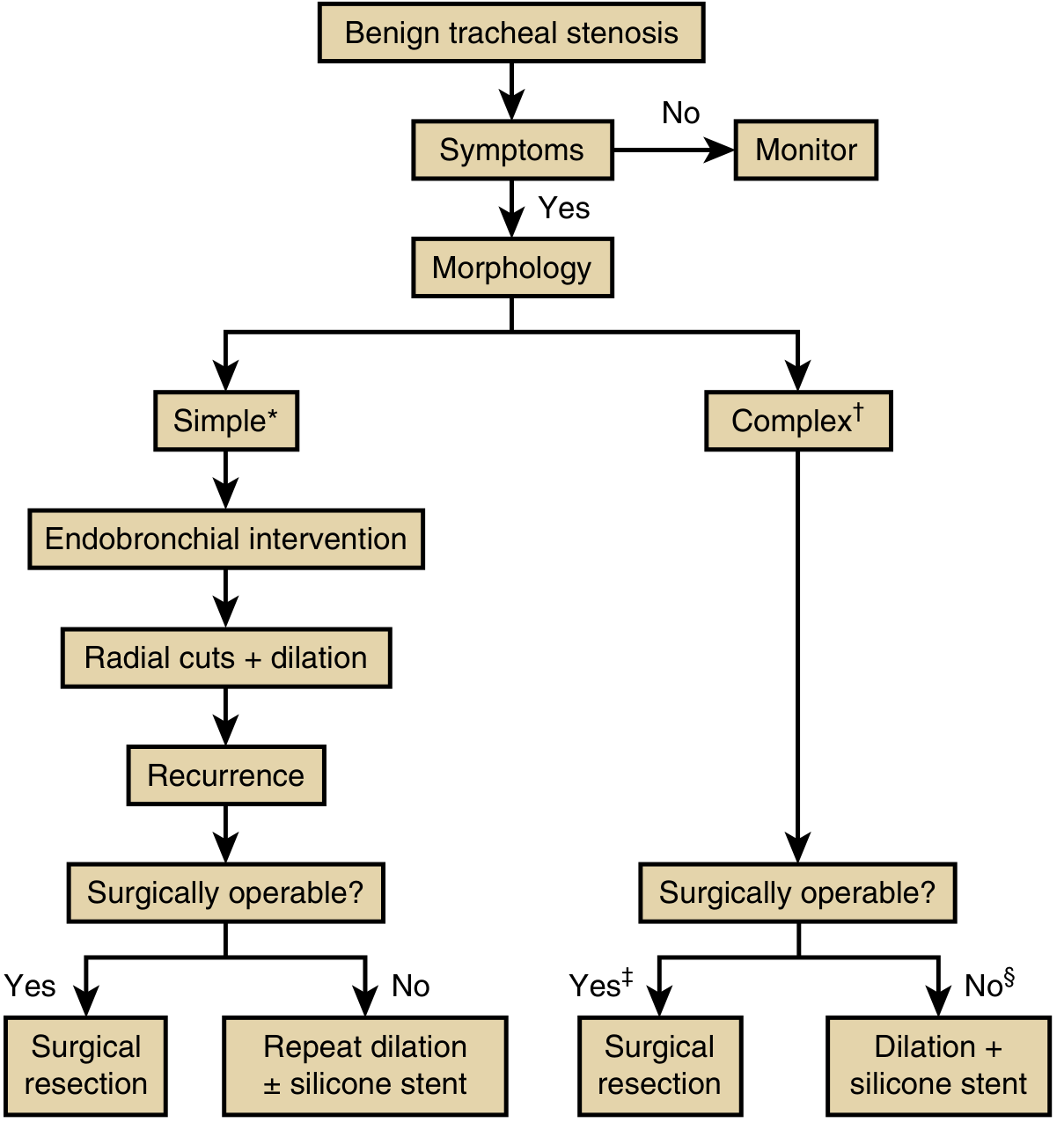
Asymptomatic patients can be monitored with serial CT and airway inspection.
Endoscopic / Bronchoscopic Interventions (simple stenosis, or bridge to surgery)
- Rigid bronchoscopic dilation - preferred; graduated pediatric-to-adult bronchoscopes serially dilate the stricture. Racemic epinephrine + steroids given for 24-48 h after to minimize edema.
- Balloon dilation - alternative for thin band stenosis or poor surgical candidates
- Radial incisions + dilation - electrocautery or laser (CO₂) radial cuts followed by dilation
- CO₂ laser excision - particularly useful for cicatricial stenosis; avoids tracheostomy; staged if circumferential (2-4 weeks apart to avoid restenosis)
- Local adjuncts: Intralesional mitomycin-C, corticosteroid injections have been studied as adjuncts to endoscopic interventions
- Silicone stent - for patients ineligible for or awaiting surgery; NOT expanding metal stents (risk of granulation tissue and erosion)
Surgical Management (complex stenosis, recurrent simple stenosis)
Tracheal resection and end-to-end anastomosis is the definitive treatment for appropriate candidates. Key points:
- Indications: Any patient with airway stenosis from tracheal injury who is a surgical candidate
- Contraindications:
- Absolute: Non-reconstructible airway (excessive length of damage), severe comorbidities, prolonged need for mechanical ventilation
- Relative: Prior radiation, active mucosal inflammation, active steroid use (must be weaned 2-4 weeks before surgery), purulent secretions
- Length limit: Up to ~4-6 cm can be safely resected; high subglottic location limits surgical options
- Anesthesia: Total intravenous anesthesia (TIVA) with remifentanil and propofol is preferred; maintains spontaneous ventilation for critical stenosis
- Technique: Meticulous lateral tracheal dissection (≤1 cm circumferentially) to preserve segmental blood supply from the inferior thyroid artery; recurrent laryngeal nerves protected in the tracheoesophageal groove; anastomosis with interrupted 4-0 Vicryl sutures (knots outside); neck kept flexed postoperatively ("guardian suture") to reduce anastomotic tension
- Laryngotracheoplasty or cricotracheal resection for subglottic involvement
- Montgomery T-tube for complex multi-site stenosis or poor surgical candidates needing long-term stenting
Inflammatory/Autoimmune Stenosis
Immune suppression (systemic) is the primary treatment; endobronchial interventions used adjunctively; surgery reserved for refractory cases.
Malignant Tracheal Stenosis
- Localized: Surgical resection (5-year survival 39-79% for squamous; adenoid cystic carcinoma - complete resection is more important than nodal staging)
- Unresectable: Chemoradiation (SqCC); radiation alone (adenoid cystic)
- Palliative: Stenting, laser debulking for critical obstruction
Postoperative Care
- Neck flexion maintained for ~1 week via "guardian suture" (chin-to-chest suture)
- Avoid neck hyperextension
- Racemic epinephrine + steroids for post-dilation edema
- Voice/swallowing assessment (VHI, DHI questionnaires) as baseline and follow-up
Recent Evidence (2024)
- A 2024 systematic review and meta-analysis (PMID 39263927) identified key risk factors for tracheal stenosis, including intubation duration and cuff management.
- A 2024 systematic review on tracheal resection (PMID 39254596) confirmed resection and anastomosis as the gold standard for postintubation stenosis.
- A 2024 meta-analysis (PMID 38967672) on local adjuncts (mitomycin-C, steroids) to endoscopic interventions for benign laryngotracheal stenosis found benefit in reducing recurrence rates.
- A 2024 systematic review on slide tracheoplasty (PMID 38885545) for congenital tracheal stenosis demonstrated excellent outcomes.
Sources: Murray & Nadel's Textbook of Respiratory Medicine | Current Surgical Therapy 14e | Cummings Otolaryngology Head and Neck Surgery | K.J. Lee's Essential Otolaryngology
Intratymapanic gentamicin injection in ent short note
Finding Sources
Finding Sources
Searching PubMed
"intratympanic gentamicin" AND "Meniere"
The Scott-Brown's section is the dedicated one. Let me read it fully:
Reading File
Reading File
Let me read the rest of the Scott-Brown's dedicated section and more of Cummings:
Reading File
Reading File
I now have comprehensive information for a complete short note. Here it is:
Intratympanic Gentamicin Injection (IT-G)
Introduction
Intratympanic gentamicin (IT-G) injection is a minimally invasive procedure that exploits the selective vestibulotoxicity of aminoglycosides to ablate or partially ablate peripheral vestibular function in the affected ear. It is primarily used in the management of refractory Meniere's disease (endolymphatic hydrops).
The technique was first described by Schuknecht in 1956-57, initially using streptomycin. Streptomycin caused unacceptable rates of profound hearing loss; gentamicin was subsequently adopted due to its relatively higher vestibulotoxicity compared to cochleotoxicity.
Mechanism of Action
Gentamicin enters the inner ear via the round window membrane (and, to a lesser extent, the oval window/annular ligament) after being deposited in the middle ear. Once in the perilymph:
- Binds NMDA receptors → excitotoxic vestibular hair cell damage
- Generates reactive oxygen species (ROS) → oxidative injury to type I vestibular hair cells (predominantly in the cristae and maculae)
- Destroys vestibular hair cell function → partial or complete chemolabyrinthectomy (also called "partial chemical labyrinthectomy")
- After ablation, the Meniere's disease vertigo attacks are no longer detected or provoke nystagmus, so the patient stops experiencing rotational vertigo
Gentamicin preferentially targets type I vestibular hair cells more than cochlear hair cells, which accounts for its relative sparing of hearing at lower doses.
Indications
- Refractory unilateral Meniere's disease - failure of medical management (dietary sodium restriction, diuretics, betahistine)
- After failed intratympanic dexamethasone (preferred first intratympanic agent)
- Tumarkin drop attacks (otolithic crises) - particularly effective in this setting
- "End-stage" Meniere's disease with disabling vertigo in an ear with significantly impaired hearing (where hearing preservation is less of a concern)
Place in stepwise management: IT-dexamethasone is offered first (Grade A recommendation, 80% long-term vertigo control, no hearing risk). IT-gentamicin is offered when dexamethasone fails (Grade A recommendation). - Scott-Brown's Otorhinolaryngology
Contraindications
- Bilateral Meniere's disease (relative - risk of bilateral vestibular loss causing oscillopsia/chronic imbalance)
- Only-hearing or better-hearing ear
- Active middle ear infection / perforated tympanic membrane (relative)
- Patients unable to undergo vestibular compensation (severe neurological comorbidity)
Preparation and Technique
| Step | Detail |
|---|---|
| Concentration | 26.7 mg/mL or 40 mg/mL gentamicin (buffered to pH 6.4 with NaHCO₃ to reduce local irritation) |
| Position | Patient supine, head turned 45° to opposite side, affected ear up |
| Anaesthesia | Topical anaesthesia of tympanic membrane (EMLA, phenol, or iontophoresis) |
| Delivery | Direct transtympanic injection with fine needle (25-27 gauge) via anteroinferior quadrant, OR through a pre-existing tympanostomy tube |
| Volume | 0.5-1 mL injected to fill middle ear |
| Post-injection | Patient remains supine/still for 20-30 minutes; avoids swallowing/nose-blowing to prevent drainage through Eustachian tube |
Dosing Schedules
Three main protocols exist:
| Protocol | Description | Notes |
|---|---|---|
| Fixed-dose | 1 injection/week for 4 weeks (or daily x 3 days) | Higher hearing loss risk; largely abandoned |
| "Titration" / as-needed | Single injection; repeat only if vertigo not controlled | Current preferred approach; minimizes hearing loss |
| Continuous micro-pump | Catheter placed through TM for continuous delivery | Research setting; not routine |
The titration (as-needed) protocol is now the standard of care. The current trend is toward a single injection followed by reassessment, with additional doses only if symptoms recur.
Efficacy
- Vertigo control (Class A + B): 87.5% (Huon et al. 2012 meta-analysis); 93.5% at 2-year follow-up in one RCT
- vs. IT-dexamethasone: Gentamicin superior for vertigo control (93.5% vs. 61% in a 2012 RCT)
- Drop attacks (Tumarkin crises): Effective - specifically noted in guidelines as an indication
- vs. vestibular nerve section: Comparable results (vestibular neurectomy ~90% vs. IT-G ~66-90% depending on protocol)
A 2025 systematic review and meta-analysis (PMID 40421807) confirmed the efficacy of IT-G for Meniere's disease. A 2024 meta-analysis (PMID 39329015) comparing IT-G vs. IT-corticosteroid found gentamicin superior for vertigo control.
Complications
| Complication | Incidence | Comment |
|---|---|---|
| Sensorineural hearing loss (SNHL) | 0-38.7% (varies by dose/schedule) | Most significant risk; with titration protocols, risk approaches natural history of Meniere's disease |
| Disequilibrium / imbalance | Common transiently | Due to unilateral vestibular hypofunction; improves with compensation |
| Visual-vestibular mismatch / oscillopsia | Uncommon | More common with complete ablation |
| Tympanic membrane perforation | Rare | From repeated injections |
| Otitis media | Rare |
With modern titration protocols, the risk of clinically significant hearing loss is comparable to the natural history of Meniere's disease itself. - Cummings Otolaryngology
Monitoring After Injection
- Head thrust test / video head impulse test (vHIT): Peripheral vestibular deficit evident after even a single dose - key objective marker of successful ablation
- Ice water caloric testing: Complete ablation of caloric response is NOT required for vertigo control; targeting complete ablation leads to unnecessary hearing loss
- Audiogram at each visit
- Patient diary of vertigo attacks (AAO-HNS classification)
Position vs. Other Ablative Options
| Option | Vertigo control | Hearing preservation | Invasiveness |
|---|---|---|---|
| IT-gentamicin | ~87-93% | Moderate risk with old protocols; low with titration | Minimally invasive |
| Vestibular nerve section | ~88-90% | Excellent | Major surgery (retro/middle fossa) |
| Labyrinthectomy | >95% | Complete loss | Surgical |
IT-G is the preferred ablative therapy because it is minimally invasive, repeatable, and office-based, reserving vestibular neurectomy for cases where IT-G fails or hearing preservation in the setting of ablation is paramount.
Sources: Scott-Brown's Otorhinolaryngology Head & Neck Surgery Vol. 2 | Cummings Otolaryngology Head and Neck Surgery | K.J. Lee's Essential Otolaryngology
This is a shared conversation. Sign in to Orris to start your own chat.