
Eyeball muscles with image
Reading File
Now let me search the library and images in parallel.
Finding Sources
Reading File
Reading File
Searching Images
rectus oblique eye muscle actions movement
I have rich textbook content and image URLs. Let me fetch the key anatomy images from the textbooks.
Loading Image
Loading Image
Reading File
Reading File
Loading Image
I have all the key content. Let me now fetch the key anatomy images directly from Kanski's and the neuroanatomy textbook.
Loading Image
Loading Image
Loading Image
Excellent! I have three outstanding anatomy images. Let me also get the CT/orbital diagram image and read the nerve supply section.
Loading Image
Reading File
I now have everything needed for a comprehensive, well-illustrated answer. Let me compile it.
Extraocular Muscles of the Eyeball
There are 6 extraocular muscles (EOMs) that control eye movement - 4 rectus muscles and 2 oblique muscles.
Anatomy Overview
Fig. 18.4A - Superior view of the orbit (Kanski's Clinical Ophthalmology):
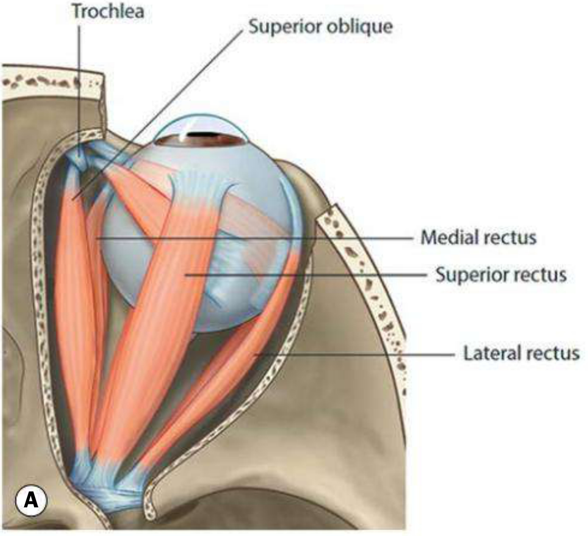
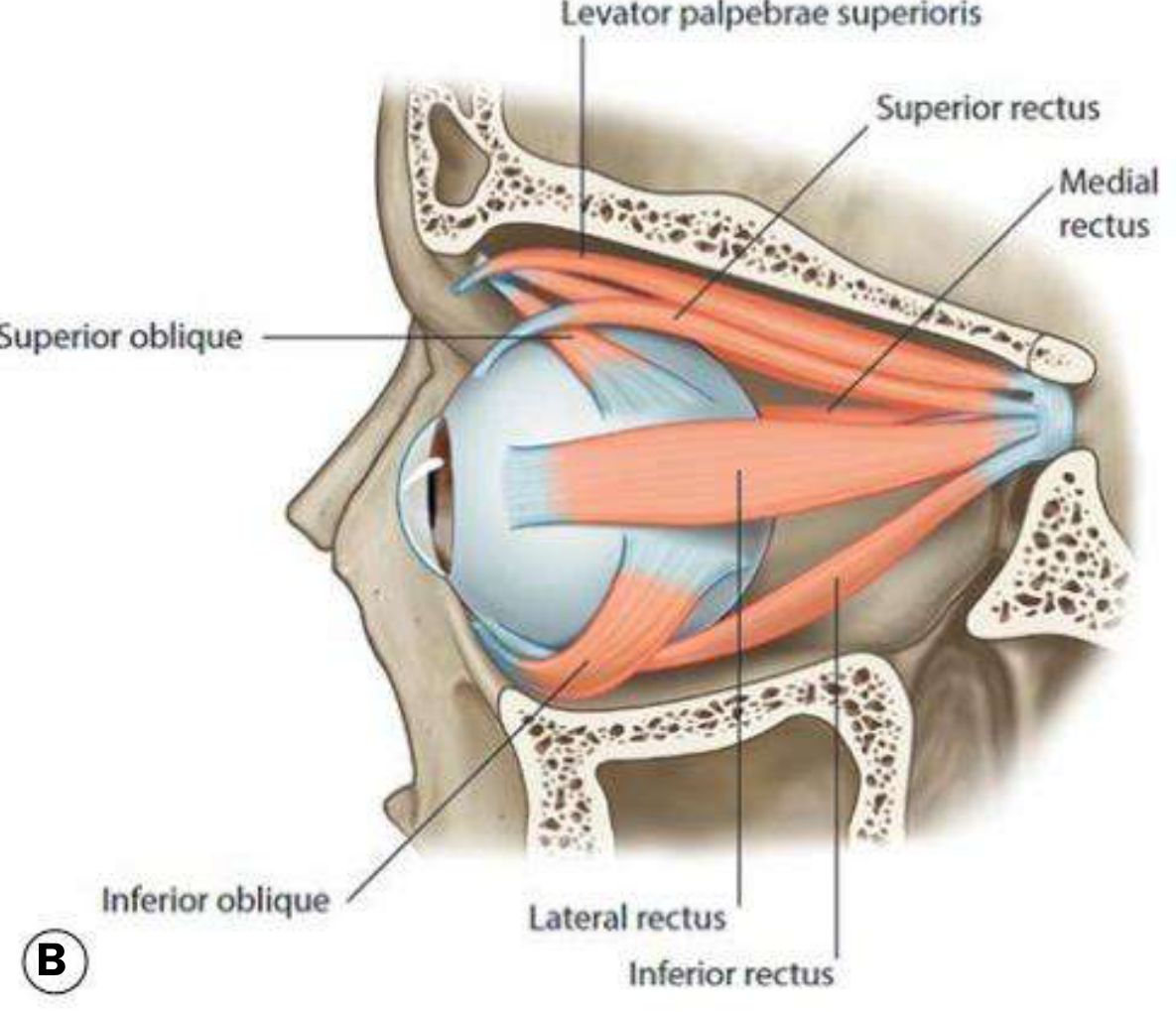
Fig. 18.4B - Lateral view of the orbit:
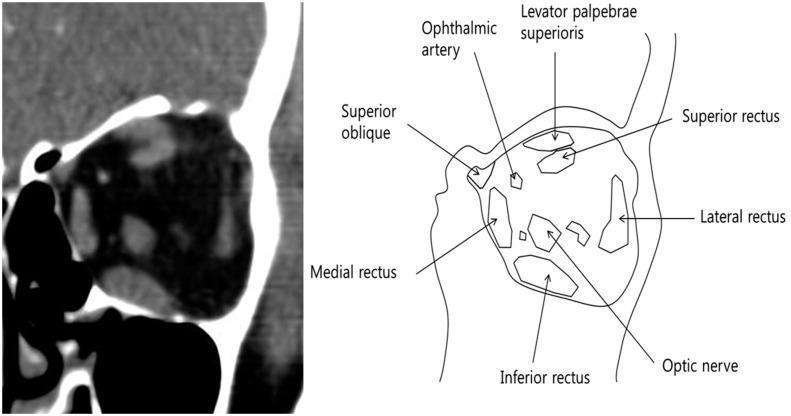
CT coronal section + diagram of the orbit:
The 6 Extraocular Muscles
All 4 rectus muscles originate from the annulus of Zinn (common tendinous ring) at the orbital apex.
1. Medial Rectus (MR)
- Origin: Annulus of Zinn
- Insertion: 5.5 mm behind the nasal limbus (closest to the limbus)
- Primary action: Adduction (moves eye nasally/inward)
- Nerve: CN III (Oculomotor)
2. Lateral Rectus (LR)
- Origin: Annulus of Zinn
- Insertion: 6.9 mm behind the temporal limbus
- Primary action: Abduction (moves eye temporally/outward)
- Nerve: CN VI (Abducens)
3. Superior Rectus (SR)
- Origin: Upper part of the annulus of Zinn
- Insertion: 7.7 mm behind the superior limbus (farthest from limbus among recti)
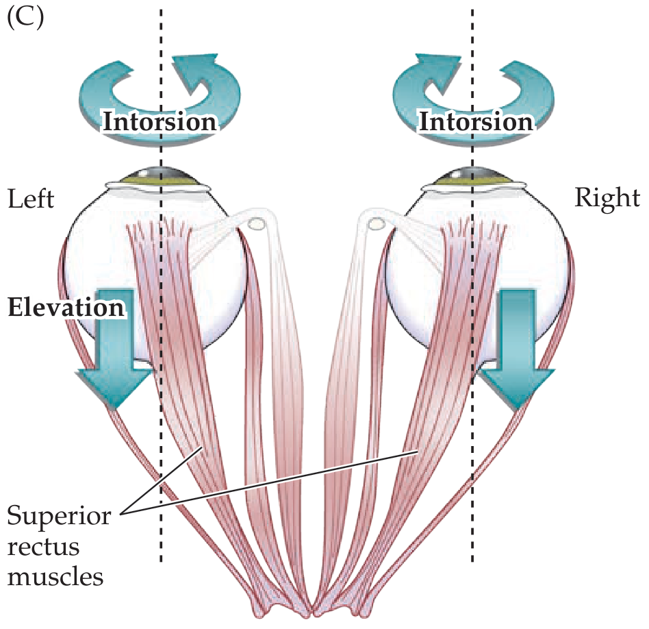
- Primary action: Elevation
- Secondary actions: Adduction + Intorsion
- Nerve: CN III (Oculomotor)
- Testing tip: The SR acts as a pure elevator when the eye is abducted 23° (visual and orbital axes align)
4. Inferior Rectus (IR)
- Origin: Lower part of the annulus of Zinn
- Insertion: 6.5 mm behind the inferior limbus
- Primary action: Depression
- Secondary actions: Adduction + Extorsion
- Nerve: CN III (Oculomotor)
- Testing tip: Acts as a pure depressor when the eye is abducted 23°
5. Superior Oblique (SO)
- Origin: Superomedial to the optic foramen (sphenoid bone)
- Course: Passes anteriorly through the trochlea (a fibrocartilaginous pulley at the superomedial orbital rim), then reflected backwards and laterally
- Insertion: Posterior upper temporal quadrant of the globe (behind the equator)
- Angle with visual axis: 51°
- Primary action: Intorsion
- Secondary actions: Depression + Abduction
- Nerve: CN IV (Trochlear)
- Testing tip: Acts as a pure depressor when the eye is adducted 51°; weakness manifests as failure of depression in adduction
6. Inferior Oblique (IO)
- Origin: Small depression just behind the orbital rim, lateral to the lacrimal sac (anterior medial orbital wall - unique: no trochlea)
- Insertion: Posterior lower temporal quadrant of the globe (near the macula)
- Angle with visual axis: 51°
- Primary action: Extorsion
- Secondary actions: Elevation + Abduction
- Nerve: CN III (Oculomotor)
- Testing tip: Acts as a pure elevator when the eye is adducted 51°
Spiral of Tillaux
The insertions of the 4 recti form the Spiral of Tillaux - an imaginary line joining their scleral insertions, progressively further from the limbus:
| Muscle | Insertion from limbus |
|---|---|
| Medial rectus | 5.5 mm |
| Inferior rectus | 6.5 mm |
| Lateral rectus | 6.9 mm |
| Superior rectus | 7.7 mm |
This is an important surgical landmark.
Nerve Supply Summary
| Muscle | Cranial Nerve |
|---|---|
| Lateral rectus | CN VI (Abducens) |
| Superior oblique | CN IV (Trochlear) |
| All others (MR, SR, IR, IO) + Levator palpebrae + ciliary + sphincter pupillae | CN III (Oculomotor) |
Memory aid: "LR6 SO4 rest 3" - Lateral Rectus = CN 6, Superior Oblique = CN 4, the remaining 4 muscles = CN 3.
Actions (Summary Table)
| Muscle | Primary | Secondary |
|---|---|---|
| Medial rectus | Adduction | - |
| Lateral rectus | Abduction | - |
| Superior rectus | Elevation | Adduction, Intorsion |
| Inferior rectus | Depression | Adduction, Extorsion |
| Superior oblique | Intorsion | Depression, Abduction |
| Inferior oblique | Extorsion | Elevation, Abduction |
Superior Rectus Actions (Intorsion + Elevation Diagram)
Muscle Pulleys
The 4 rectus muscles pass through connective tissue and smooth muscle condensations just posterior to the equator. These pulleys act as the functional origins of the rectus muscles, minimizing off-axis movements and coordinating eye movements. Pulley displacement can cause V- and A-pattern strabismus.
Sources: Kanski's Clinical Ophthalmology 10th Ed., p. 723-726; Neuroanatomy through Clinical Cases 3rd Ed., p. 594
This is a shared conversation. Sign in to Orris to start your own chat.