

45fd581d-8cb2-4ea2-9599-daa6cf8ca66a.jpeg
Tell me about it
analyse_media
Reading File
This is a Venous Blood Gas (VBG) report from a Radiometer ABL800 FLEX analyzer for Mrs. Prem Narula, dated July 29, 2026. Here is a full breakdown with clinical interpretation:
Patient & Sample
- Patient: Mrs. Prem Narula | ID: MM02865237
- Sample type: Venous whole blood
- Temperature: 37.0 °C
- Analyzer: Radiometer ABL800 FLEX
Acid-Base Status
| Parameter | Result | Reference | Status |
|---|---|---|---|
| pH | 7.417 | 7.350 - 7.450 | Normal |
| pCO₂ | 37.3 mmHg | 35.0 - 45.0 | Normal |
| pO₂ | 21.1 mmHg | 83.0 - 108 | ⬇ CRITICALLY LOW |
| HCO₃⁻ (P)c | 23.6 mmol/L | - | Normal |
| HCO₃⁻ (P,st)c | 23.0 mmol/L | - | Normal |
| Base Excess (B)c | -0.1 mmol/L | - | Normal |
| Base Excess (Ecf)c | -0.3 mmol/L | - | Normal |
| ctCO₂(P)c | 55.5 Vol% | - | - |
Interpretation: Normal pH, normal pCO₂, normal bicarbonate - no acid-base disturbance. The very low pO₂ (21.1 mmHg) is expected for venous blood - venous pO₂ is normally 30-50 mmHg in mixed venous and lower in peripheral venous. However, 21.1 is still notably low and could reflect increased oxygen extraction by tissues.
Oximetry Values
| Parameter | Result | Reference | Status |
|---|---|---|---|
| Hematocrit (Hct) | 38.6% | - | Low-normal |
| Hemoglobin (ctHb) | 12.5 g/dL | 12.0 - 16.0 | Normal (low end) |
| sO₂ | 33.9% | 95.0 - 99.0 | ⬇ LOW (venous expected) |
| FO₂Hb | 33.4% | - | - |
| FCOHb | 0.8% | - | Normal (<3%) |
| FMetHb | 0.7% | - | Normal (<1.5%) |
| FHHb (deoxy-Hb) | 65.1% | - | High (venous) |
Interpretation: The low sO₂ (33.9%) and high deoxyhemoglobin (65.1%) are consistent with venous blood. If this were arterial, sO₂ of 33.9% would be a life-threatening emergency. COHb and MetHb are both normal, ruling out carbon monoxide poisoning or methemoglobinemia.
Oxygen Status
| Parameter | Result |
|---|---|
| O₂ bound to Hb (BO₂c) | 17.2 Vol% |
| O₂ content (ctO₂c) | 5.9 Vol% |
Electrolytes - Multiple Abnormals
| Parameter | Result | Reference | Status |
|---|---|---|---|
| cNa⁺ | 124 mmol/L | 135 - 145 | ⬇ HYPONATREMIA (significant) |
| cK⁺ | 4.4 mmol/L | 3.4 - 4.5 | Normal |
| cCa²⁺ (ionized) | 1.14 mmol/L | 1.15 - 1.29 | ⬇ BORDERLINE LOW |
| cCl⁻ | 96 mmol/L | 98 - 106 | ⬇ LOW |
| Anion Gap | 4.0 mmol/L | - | Low (normal 8-12) |
Critical finding - Sodium 124 mmol/L: This represents moderate-to-severe hyponatremia (normal 135-145). This requires urgent attention. Symptoms may include nausea, headache, confusion, seizures depending on the rate of onset.
Low anion gap (4.0): A low anion gap can be seen with hypoalbuminemia, multiple myeloma (paraproteinemia), lithium toxicity, or laboratory artifact (hyperviscosity).
Metabolites
| Parameter | Result | Reference | Status |
|---|---|---|---|
| cGlu (Glucose) | 110 mg/dL | 80 - 120 | Normal |
| cLac (Lactate) | 1.4 mmol/L | 0.5 - 1.5 | Normal (upper limit) |
Calculated Values
| Parameter | Result |
|---|---|
| Barometric pressure | 731 mmHg |
| Osmolality (mOsm) | 253.5 mmol/kg |
| p50(st)c | 28.53 mmHg |
Low osmolality (253.5 mOsm/kg): Normal is 275-295 mOsm/kg. This is consistent with the hyponatremia - hypotonic hyponatremia.
Summary of Key Abnormalities
| Finding | Value | Clinical Significance |
|---|---|---|
| Hyponatremia | Na 124 mmol/L | Significant - requires urgent workup (SIADH, hypothyroidism, adrenal insufficiency, heart failure, cirrhosis, etc.) |
| Low osmolality | 253.5 mOsm/kg | Confirms hypotonic state |
| Low pO₂ | 21.1 mmHg | Expected for venous sample; reflects tissue O₂ extraction |
| Low anion gap | 4.0 mmol/L | Consider hypoalbuminemia or paraproteinemia |
| Borderline low ionized Ca²⁺ | 1.14 mmol/L | Monitor; could be clinically significant |
| Borderline low Cl⁻ | 96 mmol/L | Consistent with hyponatremia |
Clinical Priority
The most clinically urgent finding is the sodium of 124 mmol/L with serum osmolality of 253.5 mOsm/kg, indicating hypotonic hyponatremia. The treating team should:
- Assess for symptoms (confusion, seizures, headache)
- Check urine sodium and osmolality to differentiate SIADH from other causes
- Review medications (diuretics, antidepressants, antiepileptics)
- Assess volume status clinically
- Correct sodium cautiously (max 10-12 mEq/L/day to prevent osmotic demyelination syndrome)
Note: This is a venous blood gas - all pO₂ and sO₂ values should be interpreted in that context, not as arterial values.
What are the causes of hyponatremia?
Finding Sources
Searching the Web
causes of hyponatremia classification clinical
Reading File
Reading File
Reading File
Reading File
Reading File
Loading Image
Here is a structured overview of the causes of hyponatremia, organized by serum tonicity and volume status -- the standard clinical framework.
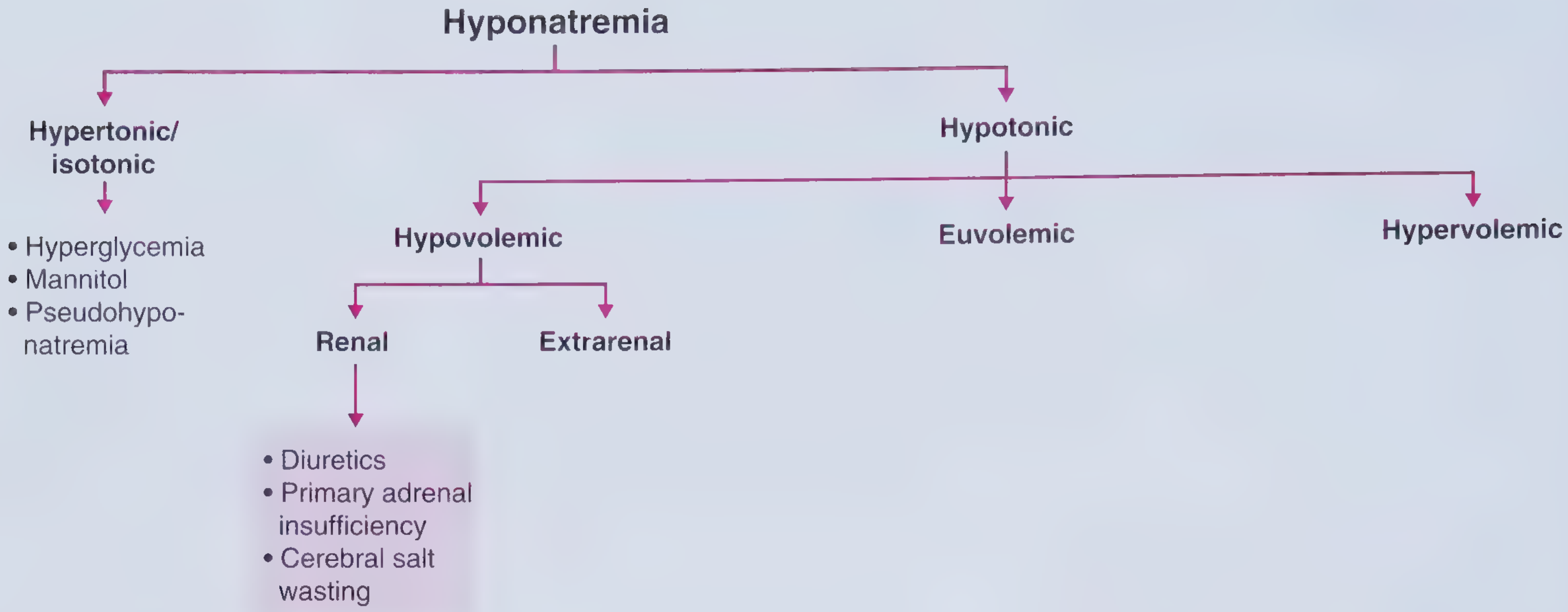
Step 1: Is the Sodium Truly Low? - Tonicity Classification
1. Hypertonic / Isotonic Hyponatremia (NOT true hyponatremia)
These are situations where measured sodium is low but total body osmolality is normal or high:
- Hyperglycemia - the most common cause. For every 100 mg/dL rise in glucose above 100, serum Na falls by ~1.6-2.4 mEq/L. Glucose draws water out of cells, diluting serum Na.
- Hypertonic mannitol - used to reduce intracranial pressure; same osmotic mechanism as glucose
- Pseudohyponatremia (isotonic) - a laboratory artifact seen with severe hyperlipidemia or hyperproteinemia (e.g. multiple myeloma). The actual sodium activity is normal.
Step 2: Hypotonic Hyponatremia (TRUE hyponatremia)
The key diagnostic step is assessing volume status (hypovolemic, euvolemic, or hypervolemic).
A. Hypovolemic Hyponatremia
(Total body water falls, but sodium loss is proportionally greater)
Renal causes (urine Na typically >20 mEq/L):
- Thiazide diuretics - most common renal cause. Thiazides block NaCl reabsorption in the distal convoluted tubule, impairing urinary dilution, and also stimulate vasopressin release via volume depletion.
- Primary adrenal insufficiency (Addison's disease) - aldosterone deficiency causes renal Na wasting. Clue: hyponatremia + hyperkalemia + hypotension.
- Cerebral salt wasting - occurs with subarachnoid hemorrhage or other CNS injury; renal Na wasting via poorly understood CNS-mediated mechanism. Important to distinguish from SIADH (treatment is opposite).
Extrarenal causes (urine Na typically <20 mEq/L):
- Vomiting / diarrhea / GI fistulas
- Excessive sweating
- Third-space losses (burns, pancreatitis)
B. Euvolemic Hyponatremia
(Normal total body sodium, excess free water)
Vasopressin-dependent causes:
- SIADH (Syndrome of Inappropriate ADH secretion) - the most common cause of hyponatremia overall. Vasopressin is secreted despite normal or low osmolality. Causes include:
- Medications: antidepressants (SSRIs, TCAs), antipsychotics, antiepileptics (carbamazepine, oxcarbazepine), NSAIDs, opioids, cyclophosphamide
- Pulmonary disease: pneumonia, TB, lung abscess, mechanical ventilation
- Malignancy: small cell lung cancer (ectopic ADH), thymoma, lymphoma
- CNS disease: meningitis, encephalitis, stroke, brain tumor, trauma
- Pain, nausea, stress
- Hypothyroidism (severe / myxedema coma) - reduced cardiac output lowers renal perfusion, triggering baroreceptor-mediated vasopressin release
- Secondary adrenal insufficiency - hypocortisolism increases CRH, which stimulates vasopressin secretion (note: unlike primary adrenal insufficiency, no aldosterone deficiency, so patients are euvolemic)
- Reset osmostat - the osmotic set-point for vasopressin release is shifted downward (seen in pregnancy, malnutrition, chronic illness)
Vasopressin-independent causes:
- Primary polydipsia - excessive compulsive water drinking overwhelms renal excretory capacity. Most common in schizophrenia. Water intake must exceed ~16 L/day to cause hyponatremia in a normal person.
- Beer potomania / "tea and toast" syndrome - very low solute intake (low protein, high fluid) limits the kidney's ability to excrete free water. As few as 14 cans of beer/day can cause hyponatremia.
C. Hypervolemic Hyponatremia
(Total body water is increased more than total body sodium - edematous states)
Even though the body is fluid-overloaded, the effective arterial blood volume (EABV) is low, which drives vasopressin release.
Renal causes:
- Renal failure (acute or chronic) - impaired ability to dilute urine; minimum urine osmolality may be as high as 200-250 mOsm/kg
- Nephrotic syndrome - when albumin falls below 2 g/dL, oncotic pressure drops, leading to intravascular depletion and vasopressin stimulation
Extrarenal causes:
- Congestive heart failure - poor forward flow reduces EABV, triggering vasopressin release and renal Na/water retention
- Cirrhosis - portal hypertension causes splanchnic vasodilation, reducing EABV; compensatory vasopressin and RAAS activation retains water disproportionately to sodium
Quick Reference Summary
| Category | Common Causes |
|---|---|
| Hypertonic/Isotonic | Hyperglycemia, mannitol, pseudohyponatremia |
| Hypovolemic | Thiazides, adrenal insufficiency, GI losses, cerebral salt wasting |
| Euvolemic | SIADH (most common), hypothyroidism, secondary adrenal insufficiency, polydipsia, beer potomania |
| Hypervolemic | Heart failure, cirrhosis, renal failure, nephrotic syndrome |
Relating to Mrs. Prem Narula's report: Her Na of 124 with osmolality of 253.5 mOsm/kg confirms hypotonic hyponatremia. The next step is to assess her volume status and check urine sodium + osmolality to narrow toward SIADH, hypothyroidism, adrenal insufficiency, or another cause.
- Frameworks for Internal Medicine, p. 508-513
This is a shared conversation. Sign in to Orris to start your own chat.