
Tell me about the leprosy in detail microbiology aspect exam oriented
Reading File
Now let me search the medical library for leprosy microbiology content in parallel:
Finding Sources
Finding Sources
Excellent! Now let me read the key microbiology sources in parallel:
Reading File
Reading File
Reading File
Let me now read the Sherris microbiology content more (clinical + lab aspects) and the Robbins morphology section, plus a dedicated microbiology textbook:
Reading File
Reading File
Finding Sources
Now let me get the Harrison's content on leprosy (clinical + diagnosis) and the Park's content on lepromin test, plus fetch key images:
Reading File
Reading File
Loading Image
Loading Image
Now let me get the Harrison's classification/diagnosis details and the Sherris treatment section:
Reading File
Reading File
I now have comprehensive information from multiple authoritative textbooks. Let me compile the detailed exam-oriented answer.
Leprosy (Hansen's Disease) - Detailed Microbiology (Exam-Oriented)
1. THE ORGANISM - Mycobacterium leprae
| Feature | Detail |
|---|---|
| Kingdom/Type | Gram-positive (by cell wall chemistry), acid-fast bacillus |
| Morphology | Straight or slightly curved rod, 1-8 µm long, 0.3-0.5 µm wide |
| Arrangement | Found in clumps called "globi" inside macrophages (lepromatous form) |
| Culture | Cannot be cultured in vitro (absolute obligate intracellular pathogen) - classic exam fact |
| Animal models | Grows in nine-banded armadillo (ideal model, 32-34°C body temp); also in mouse foot pads and nude mice |
| Temperature | Replicates best at 32-34°C (body surface temperature) - explains distribution on cool skin, nose, ears, testes |
| Staining | Acid-fast bacillus - but uses Fite stain (not standard Ziehl-Neelsen) because M. leprae is more sensitive to decolorization than M. tuberculosis |
| Doubling time | ~12-14 days - the slowest dividing bacterium known |
| Cell wall | Contains PGL-1 (phenolic glycolipid-1) - key virulence factor; enables invasion of Schwann cells and binding to laminin of peripheral nerve axons |
| Species note | M. lepromatosis (proposed 2008) - rarely causes diffuse lepromatous leprosy (mainly Mexico, Caribbean) |
2. EPIDEMIOLOGY
- Reservoir: Humans (primary); nine-banded armadillos (in Southern USA, South America); wild squirrels and certain primates
- Transmission: Nasal droplets from untreated multibacillary cases (primary route); skin contact through wounds/tattoos possible; NOT highly contagious
- Incubation period: Average 2-7 years; can be up to 40 years (longest incubation period of any bacterial disease)
- Endemic regions: India, Brazil, Indonesia account for ~80% of global burden
- Current burden: ~200,000 new cases/year globally (down from 5.2 million in 1985 due to MDT)
- WHO elimination target: <1 case per 10,000 population - achieved globally in 2000
- Children: ~8% of new cases; rare in children <5 years; age distribution bimodal (teens and adults)
3. PATHOGENESIS
Entry and Primary Target
- M. leprae enters via respiratory tract (primary) or skin breach
- Disseminates through blood and lymphatics
- Primary target = Schwann cells (glial cells of peripheral nervous system)
- PGL-1 + laminin-binding protein mediate invasion of Schwann cells and binding to peripheral nerve basal lamina
- This causes demyelination of peripheral sensory nerves → anesthesia
Virulence Mechanisms (Exam High-Yield)
- Cannot be cultured in vitro - absolute obligate intracellular
- Secretes no toxins - virulence is through cell wall components
- Inhibits mitochondrial energy metabolism to evade host immune response
- BCG vaccination offers partial cross-protection (similar cell wall to M. tuberculosis)
The Immunologic Spectrum - KEY EXAM CONCEPT
The entire spectrum of leprosy depends on the host Th1 vs Th2 response:
| Feature | Tuberculoid (TT) | Lepromatous (LL) |
|---|---|---|
| CMI | Strong Th1 | Weak Th1 / Th2 predominant |
| CD4+ T cells | Abundant | Scarce |
| CD8+ T cells | Few | Numerous |
| Cytokines | IL-2, IFN-γ (Th1) | IL-4, IL-5, IL-10 (Th2) |
| Bacterial load | Paucibacillary (few/no AFB) | Multibacillary (many AFB) |
| Granulomas | Well-formed, with giant cells | Poorly formed / absent |
| Macrophages | Epithelioid, activated (M1) | Foamy (lipid-laden), deactivated |
| Lepromin test | Positive | Negative |
| Antibodies | Low | High (but non-protective - form immune complexes) |
| Contagiousness | Low (non-contagious) | High (contagious) |
4. RIDLEY-JOPLING CLASSIFICATION (1962) - Exam Classic
Five overlapping categories on a spectrum:
TT ←→ BT ←→ BB ←→ BL ←→ LL
(tuberculoid)         (lepromatous)
Strong CMI ←————————→ Weak CMI
- TT = Tuberculoid (polar, stable)
- BT = Borderline tuberculoid
- BB = Mid-borderline (unstable, rare - rapidly shifts toward BL)
- BL = Borderline lepromatous
- LL = Lepromatous (polar, stable)
- IL = Indeterminate leprosy (early, not yet classified)
WHO Simplified Classification (for field use):
| WHO Type | Definition | Skin Lesions | Nerves |
|---|---|---|---|
| Paucibacillary (PB) | Smear negative at all sites | 1-5 lesions | ≤1 nerve trunk |
| Multibacillary (MB) | Smear positive at any site | >5 lesions | >1 nerve trunk |
5. MORPHOLOGY / HISTOPATHOLOGY
Tuberculoid Leprosy
- Skin: Flat red lesions → enlarge with indurated, elevated, hyperpigmented margins and depressed, pale, hairless centers (central healing)
- Histology: Well-formed granulomas - epithelioid cells + Langhans giant cells + lymphocyte infiltration (resembles TB granuloma)
- Nerves: Enclosed within granulomatous reaction; small peripheral twigs can be completely destroyed → sensory loss
- AFB: Almost none visible → "paucibacillary"
- The presence of granulomas + absence of bacteria = reflects strong T-cell immunity
Lepromatous Leprosy
- Skin: Macular/papular/nodular lesions on face, ears, wrists, elbows, knees; diffuse symmetric thickening
- Facies: "Leonine facies" - nodular lesions coalesce giving lion-like appearance; loss of eyebrows (madarosis)
- Histology: Large aggregates of lipid-laden foamy macrophages = "Lepra cells" (Virchow cells) filled with masses ("globi") of AFB
- Nerves: Symmetric invasion of ulnar and peroneal nerves by mycobacteria; minimal inflammation despite massive bacillary load
- Other sites: Testes (destruction of seminiferous tubules → sterility); anterior eye; upper airways; lymph nodes (paracortical foamy macrophages)
- AFB: Abundant → "multibacillary"
- Vital organs/CNS usually spared (temperature too high for M. leprae)
Histopathology Images (Robbins Pathology):
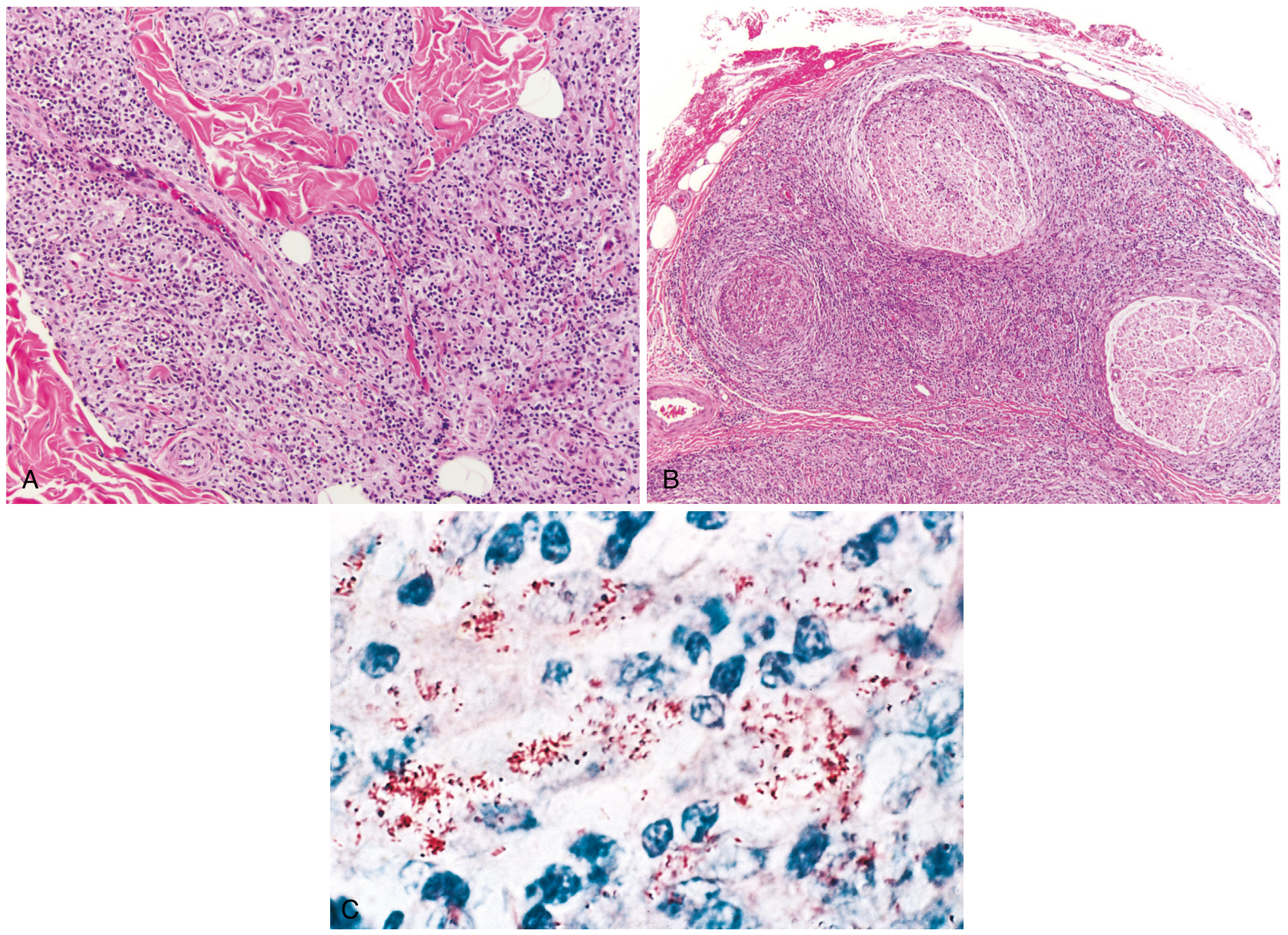
(A) Dense dermal macrophage infiltration surrounding adnexa, vessels, and nerves in lepromatous leprosy; (B) Dense chronic lymphocytic and macrophage infiltration into large nerve bundles in tuberculoid leprosy; (C) Acid-fast bacilli (red) within macrophages - lepromatous form
Clinical Lepromatous Leprosy:
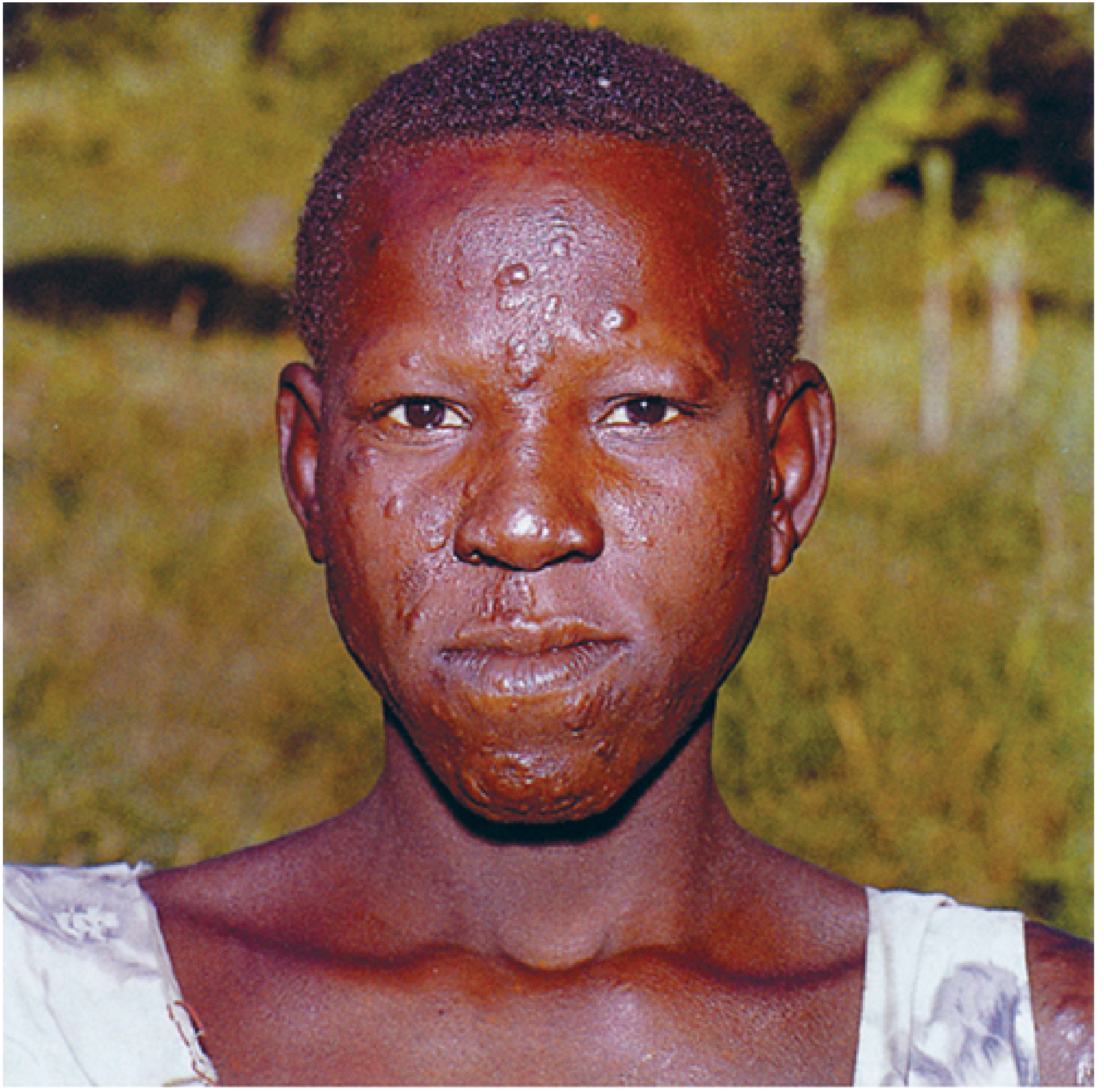
Lepromatous leprosy - note cutaneous plaques, infiltrates, and loss of eyebrows (madarosis)
6. CLINICAL FEATURES - Cardinal Signs (WHO)
Diagnosis requires ONE or more of these:
- Hypopigmented/erythematous skin patch with definite loss of sensation
- Thickened peripheral nerve (with or without tenderness)
- Positive slit-skin smear for AFB (Fite stain)
7. LABORATORY DIAGNOSIS
| Test | Details |
|---|---|
| Slit-skin smear | Smear from ear lobe, nasal mucosa, or skin lesion; Fite stain (modified AFB); use this not ZN - M. leprae is more acid-labile |
| Bacteriological Index (BI) | Logarithmic scale 0-6+ for bacillary load on slit-skin smear |
| Morphological Index (MI) | % of solid-staining (viable) bacilli |
| Skin biopsy | Full-thickness biopsy showing granulomas (tuberculoid) or foamy macrophages with AFB (lepromatous) |
| Culture | NOT POSSIBLE in vitro - classic exam question |
| Animal inoculation | Mouse foot pad; nine-banded armadillo |
| Lepromin (Mitsuda) test | Skin test using killed M. leprae; NOT a diagnostic test; measures CMI status |
| PCR | Detects M. leprae DNA; useful for paucibacillary and doubtful cases |
| Anti-PGL-1 antibody (ELISA) | Detects antibodies to phenolic glycolipid-1; elevated in lepromatous; not useful for paucibacillary |
8. LEPROMIN (MITSUDA) TEST - Exam High-Yield
What it is: Intradermal injection of heat-killed M. leprae (lepromin antigen)
Two reactions:
| Reaction | Timing | Size (positive) | Significance |
|---|---|---|---|
| Fernandez reaction (early) | 48-72 hours | >10 mm erythema | Measures delayed hypersensitivity (DTH); similar to tuberculin |
| Mitsuda reaction (late) | 3-4 weeks | >3-5 mm nodule | Measures cell-mediated immunity (CMI); more clinically significant |
Key facts:
- NOT a diagnostic test (positive in healthy non-cases; negative in lepromatous)
- Used to classify leprosy and assess prognosis (CMI status)
- Positive in TT, negative in LL
- BCG vaccination can convert lepromin-negative to positive
- First 6 months of life: most children are lepromin-negative
9. IMMUNITY
| Aspect | Detail |
|---|---|
| Primary immunity | T-cell mediated (CMI) - not humoral |
| Th1 → tuberculoid | IL-2, IFN-γ produced → activates macrophages (M1) → kills bacilli |
| Th2/regulatory T cells → lepromatous | Weak CMI; humoral response predominates; antibodies non-protective |
| IFN-γ role | Same as in TB - mobilizes effective macrophage response; low bacterial burden |
| Immune complexes | In lepromatous: antibody + free antigens → erythema nodosum leprosum (ENL), vasculitis, glomerulonephritis (Type III reaction) |
| BCG vaccination | Provides partial cross-protection against M. leprae |
10. LEPROSY REACTIONS (Immunological Complications)
| Reaction | Type | Form | Mechanism |
|---|---|---|---|
| Type 1 (Reversal reaction) | Delayed hypersensitivity (Type IV) | Borderline (BT, BB, BL) | Sudden increase in CMI → inflammation in existing lesions and nerves |
| Type 2 - ENL (Erythema Nodosum Leprosum) | Immune complex (Type III) | Lepromatous/BL | Antigen-antibody complexes → complement activation → tender nodules, fever, neuritis |
11. TREATMENT (WHO MDT - Multi-Drug Therapy)
| Regimen | Disease | Drugs | Duration |
|---|---|---|---|
| Paucibacillary | 1-5 lesions | Dapsone + monthly Rifampicin | 6 months |
| Multibacillary | >5 lesions | Dapsone + Clofazimine + monthly Rifampicin | 12 months |
Mechanisms:
- Dapsone (sulfone): Blocks PABA metabolism in M. leprae (like sulfonamides in bacteria)
- Rifampicin: Inhibits RNA polymerase - most bactericidal drug
- Clofazimine: Binds DNA; also anti-inflammatory
12. QUICK-FIRE EXAM MNEMONICS
"CAFE" for cardinal signs: Cutaneous patch, Anesthesia, Fiber (nerve thickening), Evidence (AFB smear positive)
Tuberculoid vs Lepromatous - remember "PALE vs FULL":
- Tuberculoid = Pauci-bacillary, Anesthetic lesions, Low organisms, Epitheloid granulomas
- Lepromatous = Foamy macrophages, Unlimited bacteria, Leonine facies, Loss of eyebrows
Lepromin test: NOT for Diagnosis, YES for Classification and Prognosis
13. KEY EXAM FACTS SUMMARY
| Fact | Answer |
|---|---|
| Causative agent | Mycobacterium leprae |
| Cannot be cultured | In vitro - only animal models (armadillo, mouse foot pad) |
| Primary target in nerve | Schwann cells |
| Optimal temperature | 32-34°C |
| Slowest dividing bacterium | M. leprae (~12-14 day doubling time) |
| Stain used | Fite stain (not ZN) |
| Smear from | Ear lobe / nasal mucosa / slit-skin |
| Lepromin test is | NOT diagnostic - measures CMI, used for classification |
| Paucibacillary treatment | Dapsone + Rifampicin × 6 months |
| Multibacillary treatment | Dapsone + Clofazimine + Rifampicin × 12 months |
| Lepra cells | Foamy macrophages in lepromatous leprosy (Virchow cells) |
| Globi | Masses of AFB inside macrophages |
| Longest incubation | Up to 40 years (average 2-7 years) |
| Leonine facies | Lepromatous leprosy |
| ENL reaction type | Type III (immune complex) |
| Type 1 reaction type | Type IV (DTH) - borderline forms |
Sources: Sherris & Ryan's Medical Microbiology 8e | Robbins, Cotran & Kumar Pathologic Basis of Disease | Harrison's Principles of Internal Medicine 22e | Park's Preventive & Social Medicine
This is a shared conversation. Sign in to Orris to start your own chat.