
Miliary TB
miliary tuberculosis
WHO tuberculosis consolidated guidelines disseminated miliary tuberculosis diagnosis treatment
miliary tuberculosis chest radiograph diffuse bilateral 1-3 mm nodules
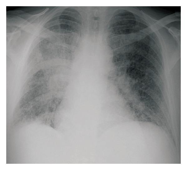
This diagnostic image is a posteroanterior (PA) chest radiograph demonstrating significant pulmonary pathology. The primary finding is a widespread, bilateral miliary pattern characterized by numerous, small (1–3 mm), well-defined nodular opacities distributed throughout both lung fields. There is a noticeable higher concentration of these nodules in the lower lung zones compared to the apices. Additionally, a large, dense, ill-defined focal opacity is visible in the right lower lobe, consistent with a progressive consolidation or mass-like lesion. The combination of a localized consolidated lesion and diffuse miliary dissemination in a patient with underlying immunosuppression (such as CLL) is highly suggestive of hematogenous spread of infection, like miliary tuberculosis or fungal diseases such as blastomycosis. The mediastinal shadow appears slightly widened, and the cardiac silhouette is within normal limits. This image serves as a classic educational example of miliary lung disease and rapid radiological progression of opportunistic pulmonary infections.
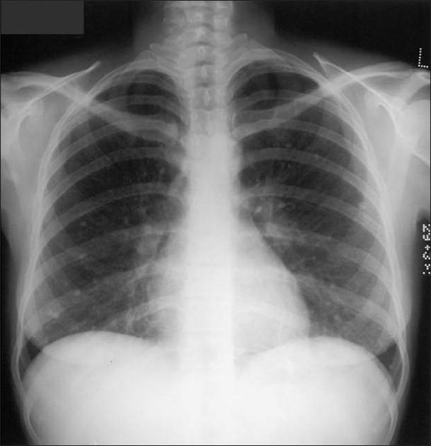
This diagnostic image is a posteroanterior (PA) chest radiograph of an adult, demonstrating multiple small, high-density calcific micronodules. These nodules, measuring approximately 1–3 mm in diameter, are diffusely and randomly distributed throughout both lung fields (bilateral involvement). The nodules exhibit a punctate morphology, characteristic of dystrophic calcification. In this clinical context, these findings represent late sequelae of healed varicella (chickenpox) pneumonia. The mediastinal contour is normal, with no evidence of hilar or mediastinal lymphadenopathy, which helps distinguish this from other granulomatous diseases like histoplasmosis or tuberculosis. The heart size is within normal limits, the pleura appears clear with no effusions, and the visualized bony structures of the thorax, including the ribs and clavicles, are intact. This image serves as a classic educational example of diffuse miliary-pattern calcification resulting from a prior viral pulmonary infection.
CDC disseminated miliary tuberculosis diagnosis treatment
Miliary tuberculosis
Who is at risk?
- Infants and young children
- HIV or other severe immunosuppression
- Diabetes, malnutrition, chronic kidney disease
- Anti-TNF therapy, steroids, transplant recipients
- Recent primary TB infection or untreated TB
Clinical features
- Persistent fever, night sweats, weight loss, anorexia, fatigue
- Cough, dyspnea, hypoxemia
- Hepatomegaly, splenomegaly, lymphadenopathy
- Pancytopenia or abnormal liver tests from marrow/liver involvement
- May involve meninges, eyes, bones, kidneys, adrenals, and other organs
Diagnosis
- Chest X-ray: diffuse, randomly distributed bilateral 1-3 mm nodules. This classic pattern may be absent early.
- CT chest: detects micronodules earlier and more sensitively.
- Obtain microbiologic confirmation wherever possible:
- Sputum or induced sputum for AFB smear, NAAT/Xpert, culture, and drug-susceptibility testing
- Mycobacterial blood culture in disseminated disease
- Tissue/fluid sampling from accessible affected sites, such as lymph node, liver, bone marrow, or pleural fluid
- Test for HIV and assess for organ involvement.
- Lumbar puncture and brain imaging are considered when there are neurologic symptoms/signs, altered mental status, severe headache, or concern for TB meningitis.
Treatment
- isoniazid
- rifampicin/rifampin
- pyrazinamide
- ethambutol
Complications and prognosis
- Acute respiratory distress syndrome
- Septic shock and multiorgan failure
- TB meningitis
- Adrenal insufficiency
- Disseminated intravascular coagulation
- High mortality, especially when diagnosis and treatment are delayed