
Venous ulcer case discussion surgery for mbbs graduate
Reading File
Now let me search the medical library for venous ulcer content in parallel:
Finding Sources
Finding Sources
Finding Sources
Excellent - found great sources. Now let me read the key sections in parallel:
Reading File
Reading File
Finding Sources
Excellent content found. Let me read more of the S Das section on venous ulcers and Bailey & Love pathophysiology/management sections in parallel:
Reading File
Reading File
Reading File
Now let me get more of the treatment section from Bailey & Love and S Das:
Reading File
Reading File
Let me get the treatment section from S Das and the Bailey & Love surgical management section:
Reading File
Reading File
Now let me get the investigations and treatment of venous ulcers from S Das and the Bailey & Love management section:
Reading File
Finding Sources
Reading File
Now let me fetch a key clinical image and get the S Das treatment section:
Loading Image
Finding Sources
I now have comprehensive content from multiple authoritative textbooks. Let me compile the full case discussion:
Venous Ulcer - Case Discussion for MBBS Surgery
MODEL CASE PRESENTATION
Patient: A 52-year-old female presents with a painless ulcer on the medial aspect of the lower left leg for 8 months. She has a history of multiple pregnancies, prolonged standing at work, and a previous episode of deep vein thrombosis (DVT) 5 years ago. The ulcer is not improving despite home dressings.
1. DEFINITION
A venous ulcer (also called varicose ulcer, gravitational ulcer, or post-thrombotic ulcer) is a chronic, non-healing ulcer of the lower limb caused by ambulatory venous hypertension in the lower third of the leg.
- S Das Manual of Clinical Surgery, 13th Ed. - It is the "abnormal venous hypertension in the lower third of the leg" that forms the basic cause.
2. ETIOLOGY AND PREDISPOSING FACTORS
Causes of venous hypertension:
- Superficial venous incompetence - primary varicose veins (sapheno-femoral or sapheno-popliteal junction incompetence)
- Perforator vein incompetence - especially the Cockett perforators (ankle perforators connecting posterior tibial vein to posterior arch vein)
- Deep venous obstruction/incompetence - post-thrombotic destruction of deep vein valves (most important)
Risk factors:
- Female sex (women > men markedly)
- Age 40-60 years
- Obesity
- Multiple pregnancies
- Prolonged standing
- Previous DVT / post-thrombotic syndrome
- Family history of varicose veins
3. PATHOPHYSIOLOGY
Ambulatory venous hypertension is the only accepted underlying cause.
- Normal mechanism: Calf muscle pump + competent valves lower venous pressure during walking
- Valve failure: Damaged valves (from DVT, primary incompetence) cause reversal of flow through incompetent perforators
- Venous hypertension in the gaiter region (ankle skin) results
- Pericapillary fibrin cuffs form (from protein leakage), acting as barriers to O2 and nutrient diffusion
- Leukocyte trapping - WBCs trapped in capillaries release proteolytic enzymes and reactive oxygen species
- Fibroblast senescence, growth factor inhibition, mast cell degranulation - all impair healing
- Haemosiderin deposition from RBC extravasation - key factor in ulcer development
- Ultimately: skin necrosis and ulceration
(Bailey & Love's Short Practice of Surgery, 28th Ed., p. 1061)
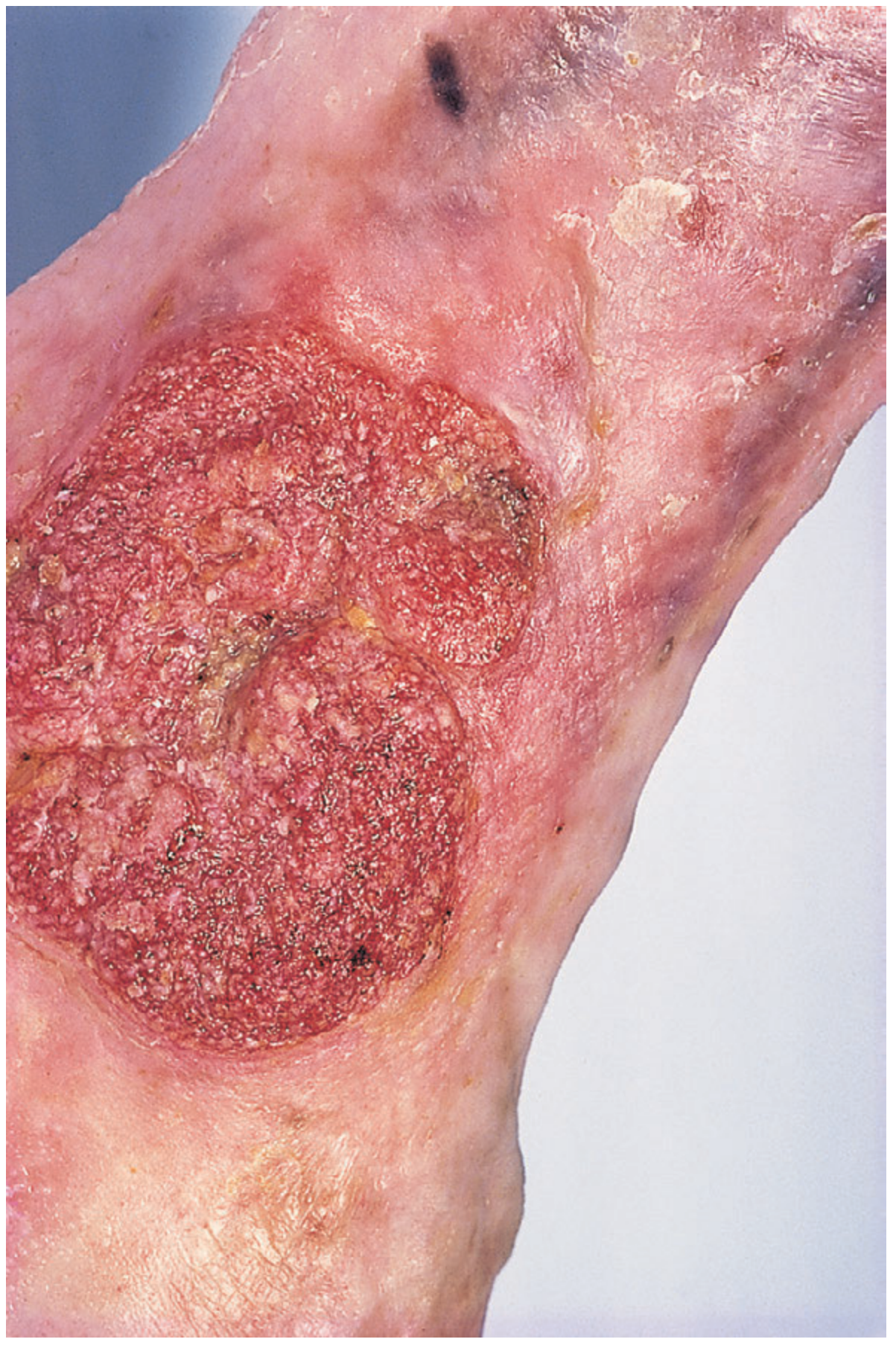
4. CLINICAL FEATURES
Site
- Lower third of the medial leg (gaiter area) - between calf muscles and ankle
- Never above the junction of the middle and upper thirds of the leg
- Majority on the medial side at the site of Cockett perforators
- Extension to foot or upper calf is uncommon and should raise suspicion of another diagnosis
Appearance of Ulcer
| Feature | Description |
|---|---|
| Edge | Sloping (shelving), pale purple-blue, thin blue margin of growing epithelium |
| Floor | Pale granulation tissue with variable slough; seropurulent discharge |
| Base | Shallow, flat; never penetrates the deep fascia; fixed to deeper structures |
| Shape | Any shape or size |
| Depth | Superficial (does not expose tendons) |
Surrounding Skin (Signs of Chronic Venous Hypertension)
- Haemosiderosis / hyperpigmentation - from haemosiderin (iron) and melanin deposits
- Lipodermatosclerosis (LDS) - fibrosis, thickening, and induration of subcutaneous tissue; gives "inverted champagne bottle" appearance
- Venous eczema - itchy, scaly skin
- Varicose veins - may or may not be present proximally
- Atrophie blanche - white scarred patches with telangiectasia
- Scars of previous ulcers
Symptoms
- Painless when chronic (key feature!)
- Initially painful when acute; pain settles as it becomes chronic
- Surrounding skin may be tender
- Aching, heaviness, swelling - worse at end of day, better on elevation
Regional lymph nodes
- Inguinal nodes enlarged only if the ulcer is infected
5. CEAP CLASSIFICATION
(Clinical-Etiology-Anatomy-Pathophysiology)
| Class | Description |
|---|---|
| C0 | No visible/palpable venous disease |
| C1 | Telangiectasia or reticular veins |
| C2 | Varicose veins |
| C3 | Oedema |
| C4a | Pigmentation or eczema |
| C4b | Lipodermatosclerosis or atrophie blanche |
| C5 | Healed venous ulcer |
| C6 | Active venous ulcer |
Modifiers: s = symptomatic, a = asymptomatic, r = recurrent.
6. DIFFERENTIAL DIAGNOSIS
| Ulcer Type | Key Differentiating Features |
|---|---|
| Venous | Medial gaiter area, painless when chronic, sloping edge, does NOT penetrate deep fascia, pigmentation, LDS |
| Arterial (ischaemic) | Painful, punched-out edge, deep (may expose tendons), pale/necrotic floor, absent pulses, intermittent claudication, toes discolored, any site |
| Neuropathic (diabetic) | Plantar surface over pressure points, painless due to neuropathy, "punched-out", peripheral neuropathy signs |
| Martorell's (hypertensive) | Posterior/lateral calf, severe pain, all foot pulses present, hypertensive patient, punched-out |
| Malignant (SCC / Marjolin's) | Raised/everted/rolled edge on a chronic venous ulcer, hard base, raised suspicious edge - biopsy mandatory |
| Bazin's (erythrocyanosis frigida) | Young women, cold-sensitive, multiple small ulcers, chilblains |
| Tropical ulcer | Infected, painful initially, undermined edges, Vincent's organisms |
(S Das Manual of Clinical Surgery, 13th Ed.)
7. INVESTIGATIONS
Mandatory
-
ABPI (Ankle-Brachial Pressure Index) - Doppler measurement
- ABPI >0.8: Pure venous ulcer - full compression safe
- ABPI 0.5-0.8: Mixed arterial-venous ulcer - modified compression (30 mmHg)
- ABPI <0.5 or ankle pressure <60 mmHg: Revascularization required BEFORE compression
- 15-30% of venous ulcers have concurrent arterial disease (mixed ulcer)
-
Duplex ultrasonography - Assessment of deep and superficial venous system
- Identifies reflux sites (saphenofemoral junction, saphenopopliteal junction, perforators)
- Confirms or excludes DVT
- Guides surgical planning
Ancillary (For Atypical/Non-Healing Ulcers)
- Full blood count (FBC) - anaemia (sickle cell), polycythaemia
- Blood glucose - diabetes
- ESR / CRP - inflammatory causes
- Sickle cell test (if appropriate ethnicity)
- Antibody screen - rheumatoid factor (rheumatoid ulcer)
- Biopsy - if edge is raised, everted, or ulcer is suspicious for malignancy (Marjolin's ulcer)
(Bailey & Love's, 28th Ed., p. 1062)
8. MANAGEMENT
Keystone Principle
Reduce ambulatory venous hypertension - through compression AND ablation of venous incompetence.
Best results come from specialist multidisciplinary ulcer services.
A. CONSERVATIVE (Non-operative)
1. Wound Care / Dressings
- Clean the ulcer, remove slough
- Non-adherent primary dressing
- Manage exudate
- Antibiotics only if cellulitis is present (NOT routinely - they do NOT speed healing without infection)
2. Leg Elevation
- Elevate the foot of the bed
- Reduces venous hypertension and oedema
3. Compression Therapy - THE MAINSTAY
Two best regimes (equally effective):
- Four-layer compression bandaging:
- Layer 1: Orthopaedic wool (distributes pressure, absorbs exudate)
- Layer 2: Cotton crepe (smooths wool)
- Layer 3: Elastic bandage (1/3 of interface pressure)
- Layer 4: Cohesive bandage (2/3 of interface pressure - stiffness)
- Target interface pressure: 35-40 mmHg
- Two-layer compression hosiery - equally effective, used when ABPI is adequate
4. Pharmacological Adjuncts
- Pentoxifylline - increases microvascular perfusion (reduces plasma viscosity + cytokine inhibition); useful adjunct to compression
- Horse chestnut seed extract (Aescin) - safe adjunct for venous hypertension; improves symptoms, reduces leg volume
- Aspirin (antiplatelet) - some evidence as adjunct
B. OPERATIVE / INTERVENTIONAL
Principle: Ablation of superficial venous incompetence accelerates healing and reduces recurrence - referral to vascular surgeon should NOT be delayed.
1. Superficial Venous Ablation/Surgery
- Endovenous Laser Ablation (EVLA) or Radiofrequency Ablation (RFA) - preferred (minimally invasive)
- High ligation + stripping of incompetent saphenous vein
- The ESCHAR trial showed: compression + surgery reduced ulcer recurrence significantly vs. compression alone (but did NOT accelerate primary healing in isolation)
- Early endovenous ablation nearly halves time to healing and is cost-effective (Bailey & Love, p. 1063)
2. Perforator Surgery
- SEPS (Subfascial Endoscopic Perforator Surgery) - minimally invasive perforator ligation
- Classical open Linton procedure (1938) abandoned due to high wound complications
- SEPS technique: Patient supine, leg elevated 45-60°, Esmarch bandage + thigh tourniquet applied, two small proximal medial incisions, laparoscopic trocars, CO2 insufflation of subfascial space, perforators doubly clipped and divided, compression bandage for 5 days post-op
- Ulcer healing in 88% at 1 year in North American registry (146 patients)
- Ulcer recurrence: 16% at 1 year, 28% at 2 years
- SEPS alone: controversial - no advantage over superficial venous surgery + compression alone in primary analysis
- Often combined with saphenous ablation (72% of cases)
(Schwartz's Principles of Surgery, 11th Ed.)
3. Skin Grafting (for Large/Non-Healing Ulcers)
- Pinch grafting - simple, outpatient procedure
- Split skin graft (SSG) / mesh graft - for larger ulcers after preparation of wound bed
- Results: Good early healing, moderate long-term results (50% healed at 5 years)
- Apligraf (bilayered living skin construct) - 63% healed at 6 months vs. 49% with compression alone; median healing 61 vs. 181 days; most benefit for large (>1000 mm²) or longstanding (>6 months) ulcers
4. Deep Venous Reconstruction
- For post-thrombotic disease with deep venous obstruction
- Options: venous bypass, valve reconstruction/transplantation
- Reserved for highly selected patients failing all other treatment
C. PREVENTION OF RECURRENCE
Once healed:
- Treat superficial venous incompetence if not already done
- Class 2 below-knee graduated compression stockings - worn for life
- Indicated for: residual reflux, deep venous occlusion, recurrent ulceration
- Even with successful ablation/stockings: 20-30% reulceration by 5 years
- Highest risk of reulceration: post-thrombotic limb
(Bailey & Love's Short Practice of Surgery, 28th Ed., p. 1063)
9. COMPLICATIONS OF VENOUS ULCER
- Infection / Cellulitis - inguinal lymphadenopathy
- Marjolin's Ulcer - squamous cell carcinoma arising from the edge of a long-standing venous ulcer
- Key features: raised, everted edge; hard base; painless; radioresistant (due to fibrosis)
- Lymphatic metastasis is unusual (lymphatics destroyed by chronic disease)
- Biopsy is mandatory whenever the edge changes character
- Contact dermatitis - from topical preparations
- Osteomyelitis - rare, from deep penetration
- Psychological morbidity and impaired quality of life
10. PROGNOSIS
- Nearly all venous ulcers can be healed with proper compression and venous treatment
- 20-30% recurrence rate by 5 years even in compliant patients
- Worst prognosis: post-thrombotic limbs
- Venous disease accounts for ~85% of all chronic lower limb ulcers in developed countries
- Community prevalence: 0.1-0.3% (2-4% in elderly)
- Dressings alone account for 1-3% of western healthcare expenditure
QUICK EXAM REVISION SUMMARY
| Point | Fact |
|---|---|
| Basic cause | Ambulatory venous hypertension |
| Site | Medial gaiter area (never above middle 1/3 of leg) |
| Edge | Sloping (shelving) |
| Floor | Pale granulation tissue |
| Pain | Painless when chronic |
| Depth | Never penetrates deep fascia |
| Surrounding skin | Pigmentation (haemosiderin), LDS, eczema |
| Investigation | ABPI + Duplex USS |
| Mainstay of treatment | Compression (35-40 mmHg) |
| Surgery | EVLA/RFA or high ligation + stripping |
| Key complication | Marjolin's ulcer (SCC) |
| Recurrence | 20-30% at 5 years |
| Life-long prevention | Class 2 compression stockings |
Sources:
- S Das - A Manual on Clinical Surgery, 13th Ed.
- Bailey and Love's Short Practice of Surgery, 28th Ed.
- Schwartz's Principles of Surgery, 11th Ed.
- Mulholland and Greenfield's Surgery, 7th Ed.
This is a shared conversation. Sign in to Orris to start your own chat.