
MBBS Essay OBG Endometrial Cancer
Endometrial Cancer
MBBS Essay - Obstetrics & Gynaecology
Definition
- Berek & Novak's Gynecology, p. 2141
- Robbins, Cotran & Kumar Pathologic Basis of Disease, p. 932
Epidemiology
- Predominantly a disease of postmenopausal women; peak incidence in the 6th-7th decade, average age at diagnosis ~60 years
- 75% of cases occur in women over 50 years of age
- Overall lifetime risk: approximately 2.8% of women
- Overall 5-year survival rate: approximately 75%
- Deaths from endometrial carcinoma rose ~2% per year from 2010 to 2014, reflecting an increasing burden
- Berek & Novak's Gynecology, p. 2141
Pathogenesis: Two Major Types
| Feature | Type I (Endometrioid) | Type II (Non-Endometrioid) |
|---|---|---|
| Frequency | 75-85% | 15-25% |
| Estrogen | Estrogen-dependent | Estrogen-independent |
| Background | Endometrial hyperplasia | Atrophic endometrium |
| Histology | Endometrioid adenocarcinoma | Serous, clear cell |
| Grade | Well-differentiated (low grade) | Poorly differentiated (high grade) |
| Prognosis | Favorable | Poor |
| Patient | Obese, younger, perimenopausal | Thin, older, postmenopausal |
| Racial predominance | Any | Disproportionate in African American, Asian women |
- Berek & Novak's Gynecology, p. 2141
Risk Factors
- Obesity - adipose tissue converts androgens to estrone (peripheral aromatization); major risk in Western populations
- Nulliparity
- Late menopause (after age 52)
- Unopposed exogenous estrogen therapy - risk increased 4 to 8 times; risk increases with duration and dose
- Tamoxifen use (weak estrogen agonist on endometrium)
- Polycystic ovarian syndrome (PCOS)
- Estrogen-secreting tumours (granulosa cell tumours)
- Type 2 diabetes mellitus, hypertension - commonly co-occur with obesity
- Lynch syndrome (Hereditary Non-Polyposis Colorectal Cancer - HNPCC) - mutations in DNA mismatch repair genes (MLH1, MSH2, MSH6, PMS2); lifetime endometrial cancer risk up to 40-60%
- Cowden syndrome (PTEN hamartoma tumour syndrome)
-
Combined oral contraceptive pill use (reduces risk by ~50%)
-
Progestogen therapy (opposes estrogenic stimulation)
-
Multiparity
-
Smoking (paradoxically reduces risk via anti-estrogenic effects - not recommended)
-
Physical activity
-
Berek & Novak's Gynecology, p. 2141; Robbins, p. 932
Molecular Pathogenesis
- Ultramutated / POLE tumours - Mutations in DNA polymerase epsilon (POLE); exceptionally high somatic mutation burden; paradoxically good prognosis
- Hypermutated / MSI (Microsatellite Instability) - Mutations or epigenetic silencing of DNA mismatch repair genes (MLH1 promoter hypermethylation most common); ~20% of sporadic tumours; associated with Lynch syndrome
- Copy number low / Non-specific molecular phenotype - Common subtype; associated with endometrioid morphology; multiple mutations upregulating PI3K/AKT pathway (PTEN, PIK3CA, KRAS, ARID1A)
- Copy number high / TP53 mutated - Aggressive tumours with serous or high-grade endometrioid morphology; TP53 mutations >90% of serous carcinomas; worst prognosis
-
PTEN - most frequently mutated gene (30-80%)
-
PIK3CA - activating mutations (~40%)
-
KRAS - (~25%)
-
ARID1A - loss of function (~33%)
-
TP53 - late event; found in ~50% of poorly differentiated tumours
-
Robbins, p. 932-933
Precursor Lesion: Endometrial Hyperplasia
- Hyperplasia without atypia - Low risk of malignant progression (<5%)
- Atypical hyperplasia (Endometrial Intraepithelial Neoplasia - EIN) - High risk; ~30% progress to carcinoma if untreated; some cases of "atypical hyperplasia" are already carcinoma on hysterectomy specimen
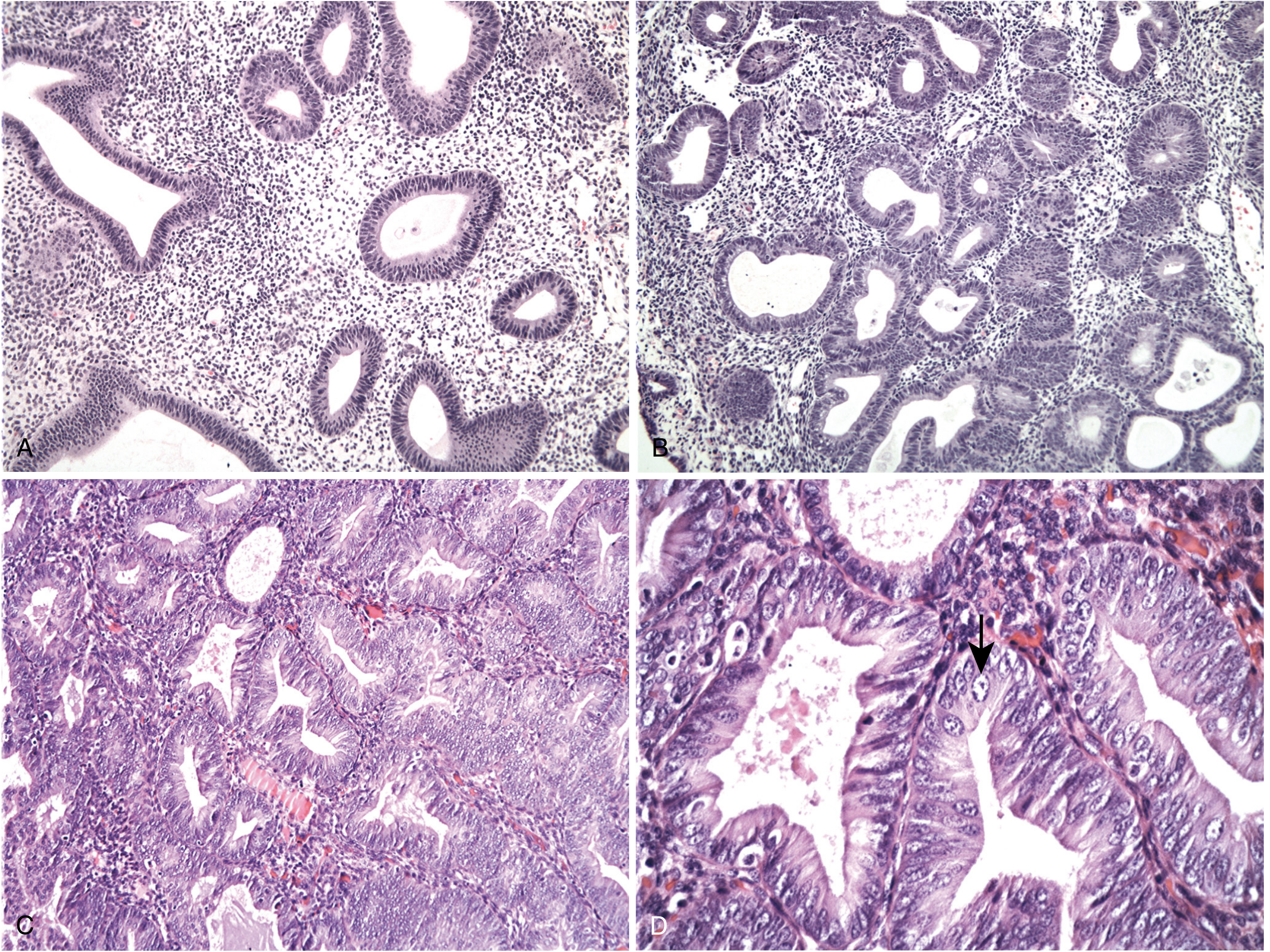
Pathology / Histological Types
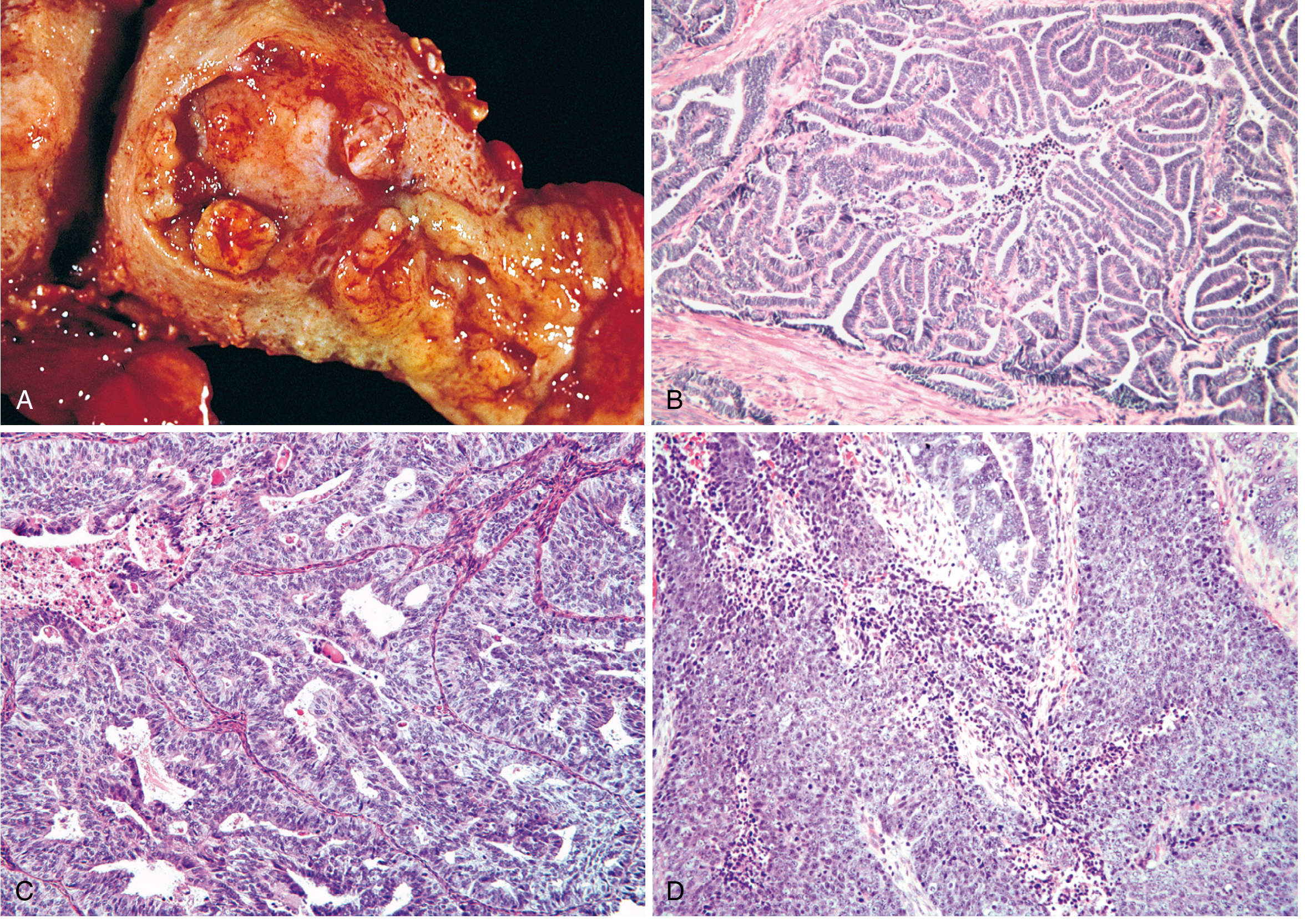
1. Endometrioid Adenocarcinoma (Most Common - 80-85%)
- Arises from endometrial hyperplasia
- Mimics proliferative endometrial glands (hence "endometrioid")
- Histologic grading (FIGO):
- Grade 1 (well-differentiated): <5% non-squamous solid growth
- Grade 2 (moderately differentiated): 6-50% solid areas
- Grade 3 (poorly differentiated): >50% solid growth pattern
- May contain foci of squamous differentiation (up to 20% of cases); graded on glandular component alone
2. Serous Carcinoma (Uterine Papillary Serous Carcinoma - UPSC) (~15%)
- Type II tumour; arises on a background of endometrial atrophy in older women
- Highly aggressive; fibrovascular papillary stalks lined by highly atypical cells with tufting
- Psammoma bodies frequently present
- TP53 mutations >90%
- Despite being <10% of endometrial cancers, accounts for >50% of all endometrial cancer deaths
- Has propensity for intraperitoneal spread even without myometrial invasion (simulates ovarian carcinoma)
- Berek & Novak's Gynecology, p. 2158
3. Clear Cell Carcinoma
- Type II; aggressive tumour; poor prognosis
- Clear cells (glycogen-rich cytoplasm) and hobnail cells
- Also arises in older, postmenopausal women without hyperestrogenism
4. Mucinous Carcinoma
- Rare; most are low-grade and well-differentiated
- Must be distinguished from endocervical carcinoma
5. Mixed Carcinomas
- Combinations of histologic types (especially endometrioid + serous); behave as aggressively as the higher-grade component
Clinical Features
Symptoms
- Postmenopausal bleeding - cardinal symptom; present in ~90% of patients
- Any postmenopausal bleeding must be investigated until cancer is excluded
- Abnormal perimenopausal bleeding - irregular, heavy
- Pelvic pressure or pain - indicates uterine enlargement or extrauterine spread
- Purulent vaginal discharge - if cervical stenosis causes hematometra/pyometra (poor prognostic sign)
- <5% are asymptomatic (detected incidentally on imaging or Pap smear)
Signs
- Uterine enlargement on bimanual palpation
- In advanced disease: adnexal mass, ascites, inguinal lymphadenopathy
- Pap smear may show malignant cells in ~50% (not a screening test)
Differential Diagnosis of Postmenopausal Bleeding
- Endometrial atrophy (most common, 60-80%)
- Endometrial polyps (2-12%)
- Exogenous estrogen therapy
- Endometrial hyperplasia
- Endometrial carcinoma
- Cervical/vaginal/vulval pathology
- Atrophic vaginitis (up to 15%)
Investigations
Step 1 - Endometrial Biopsy (Gold Standard)
- Office endometrial aspiration biopsy (Pipelle biopsy) - first-line, accepted standard
- Sensitivity for endometrial cancer: ~90-95%
- If inadequate sample or high clinical suspicion: proceed to D&C ± hysteroscopy
Step 2 - Transvaginal Ultrasound (TVS)
- Endometrial thickness:
- >4-5 mm in postmenopausal women is threshold for further investigation
- Atrophic endometrium (<4 mm) makes cancer very unlikely
- A thin endometrial stripe essentially excludes malignancy
Step 3 - Hysteroscopy + Directed Biopsy
- Direct visualization of endometrial cavity
- Best for detecting polyps and focal lesions missed by blind biopsy
- Considered gold standard for diagnosis
Further Workup (Pre-treatment):
- Complete Blood Count, LFT, RFT, blood glucose
- CA-125 (elevated in advanced disease, serous type)
- Chest X-ray, CT chest/abdomen/pelvis - for staging, lymph node assessment, metastases
- MRI pelvis - best modality for assessing depth of myometrial invasion and cervical involvement
- Cystoscopy / Proctoscopy - if bladder/bowel involvement suspected (Stage IVA)
Staging
| Stage | Description |
|---|---|
| Stage I | Confined to the uterine body |
| IA | Tumour invades <50% of myometrium |
| IB | Tumour invades ≥50% of myometrium |
| Stage II | Tumour invades cervical stroma, but not beyond uterus |
| Stage III | Local/regional spread |
| IIIA | Tumour invades uterine serosa and/or adnexa |
| IIIB | Vaginal and/or parametrial involvement |
| IIIC1 | Pelvic lymph node metastasis |
| IIIC2 | Para-aortic lymph node metastasis |
| Stage IV | Tumour invades bladder/bowel mucosa, or distant metastases |
| IVA | Bladder/bowel mucosal invasion |
| IVB | Distant metastases (lungs, liver, bone, inguinal lymph nodes) |
- Berek & Novak's Gynecology, p. 2141
Prognostic Factors
- Advanced age
- Non-endometrioid histology (serous, clear cell) or Grade 3 histology
- Deep myometrial invasion (≥50%) - Stage IB and beyond
- Lymph-vascular space invasion (LVSI)
- Large tumour size (>2 cm)
- Cervical extension (Stage II)
- Lymph node metastasis (Stage IIIC)
- Intraperitoneal spread
- Positive peritoneal cytology
- Hormone receptor negativity (ER/PR negative tumours behave more aggressively)
- Abnormal DNA ploidy (aneuploidy)
- Berek & Novak's Gynecology, p. 2142
Treatment
Surgical Treatment (Primary)
- Total Hysterectomy + Bilateral Salpingo-Oophorectomy (TAH+BSO)
- Peritoneal washings/cytology
- Lymph node assessment - pelvic and para-aortic lymph node dissection in most patients
- May be omitted in low-risk patients (Grade 1-2, Stage IA, no LVSI)
- Omentectomy and peritoneal biopsies for serous/clear cell histology
- Laparoscopic/robotic-assisted preferred over open laparotomy (comparable oncologic outcomes, faster recovery, lower morbidity)
- Open laparotomy for bulky disease or advanced stages
- High-dose progestin therapy (medroxyprogesterone acetate / megestrol acetate)
- Close surveillance with serial biopsies
- Hysterectomy recommended after childbearing is complete
Radiotherapy
- Vaginal brachytherapy - for Stage I intermediate/high-intermediate risk; reduces vaginal vault recurrence (up to 80% salvage for isolated vaginal recurrence)
- External Beam Pelvic Radiotherapy (EBRT) - Stage IB high grade, Stage II; decreases pelvic recurrence
- Extended-field radiation (para-aortic) - when para-aortic nodes involved
- Whole-abdomen radiation - occasionally for peritoneal spread (largely replaced by chemotherapy)
- Radiotherapy as primary treatment is reserved for medically unfit patients who cannot undergo surgery
Chemotherapy
- Indicated for high-risk disease (serous, clear cell, Grade 3, advanced stage)
- Standard regimen: Carboplatin + Paclitaxel (preferred over older cisplatin-doxorubicin-paclitaxel due to similar efficacy with less toxicity)
- For Stage III/IV: combined modality approach (chemotherapy + radiation)
- "Sandwich" chemotherapy-radiation-chemotherapy protocol shows improved survival in some studies
Hormonal Therapy
- Progestins (medroxyprogesterone acetate, megestrol acetate) - for recurrent/metastatic disease with ER/PR-positive tumours
- Response rates ~15-25% in recurrent disease
- Also used for fertility preservation
Treatment by Stage:
| Stage | Primary Treatment | Adjuvant |
|---|---|---|
| IA, Grade 1-2 | TAH+BSO | Observation ± vaginal brachytherapy |
| IA, Grade 3 / IB | TAH+BSO + lymph nodes | Vaginal brachytherapy ± EBRT |
| Stage II | TAH+BSO + lymph nodes | EBRT + vaginal brachytherapy |
| Stage III | TAH+BSO + debulking | Chemotherapy + radiation |
| Stage IV | Debulking surgery | Chemotherapy; palliative care |
Pattern of Spread
- Direct extension - myometrium → cervix → vagina → parametria → bladder, rectum
- Lymphatic - to pelvic (obturator, external iliac) and para-aortic lymph nodes
- Transtubal / Peritoneal - through fallopian tubes to peritoneal cavity (especially serous type)
- Haematogenous - lungs, liver, bone (late stage)
Recurrent Disease
- Vaginal recurrence most common (vault)
- Managed by: radiotherapy (if not previously irradiated), surgery, chemotherapy, hormonal therapy
- Isolated vaginal recurrences: salvage in up to 80% of patients with radiotherapy
- Berek & Novak's Gynecology, p. 2142
Uterine Sarcomas (Brief Mention)
| Type | Origin | Notes |
|---|---|---|
| Leiomyosarcoma | Smooth muscle | Most common uterine sarcoma; arises de novo, not from fibroids |
| Endometrial Stromal Sarcoma | Endometrial stroma | Low-grade; ER/PR positive; responds to hormonal therapy |
| Adenosarcoma | Mixed Mullerian | Low-grade malignant stroma + benign glands |
| Carcinosarcoma (MMMT) | Mixed | Highly aggressive; now classified as metaplastic carcinoma |
- Berek & Novak's Gynecology, p. 2142
Summary of Key Points
| Point | Detail |
|---|---|
| Most common | Malignancy of female genital tract |
| Cardinal symptom | Postmenopausal bleeding |
| Main risk factor | Unopposed estrogen stimulation |
| Commonest type | Endometrioid adenocarcinoma (80-85%) |
| Deadliest type | Serous carcinoma (<10% of cases, >50% of deaths) |
| Precursor | Atypical endometrial hyperplasia |
| Hereditary risk | Lynch syndrome (HNPCC) |
| Diagnosis | Endometrial biopsy (Pipelle) |
| Imaging | TVS (screen), MRI pelvis (staging) |
| Staging | Surgical (FIGO 2009) |
| Treatment | TAH+BSO ± lymph node dissection |
| Adjuvant | Radiotherapy ± chemotherapy |
| Overall 5-yr survival | ~75% |
- Berek & Novak's Gynecology, pp. 2141-2165
- Robbins, Cotran & Kumar: Pathologic Basis of Disease, pp. 932-934
- Goldman-Cecil Medicine (International Edition)
Pupillary light reflex pathway
Pupillary Light Reflex Pathway
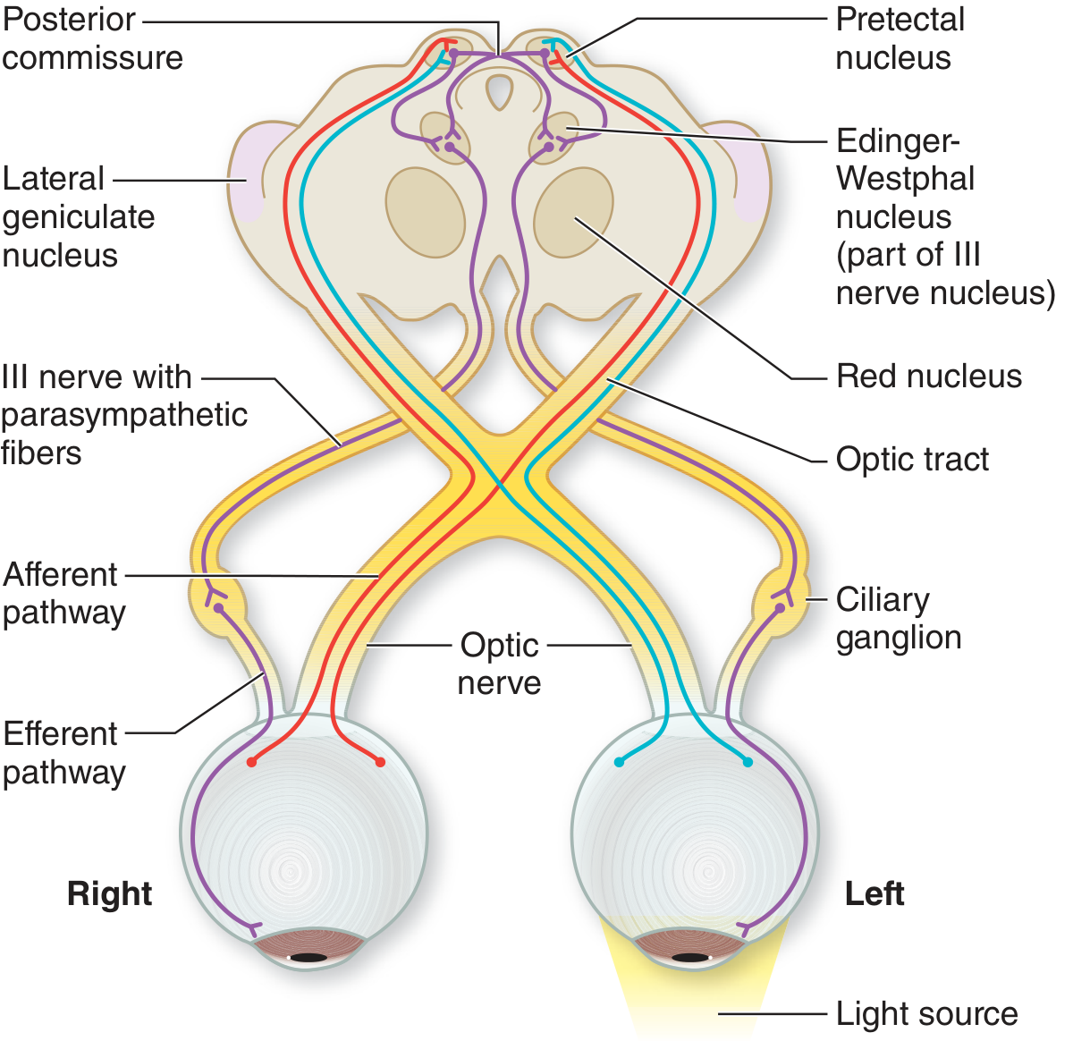
Pathway Diagram
Afferent Limb (Sensory - Optic Nerve / CN II)
Neuron 1 - Photoreceptors (Retina)
- Light stimulates rods and cones in the outer retina
- Additionally, a special population of intrinsically photosensitive retinal ganglion cells (ipRGCs) containing the photopigment melanopsin directly detect ambient light intensity
- These ipRGCs are the primary drivers of the pupillary reflex - they are irradiance detectors, not image-forming cells
- This is why pupillary reflexes are preserved even in patients with rod and cone degeneration (functionally blind patients)
Neuron 2 - Bipolar Cells (Retina)
- Signals pass from photoreceptors through bipolar cells (inner nuclear layer of retina)
Neuron 3 - Retinal Ganglion Cells → Optic Nerve → Optic Chiasm → Optic Tract
- Retinal ganglion cell axons form the optic nerve (CN II)
- At the optic chiasm: nasal fibers decussate (cross) and temporal fibers remain ipsilateral - this produces partial decussation (~53% crossed, 47% uncrossed)
- Fibers continue in the optic tract toward the midbrain
The Critical Divergence - Before the Lateral Geniculate Body
Key point: The light reflex fibers leave the optic tract just rostral (anterior) to the lateral geniculate body (LGB) and do NOT synapse there. They pass into the brachium of the superior colliculus to reach the midbrain pretectal area.This explains why lesions of the visual cortex or optic radiations (posterior to LGB) cause cortical blindness but preserve the pupillary light reflex - the reflex arc bypasses the geniculate-cortical pathway entirely.
Synapse 1 - Pretectal Nucleus (Olivary Pretectal Nucleus), Dorsal Midbrain
- Light reflex fibers synapse in the olivary pretectal nucleus at the level of the superior colliculus in the dorsal midbrain (pretectal area)
- This nucleus receives input from both eyes (due to chiasmal decussation)
Internuncial (Connecting) Neuron - Bilateral Distribution
- Axons from each pretectal nucleus pass to both Edinger-Westphal nuclei:
- Directly to the ipsilateral Edinger-Westphal nucleus
- Crossing via the posterior commissure to the contralateral Edinger-Westphal nucleus
- This bilateral projection is the anatomical basis for the consensual reflex - light in one eye produces constriction of both pupils
- Lesions of the posterior commissure (e.g., pinealoma) disrupt both light reflex pathways → fixed, slightly enlarged pupils (5-6 mm)
Efferent Limb (Motor - Oculomotor Nerve / CN III)
Neuron 4 (Preganglionic) - Edinger-Westphal Nucleus → CN III
- The Edinger-Westphal nucleus is the accessory parasympathetic nucleus of CN III; lies just dorsal to the main oculomotor nucleus, close to the midline, in the midbrain tegmentum at the level of the superior colliculus
- Preganglionic parasympathetic fibers travel in the oculomotor nerve (CN III) - they run on the superficial/outer surface of the nerve (dorsomedial aspect)
- They enter the inferior division of CN III and travel via the nerve to the inferior oblique muscle to reach the ciliary ganglion
Clinical note: Because pupilloconstrictor fibers run superficially in CN III, they are vulnerable to compression (e.g., posterior communicating artery aneurysm, uncal herniation) before the somatic motor fibers are affected. A dilated, unreactive pupil with CN III palsy strongly suggests compressive rather than ischaemic cause.
Synapse 2 - Ciliary Ganglion (Orbit)
- The ciliary ganglion lies within the muscle cone, just behind the globe, in the posterior orbit
- Preganglionic fibers synapse here on postganglionic parasympathetic neurons
- Note: Other fibers (sympathetic, sensory) pass through the ciliary ganglion without synapsing - only parasympathetic fibers synapse here
Neuron 5 (Postganglionic) - Short Ciliary Nerves → Sphincter Pupillae
- Postganglionic fibers leave the ciliary ganglion as short ciliary nerves (6-10 in number)
- These innervate the sphincter pupillae (circular smooth muscle of the iris)
- Sphincter pupillae contraction → miosis (pupil constriction)
Summary Flowchart
LIGHT stimulus (Left eye used as example)
         ↓
Retina (rods/cones + ipRGCs/melanopsin)
         ↓
Bipolar cells → Retinal ganglion cells
         ↓
Left Optic Nerve (CN II)
         ↓
Optic Chiasm (nasal fibers cross → bilateral representation)
         ↓
Both Optic Tracts
         ↓
Fibers LEAVE optic tract BEFORE lateral geniculate body
         ↓
         ↓ (via brachium of superior colliculus)
         ↓
Bilateral Pretectal Nuclei (olivary pretectal nucleus, dorsal midbrain)
         ↓
Internuncial neurons → BOTH Edinger-Westphal nuclei
(direct ipsilateral + via POSTERIOR COMMISSURE to contralateral)
         ↓                              ↓
Left Edinger-Westphal nucleus    Right Edinger-Westphal nucleus
         ↓                              ↓
Left CN III (inferior division)  Right CN III (inferior division)
(preganglionic parasympathetic)
         ↓                              ↓
Left Ciliary Ganglion            Right Ciliary Ganglion
         ↓                              ↓
Left Short Ciliary Nerves        Right Short Ciliary Nerves
         ↓                              ↓
Left Sphincter Pupillae          Right Sphincter Pupillae
         ↓                              ↓
LEFT PUPIL CONSTRICTS             RIGHT PUPIL CONSTRICTS
(Direct reflex)                   (Consensual reflex)
Direct vs Consensual Reflex Explained
| Direct Reflex | Consensual Reflex | |
|---|---|---|
| Definition | Pupil constriction in the illuminated eye | Pupil constriction in the opposite (non-illuminated) eye |
| Why it occurs | Ipsilateral efferent pathway | Contralateral distribution via posterior commissure |
| Clinical use | Tests both afferent AND efferent limb | Tests afferent of one eye + efferent of the other |
Key Anatomical Points (Exam Favourites)
| Point | Detail |
|---|---|
| Afferent nerve | CN II (Optic nerve) |
| Efferent nerve | CN III (Oculomotor nerve) |
| Reflex centre | Pretectal nucleus (olivary pretectal nucleus), dorsal midbrain |
| Parasympathetic nucleus | Edinger-Westphal nucleus (part of CN III nuclear complex) |
| Why consensual? | Internuncial neurons cross via posterior commissure to both EW nuclei |
| Critical divergence point | Fibers leave optic tract BEFORE LGB → bypass visual cortex |
| Ganglion in orbit | Ciliary ganglion (only parasympathetic synapse here) |
| Final effector | Sphincter pupillae muscle |
| Pupilloconstrictor fiber position in CN III | Superficial/dorsomedial - vulnerable to compression |
Clinical Correlations
1. Relative Afferent Pupillary Defect (RAPD) / Marcus Gunn Pupil
- Caused by optic nerve lesion (unilateral or asymmetric)
- Swinging flashlight test: when light swings to the affected eye, both pupils paradoxically dilate (weak afferent signal from diseased eye)
- Indicates lesion in: retina, optic nerve, optic chiasm, optic tract, or pretectal nucleus
2. Absolute Afferent Pupillary Defect (Amaurotic Pupil)
- Complete optic nerve lesion
- Affected eye is blind (no light perception)
- Light in affected eye → neither pupil reacts
- Light in normal eye → both pupils react normally
3. CN III Palsy (Efferent Defect)
- Pupil is dilated and unreactive (mydriasis) - loss of parasympathetic constriction
- Both direct AND consensual reflex lost in the affected eye
- Consensual reflex still intact in the other eye (afferent intact)
- Associated with ptosis and "down and out" gaze (involvement of somatic motor fibers)
4. Horner's Syndrome (Sympathetic Pathway Disruption)
- NOT a PLR pathway defect (sympathetic pathway is separate)
- Ipsilateral miosis (small pupil), ptosis, anhidrosis
- Pupil still constricts to light, but dilates poorly in dark
5. Light-Near Dissociation (Argyll Robertson Pupil)
- Pupil constricts to near (accommodation-convergence) but not to light
- Classic cause: neurosyphilis
- Lesion in the dorsal internuncial neurons connecting pretectal nucleus to Edinger-Westphal nucleus
- Near reflex centre is more ventral in midbrain, spared by dorsal lesions
6. Pinealoma / Parinaud's Syndrome
- Compresses the posterior commissure and pretectal area
- Results in bilateral fixed pupils (internuncial neurons disrupted)
- Also causes: upgaze palsy, convergence-retraction nystagmus
7. Midbrain / Pontine Lesions (Coma)
- Midbrain lesion: Fixed, mid-position (5-6 mm) pupils - bilateral EW nucleus damage
- Pontine lesion: Pinpoint reactive pupils - loss of descending sympathetic inhibition, parasympathetic preserved
- Metabolic coma: Pupils usually small but reactive (last sign to go)
What about you
- What I do well - Medical and clinical questions (I have access to authoritative textbooks like Robbins, Berek & Novak, Adams & Victor, Kanski, and more), anatomy, pharmacology, pathology, surgery, and general knowledge across most domains.
- How I work - I pull answers from real textbook sources rather than just generating text from memory. You get cited, accurate content with actual page references and images where available.
- What I'm not - I'm not a replacement for a doctor or clinical judgment. I'm a study and research tool.
- Essay-style answers for exams
- Pathway and mechanism explanations
- Pathology with histology images
- Clinical correlations and applied anatomy
Generate a Word document on pupillary light reflex for revision
mkdir -p /home/daytona/workspace/pupillary-reflex && ls /home/daytona/workspace/pupillary-reflex
~/pupillary-reflex/generate.js
const {
  Document, Packer, Paragraph, TextRun, Table, TableRow, TableCell,
  HeadingLevel, AlignmentType, BorderStyle, WidthType, ShadingType,
  VerticalAlign, PageNumber, Header, Footer, TableOfContents,
  UnderlineType, PageBreak
} = require('docx');
const fs = require('fs');

// ─── COLOUR PALETTE ───────────────────────────────────────────────
const BLUE        = "1F3864";   // dark navy heading
const LIGHT_BLUE  = "2E75B6";   // medium blue subheading
const ACCENT      = "1F4E79";   // deep blue for table header
const TABLE_HDR   = "2E75B6";   // table header fill
const TABLE_ALT   = "DEEAF1";   // alternate row
const TABLE_WHITE = "FFFFFF";
const KEY_FILL    = "FFF2CC";   // yellow highlight box
const WARN_FILL   = "FCE4D6";   // orange highlight box
const GREEN_FILL  = "E2EFDA";   // green highlight box

// ─── HELPERS ──────────────────────────────────────────────────────
function heading1(text) {
  return new Paragraph({
    heading: HeadingLevel.HEADING_1,
    spacing: { before: 360, after: 120 },
    children: [new TextRun({ text, bold: true, color: BLUE, size: 32 })]
  });
}

function heading2(text) {
  return new Paragraph({
    heading: HeadingLevel.HEADING_2,
    spacing: { before: 280, after: 80 },
    children: [new TextRun({ text, bold: true, color: LIGHT_BLUE, size: 26 })]
  });
}

function heading3(text) {
  return new Paragraph({
    heading: HeadingLevel.HEADING_3,
    spacing: { before: 200, after: 60 },
    children: [new TextRun({ text, bold: true, color: ACCENT, size: 24 })]
  });
}

function body(text, options = {}) {
  return new Paragraph({
    spacing: { before: 60, after: 60, line: 320 },
    children: [new TextRun({ text, size: 22, ...options })]
  });
}

function bold(text) {
  return new Paragraph({
    spacing: { before: 60, after: 60, line: 320 },
    children: [new TextRun({ text, bold: true, size: 22 })]
  });
}

function bullet(text, level = 0) {
  const indent = level === 0 ? {} : { indent: { left: 720 * (level + 1) } };
  return new Paragraph({
    bullet: { level },
    spacing: { before: 40, after: 40, line: 300 },
    ...indent,
    children: [new TextRun({ text, size: 22 })]
  });
}

function bulletMixed(runs) {
  return new Paragraph({
    bullet: { level: 0 },
    spacing: { before: 40, after: 40, line: 300 },
    children: runs
  });
}

function spacer(n = 1) {
  return Array.from({ length: n }, () =>
    new Paragraph({ children: [new TextRun({ text: '' })], spacing: { before: 0, after: 0 } })
  );
}

function divider() {
  return new Paragraph({
    border: { bottom: { style: BorderStyle.SINGLE, size: 6, color: LIGHT_BLUE } },
    spacing: { before: 120, after: 120 },
    children: [new TextRun('')]
  });
}

function highlightBox(lines, fillColor = KEY_FILL, borderColor = "FFC000") {
  return new Table({
    width: { size: 100, type: WidthType.PERCENTAGE },
    borders: {
      top: { style: BorderStyle.SINGLE, size: 8, color: borderColor },
      bottom: { style: BorderStyle.SINGLE, size: 8, color: borderColor },
      left: { style: BorderStyle.SINGLE, size: 8, color: borderColor },
      right: { style: BorderStyle.SINGLE, size: 8, color: borderColor },
    },
    rows: [new TableRow({
      children: [new TableCell({
        shading: { fill: fillColor, type: ShadingType.CLEAR },
        margins: { top: 100, bottom: 100, left: 200, right: 200 },
        children: lines.map(l => new Paragraph({
          spacing: { before: 40, after: 40, line: 300 },
          children: typeof l === 'string'
            ? [new TextRun({ text: l, size: 21 })]
            : l
        }))
      })]
    })]
  });
}

function tableRow(cells, isHeader = false, fillColor = TABLE_WHITE) {
  return new TableRow({
    tableHeader: isHeader,
    children: cells.map((cell, i) => new TableCell({
      shading: { fill: isHeader ? TABLE_HDR : fillColor, type: ShadingType.CLEAR },
      verticalAlign: VerticalAlign.CENTER,
      margins: { top: 80, bottom: 80, left: 120, right: 120 },
      children: [new Paragraph({
        alignment: AlignmentType.LEFT,
        spacing: { before: 0, after: 0 },
        children: [new TextRun({
          text: cell,
          bold: isHeader,
          color: isHeader ? "FFFFFF" : "000000",
          size: 20
        })]
      })]
    }))
  });
}

function makeTable(headers, rows) {
  return new Table({
    width: { size: 100, type: WidthType.PERCENTAGE },
    rows: [
      tableRow(headers, true),
      ...rows.map((r, i) => tableRow(r, false, i % 2 === 0 ? TABLE_WHITE : TABLE_ALT))
    ]
  });
}

// ─── TITLE PAGE ───────────────────────────────────────────────────
const titleSection = [
  ...spacer(4),
  new Paragraph({
    alignment: AlignmentType.CENTER,
    spacing: { before: 0, after: 200 },
    children: [new TextRun({ text: "PUPILLARY LIGHT REFLEX", bold: true, color: BLUE, size: 56 })]
  }),
  new Paragraph({
    alignment: AlignmentType.CENTER,
    spacing: { before: 0, after: 120 },
    children: [new TextRun({ text: "Complete Revision Notes", color: LIGHT_BLUE, size: 32, italics: true })]
  }),
  new Paragraph({
    alignment: AlignmentType.CENTER,
    spacing: { before: 0, after: 80 },
    children: [new TextRun({ text: "Anatomy  |  Physiology  |  Clinical Correlations", color: "555555", size: 24 })]
  }),
  new Paragraph({
    alignment: AlignmentType.CENTER,
    spacing: { before: 0, after: 80 },
    children: [new TextRun({ text: "MBBS  –  Ophthalmology / Neuroanatomy", color: "555555", size: 22 })]
  }),
  divider(),
  new Paragraph({
    alignment: AlignmentType.CENTER,
    spacing: { before: 80, after: 40 },
    children: [new TextRun({ text: "Sources: Adams & Victor's Principles of Neurology  |  Kanski's Clinical Ophthalmology", color: "888888", size: 18, italics: true })]
  }),
  new Paragraph({
    alignment: AlignmentType.CENTER,
    spacing: { before: 0, after: 40 },
    children: [new TextRun({ text: "Localization in Clinical Neurology  |  Plum & Posner's Stupor & Coma  |  Neuroscience: Exploring the Brain", color: "888888", size: 18, italics: true })]
  }),
  new Paragraph({ children: [new PageBreak()] })
];

// ─── SECTION 1: DEFINITION & OVERVIEW ─────────────────────────────
const sec1 = [
  heading1("1.  Definition & Overview"),
  divider(),
  body("The pupillary light reflex (PLR) is the constriction of the pupil in response to light stimulation of the retina. It is a brain stem reflex that does not involve the cerebral cortex."),
  ...spacer(1),
  body("Key characteristics:"),
  bullet("Consensual: light in ONE eye causes constriction of BOTH pupils"),
  bullet("Mediated by CN II (afferent) and CN III (efferent)"),
  bullet("Reflex centre: Pretectal nucleus (olivary pretectal nucleus), dorsal midbrain"),
  bullet("Bypasses the lateral geniculate body and visual cortex"),
  bullet("Most resistant of all nervous system responses to metabolic insult"),
  ...spacer(1),
  highlightBox([
    "★  KEY EXAM POINT",
    "The afferent fibres leave the optic tract BEFORE the lateral geniculate body.",
    "This is why cortical blindness (damage to visual cortex / optic radiations) PRESERVES the pupillary light reflex.",
    "The reflex arc is: Retina → CN II → Pretectal nucleus → Edinger-Westphal nucleus → CN III → Ciliary ganglion → Short ciliary nerves → Sphincter pupillae"
  ], KEY_FILL, "FFC000"),
  new Paragraph({ children: [new PageBreak()] })
];

// ─── SECTION 2: COMPLETE PATHWAY ──────────────────────────────────
const sec2 = [
  heading1("2.  Complete Pathway – Step by Step"),
  divider(),

  heading2("2.1  Afferent Limb (Sensory – CN II)"),

  heading3("Step 1 – Retinal Photoreceptors"),
  bullet("Light stimulates rods and cones in the outer retina"),
  bullet("Intrinsically photosensitive Retinal Ganglion Cells (ipRGCs) containing melanopsin also directly detect ambient light intensity"),
  bullet("ipRGCs are irradiance detectors – NOT image-forming cells"),
  bulletMixed([
    new TextRun({ text: "Clinical significance: ", bold: true, size: 22 }),
    new TextRun({ text: "Pupillary light reflexes are preserved even in patients with rod and cone degeneration (functionally blind), because ipRGCs are intact", size: 22 })
  ]),

  heading3("Step 2 – Bipolar Cells (Inner Nuclear Layer of Retina)"),
  bullet("Signals pass from photoreceptors through bipolar cells"),

  heading3("Step 3 – Retinal Ganglion Cells → Optic Nerve → Optic Chiasm → Optic Tract"),
  bullet("Retinal ganglion cell axons converge to form the optic nerve (CN II)"),
  bullet("At the optic chiasm: nasal fibres decussate (cross), temporal fibres remain ipsilateral (~53% crossed, 47% uncrossed)"),
  bullet("Fibres continue in the optic tract toward the midbrain"),

  ...spacer(1),
  highlightBox([
    "★  CRITICAL DIVERGENCE POINT",
    "Pupillary light reflex fibres LEAVE the optic tract just ROSTRAL (anterior) to the lateral geniculate body (LGB).",
    "They do NOT synapse in the LGB.",
    "They pass via the brachium of the superior colliculus to reach the pretectal nucleus.",
    "Result: Lesions of the visual cortex or optic radiations → cortical blindness but INTACT pupillary light reflex."
  ], KEY_FILL, "FFC000"),
  ...spacer(1),

  heading3("Step 4 – Pretectal Nucleus (Olivary Pretectal Nucleus), Dorsal Midbrain"),
  bullet("Pupillary fibres synapse in the olivary pretectal nucleus at the level of the superior colliculus"),
  bullet("This nucleus receives bilateral input (due to partial chiasmal decussation)"),

  heading2("2.2  Internuncial Neurons – Bilateral Distribution"),
  bullet("Axons from each pretectal nucleus pass to BOTH Edinger-Westphal (EW) nuclei:"),
  bullet("(a)  Directly to the ipsilateral EW nucleus", 1),
  bullet("(b)  Crossing via the posterior commissure to the contralateral EW nucleus", 1),
  ...spacer(1),
  highlightBox([
    "This bilateral distribution is the anatomical basis for the CONSENSUAL reflex.",
    "Lesion of the posterior commissure (e.g., pinealoma) → BILATERAL fixed, slightly large pupils (5–6 mm)."
  ], WARN_FILL, "FF0000"),
  ...spacer(1),

  heading2("2.3  Efferent Limb (Motor – CN III) – Two-Neuron Parasympathetic Pathway"),

  heading3("Neuron 1 (Preganglionic): Edinger-Westphal Nucleus → CN III"),
  bullet("Edinger-Westphal (EW) nucleus = accessory parasympathetic nucleus of CN III"),
  bullet("Located just DORSAL to the main oculomotor nucleus, close to the midline, in the midbrain tegmentum"),
  bullet("Preganglionic parasympathetic fibres travel in CN III on its SUPERFICIAL / DORSOMEDIAL surface"),
  bullet("Enter the inferior division of CN III → travel via the nerve to the inferior oblique → reach the ciliary ganglion"),
  ...spacer(1),
  highlightBox([
    "★  WHY COMPRESSION AFFECTS PUPIL FIRST:",
    "Pupilloconstrictor fibres run SUPERFICIALLY in CN III.",
    "Compression (e.g., posterior communicating artery aneurysm, uncal herniation) affects the outer fibres first → dilated unreactive pupil BEFORE somatic motor loss.",
    "Ischaemic CN III palsy (e.g., diabetes) affects the central fibres → somatic motor paresis with PUPIL-SPARING."
  ], WARN_FILL, "FF4500"),
  ...spacer(1),

  heading3("Synapse 2 – Ciliary Ganglion (Orbit)"),
  bullet("Located within the muscle cone, just BEHIND the globe, in the posterior orbit"),
  bullet("Preganglionic fibres synapse here on postganglionic parasympathetic neurons"),
  bullet("Other fibres (sympathetic, sensory) pass through WITHOUT synapsing – only parasympathetic fibres synapse here"),

  heading3("Neuron 2 (Postganglionic): Short Ciliary Nerves → Sphincter Pupillae"),
  bullet("Postganglionic fibres leave the ciliary ganglion as short ciliary nerves (6–10 in number)"),
  bullet("Innervate the sphincter pupillae (circular smooth muscle of the iris)"),
  bullet("Sphincter pupillae contraction → MIOSIS (pupil constriction)"),

  new Paragraph({ children: [new PageBreak()] })
];

// ─── SECTION 3: SUMMARY TABLE ─────────────────────────────────────
const sec3 = [
  heading1("3.  Pathway Summary Table"),
  divider(),
  makeTable(
    ["Step", "Structure", "Notes"],
    [
      ["1", "Retina (rods/cones + ipRGCs)", "melanopsin in ipRGCs; preserved in rod/cone degeneration"],
      ["2", "Bipolar cells", "Inner nuclear layer of retina"],
      ["3", "Retinal ganglion cells → Optic nerve", "Axons form CN II"],
      ["4", "Optic chiasm", "Nasal fibres cross; 53:47 ratio"],
      ["5", "Optic tract", "Bilateral representation"],
      ["6", "Fibres leave optic tract (before LGB)", "Critical divergence point; via brachium of SC"],
      ["7", "Pretectal nucleus (olivary)", "Dorsal midbrain; first synapse"],
      ["8", "Internuncial neurons via posterior commissure", "Bilateral EW nucleus distribution"],
      ["9", "Edinger-Westphal nucleus (bilateral)", "Midbrain; preganglionic parasympathetic origin"],
      ["10", "CN III (inferior division)", "Superficial fibres; pupilloconstrictor"],
      ["11", "Ciliary ganglion (orbit)", "Second synapse; only parasympathetic fibres synapse"],
      ["12", "Short ciliary nerves", "Postganglionic parasympathetic"],
      ["13", "Sphincter pupillae", "MIOSIS (constriction)"],
    ]
  ),
  ...spacer(2),
  new Paragraph({ children: [new PageBreak()] })
];

// ─── SECTION 4: DIRECT vs CONSENSUAL ─────────────────────────────
const sec4 = [
  heading1("4.  Direct vs Consensual Reflex"),
  divider(),
  makeTable(
    ["Feature", "Direct Reflex", "Consensual Reflex"],
    [
      ["Definition", "Constriction of illuminated eye's pupil", "Constriction of opposite eye's pupil"],
      ["Afferent", "Ipsilateral optic nerve", "Ipsilateral optic nerve"],
      ["Efferent", "Ipsilateral CN III", "Contralateral CN III"],
      ["Mechanism", "Ipsilateral EW nucleus activation", "Crossing via posterior commissure"],
      ["Clinical use", "Tests afferent + efferent (same side)", "Tests afferent (one side) + efferent (other side)"],
    ]
  ),
  ...spacer(2),

  heading2("Practical Testing – Swinging Flashlight Test"),
  body("Alternately swing a bright light from one eye to the other at ~1-second intervals."),
  bullet("Normal: both pupils constrict equally when either eye is illuminated"),
  bullet("RAPD present: when light swings to the affected eye, both pupils dilate (paradoxically) due to weak afferent signal"),
  ...spacer(1),
  new Paragraph({ children: [new PageBreak()] })
];

// ─── SECTION 5: ANATOMICAL KEY POINTS ────────────────────────────
const sec5 = [
  heading1("5.  Key Anatomical Points"),
  divider(),
  makeTable(
    ["Point", "Detail"],
    [
      ["Afferent nerve", "CN II (Optic nerve)"],
      ["Efferent nerve", "CN III (Oculomotor nerve)"],
      ["Reflex centre", "Pretectal nucleus (olivary pretectal nucleus), dorsal midbrain"],
      ["Parasympathetic nucleus", "Edinger-Westphal nucleus (part of CN III nuclear complex)"],
      ["Why consensual?", "Internuncial neurons cross via posterior commissure to both EW nuclei"],
      ["Critical divergence", "Fibres leave optic tract BEFORE LGB → bypass visual cortex"],
      ["Ganglion in orbit", "Ciliary ganglion (only parasympathetic synapse here)"],
      ["Final effector", "Sphincter pupillae muscle (circular smooth muscle)"],
      ["Pupilloconstrictor position in CN III", "Superficial / dorsomedial – vulnerable to compression"],
      ["ipRGCs", "Intrinsically photosensitive retinal ganglion cells; contain melanopsin; drive PLR"],
    ]
  ),
  ...spacer(2),
  new Paragraph({ children: [new PageBreak()] })
];

// ─── SECTION 6: CLINICAL CORRELATIONS ────────────────────────────
const sec6 = [
  heading1("6.  Clinical Correlations"),
  divider(),

  heading2("6.1  Relative Afferent Pupillary Defect (RAPD) – Marcus Gunn Pupil"),
  highlightBox([
    "Cause: Optic nerve lesion (unilateral or asymmetric)",
    "Test: Swinging Flashlight Test",
    "Finding: Light swings to affected eye → both pupils PARADOXICALLY DILATE",
    "Explanation: Weak afferent signal from diseased eye → EW nucleus less stimulated → pupils dilate",
    "Lesion sites: retina, optic nerve, optic chiasm, optic tract, pretectal nucleus",
    "Note: RAPD is NEVER caused by a dense cataract (media opacity does NOT produce RAPD)"
  ], GREEN_FILL, "00B050"),
  ...spacer(1),

  heading2("6.2  Absolute Afferent Pupillary Defect (Amaurotic Pupil)"),
  bullet("Cause: Complete optic nerve lesion"),
  bullet("Affected eye: completely blind (no light perception)"),
  bullet("Light in affected eye → NEITHER pupil reacts"),
  bullet("Light in normal eye → BOTH pupils react normally"),
  bullet("Near reflex: normal in both eyes"),
  ...spacer(1),

  heading2("6.3  CN III Palsy – Efferent Defect"),
  bullet("Pupil is DILATED and UNREACTIVE (mydriasis) – loss of parasympathetic constriction"),
  bullet("Both direct AND consensual reflex lost in the affected eye"),
  bullet("Consensual reflex still intact in the other eye (afferent is intact)"),
  bullet("Associated with: ptosis, 'down and out' gaze (exotropia + hypotropia)"),
  bullet("Compressive CN III (aneurysm, herniation): pupil involved FIRST"),
  bullet("Ischaemic CN III (diabetes, hypertension): pupil SPARED"),
  ...spacer(1),

  heading2("6.4  Horner's Syndrome – Sympathetic Pathway Disruption"),
  highlightBox([
    "NOT a PLR pathway defect (sympathetic pathway is separate).",
    "Features: Miosis (small pupil), Ptosis (partial), Anhidrosis (if pre-ganglionic), Enophthalmos (apparent)",
    "Pupil still CONSTRICTS to light, but DILATES poorly in the dark.",
    "Causes: Pancoast tumour, carotid artery dissection, lateral medullary syndrome, cervical cord lesions"
  ], TABLE_ALT, LIGHT_BLUE),
  ...spacer(1),

  heading2("6.5  Light-Near Dissociation (Argyll Robertson Pupil)"),
  bullet("Pupil constricts to NEAR (accommodation-convergence) but NOT to LIGHT"),
  bullet("Classic causes: Neurosyphilis, Parinaud's syndrome, aberrant CN III regeneration, diabetes"),
  bullet("Lesion: Dorsal internuncial neurons in pretectal area (connecting pretectal nucleus to EW nucleus)"),
  bullet("Near reflex centre is more VENTRAL in midbrain – spared by dorsal lesions"),
  bullet("Pupils typically small, irregular, bilateral in Argyll Robertson"),
  ...spacer(1),

  heading2("6.6  Parinaud's Syndrome / Pinealoma"),
  bullet("Compresses posterior commissure and pretectal area"),
  bullet("Results: bilateral fixed pupils (internuncial neurons disrupted)"),
  bullet("Also: upgaze palsy, convergence-retraction nystagmus, pseudo-Argyll Robertson pupils"),
  ...spacer(1),

  heading2("6.7  Pupillary Findings in Coma (Localisation Value)"),
  makeTable(
    ["Lesion Level", "Pupil Size", "Reactivity", "Clinical Significance"],
    [
      ["Metabolic coma", "Small (bilateral)", "Reactive", "Last sign to go in metabolic causes"],
      ["Diencephalon", "Small (bilateral)", "Reactive", "Also seen in metabolic coma – limited localising value"],
      ["Midbrain", "Mid-position 5–6 mm", "Fixed", "Bilateral EW nucleus damage; posterior commissure lesion"],
      ["Pons", "Pinpoint (bilateral)", "Reactive", "Pontine haemorrhage; loss of sympathetic, parasympathetic intact"],
      ["Uncal herniation (CN III)", "Unilateral dilated", "Fixed", "Compressive CN III; ominous sign"],
      ["Horner's (sympathetic)", "Unilateral small", "Reactive", "Ipsilateral ptosis + anhidrosis"],
    ]
  ),
  ...spacer(1),
  new Paragraph({ children: [new PageBreak()] })
];

// ─── SECTION 7: NEAR REFLEX ───────────────────────────────────────
const sec7 = [
  heading1("7.  Near Reflex (Accommodation-Convergence Reflex)"),
  divider(),
  body("The near reflex is a SYNKINESIS (not a true reflex) activated when gaze shifts from distant to near target."),
  ...spacer(1),
  body("It comprises THREE components:"),
  bullet("Accommodation – lens thickening (ciliary muscle contraction)"),
  bullet("Convergence – medial rectus contraction"),
  bullet("Miosis – sphincter pupillae contraction"),
  ...spacer(1),
  body("The final pathways are IDENTICAL to the light reflex (CN III → ciliary ganglion → short ciliary nerves)."),
  body("The near reflex centre is more VENTRAL in the midbrain than the pretectal nucleus for the light reflex."),
  ...spacer(1),
  highlightBox([
    "IMPORTANT: There is no condition in which the light reflex is PRESENT but the near response is ABSENT.",
    "However, the converse IS possible (Light-Near Dissociation = near present, light absent) –",
    "seen in Argyll Robertson pupil (neurosyphilis), Parinaud's syndrome."
  ], KEY_FILL, "FFC000"),
  ...spacer(1),
  new Paragraph({ children: [new PageBreak()] })
];

// ─── SECTION 8: QUICK REVISION BOX ───────────────────────────────
const sec8 = [
  heading1("8.  Quick Revision – High-Yield Summary"),
  divider(),
  highlightBox([
    "AFFERENT: Retina → Bipolar cells → Retinal ganglion cells → Optic nerve (CN II) → Optic chiasm → Optic tract",
    "↓ (fibres leave BEFORE LGB via brachium of superior colliculus)",
    "→ Pretectal nucleus (olivary, dorsal midbrain)  ← FIRST SYNAPSE",
    "→ Internuncial neurons → BOTH EW nuclei (via posterior commissure for contralateral)",
    "",
    "EFFERENT: EW nucleus → CN III (superficial fibres) → Ciliary ganglion (orbit)  ← SECOND SYNAPSE",
    "→ Short ciliary nerves → Sphincter pupillae → MIOSIS",
    "",
    "CONSENSUAL reflex = bilateral EW distribution via posterior commissure",
    "PUPIL SPARING CN III palsy = ischaemic (central fibres affected first)",
    "RAPD = afferent limb lesion; detected by swinging flashlight test",
    "Argyll Robertson = light-near dissociation; neurosyphilis",
    "Cortical blindness = INTACT pupillary light reflex (reflex bypasses cortex)"
  ], GREEN_FILL, "00B050"),
  ...spacer(2),

  heading2("Nerve – Nucleus Quick Reference"),
  makeTable(
    ["Component", "Nerve / Structure", "Location"],
    [
      ["Afferent", "CN II – Optic nerve", "Retina to pretectal nucleus"],
      ["1st synapse", "Olivary pretectal nucleus", "Dorsal midbrain (pretectum)"],
      ["Internuncial", "Via posterior commissure", "Dorsal midbrain"],
      ["Pre-ganglionic parasympathetic", "Edinger-Westphal nucleus → CN III", "Midbrain tegmentum"],
      ["2nd synapse", "Ciliary ganglion", "Posterior orbit (muscle cone)"],
      ["Post-ganglionic", "Short ciliary nerves", "Orbit to iris"],
      ["Effector", "Sphincter pupillae", "Iris (circular smooth muscle)"],
    ]
  ),
  ...spacer(2),

  heading2("Sources"),
  body("• Adams and Victor's Principles of Neurology, 12th Edition", { italics: true }),
  body("• Kanski's Clinical Ophthalmology: A Systematic Approach, 10th Edition", { italics: true }),
  body("• Localization in Clinical Neurology, 8th Edition", { italics: true }),
  body("• Plum and Posner's Diagnosis and Treatment of Stupor and Coma", { italics: true }),
  body("• Neuroscience: Exploring the Brain, 5th Edition", { italics: true }),
];

// ─── ASSEMBLE DOCUMENT ────────────────────────────────────────────
const doc = new Document({
  creator: "Orris Medical AI",
  title: "Pupillary Light Reflex – Complete Revision Notes",
  description: "MBBS Revision: Pupillary Light Reflex Pathway, Clinical Correlations",
  styles: {
    default: {
      document: {
        run: { font: "Calibri", size: 22, color: "222222" }
      }
    }
  },
  sections: [{
    properties: {
      page: {
        margin: { top: 1080, bottom: 1080, left: 1080, right: 1080 }
      }
    },
    headers: {
      default: new Header({
        children: [new Paragraph({
          alignment: AlignmentType.RIGHT,
          border: { bottom: { style: BorderStyle.SINGLE, size: 4, color: LIGHT_BLUE } },
          children: [
            new TextRun({ text: "Pupillary Light Reflex  |  MBBS Revision Notes", color: LIGHT_BLUE, size: 18, italics: true })
          ]
        })]
      })
    },
    footers: {
      default: new Footer({
        children: [new Paragraph({
          alignment: AlignmentType.CENTER,
          border: { top: { style: BorderStyle.SINGLE, size: 4, color: LIGHT_BLUE } },
          children: [
            new TextRun({ text: "Page ", size: 18, color: "888888" }),
            new TextRun({ children: [PageNumber.CURRENT], size: 18, color: "888888" }),
            new TextRun({ text: " of ", size: 18, color: "888888" }),
            new TextRun({ children: [PageNumber.TOTAL_PAGES], size: 18, color: "888888" }),
            new TextRun({ text: "   |   Generated by Orris Medical AI", size: 18, color: "888888" })
          ]
        })]
      })
    },
    children: [
      ...titleSection,
      ...sec1,
      ...sec2,
      ...sec3,
      ...sec4,
      ...sec5,
      ...sec6,
      ...sec7,
      ...sec8
    ]
  }]
});

Packer.toBuffer(doc).then(buf => {
  fs.writeFileSync('/home/daytona/workspace/pupillary-reflex/Pupillary_Light_Reflex_Revision.docx', buf);
  console.log('SUCCESS: Document written.');
}).catch(err => {
  console.error('ERROR:', err);
  process.exit(1);
});
cd /home/daytona/workspace/pupillary-reflex && node generate.js 2>&1
python /home/daytona/skills/docx/scripts/office/validate.py /home/daytona/workspace/pupillary-reflex/Pupillary_Light_Reflex_Revision.docx 2>&1
cd /home/daytona/workspace/pupillary-reflex && ls -lh Pupillary_Light_Reflex_Revision.docx && python3 -c " import zipfile, sys try: with zipfile.ZipFile('Pupillary_Light_Reflex_Revision.docx') as z: names = z.namelist() print('ZIP OK, files:', len(names)) for n in names[:10]: print(' ', n) except Exception as e: print('ERROR:', e) sys.exit(1) "
Pupillary Light Reflex Revision
Word Document · DOCX
| Section | Content |
|---|---|
| 1 | Definition & Overview - key exam point on LGB bypass |
| 2 | Complete Pathway Step-by-Step - afferent, internuncial, efferent neurons |
| 3 | Pathway Summary Table - all 13 steps at a glance |
| 4 | Direct vs Consensual Reflex - comparison table + swinging flashlight test |
| 5 | Key Anatomical Points - exam favourites quick reference |
| 6 | Clinical Correlations - RAPD, CN III palsy, Horner's, Argyll Robertson, coma pupils |
| 7 | Near Reflex & Light-Near Dissociation |
| 8 | High-Yield Quick Revision Summary Box |