
Venules
Reading File
Searching the Web
venules anatomy physiology microcirculation overview
Searching PubMed
venules microcirculation
Finding Sources
Finding Sources
Reading File
Reading File
Venules are small blood vessels that carry blood from capillary beds toward progressively larger veins.
Types and structure
-
Postcapillary venules
- First vessels after capillaries.
- About 15-20 μm in diameter.
- Thin wall: endothelium, basal lamina, and pericytes, without a true smooth-muscle tunica media.
-
Collecting venules
- Formed by convergence of postcapillary venules.
- Larger lumen and increasing numbers of contractile cells.
-
Muscular venules
- Up to approximately 0.1 mm in diameter.
- Have a thin tunica adventitia and one or two layers of smooth muscle forming a tunica media.
Key functions
- Drain blood from capillaries into veins.
- Serve as the principal site of leukocyte adhesion and transmigration into tissues, especially during infection or inflammation.
- Inflammatory mediators such as histamine increase permeability of postcapillary venules, causing fluid leakage and edema.
- In lymph nodes and other secondary lymphoid organs, specialized postcapillary venules called high endothelial venules (HEVs) enable lymphocytes to enter lymphoid tissue from the bloodstream.
A distinguishing histologic feature is a large lumen with a wall that is thin relative to its diameter.
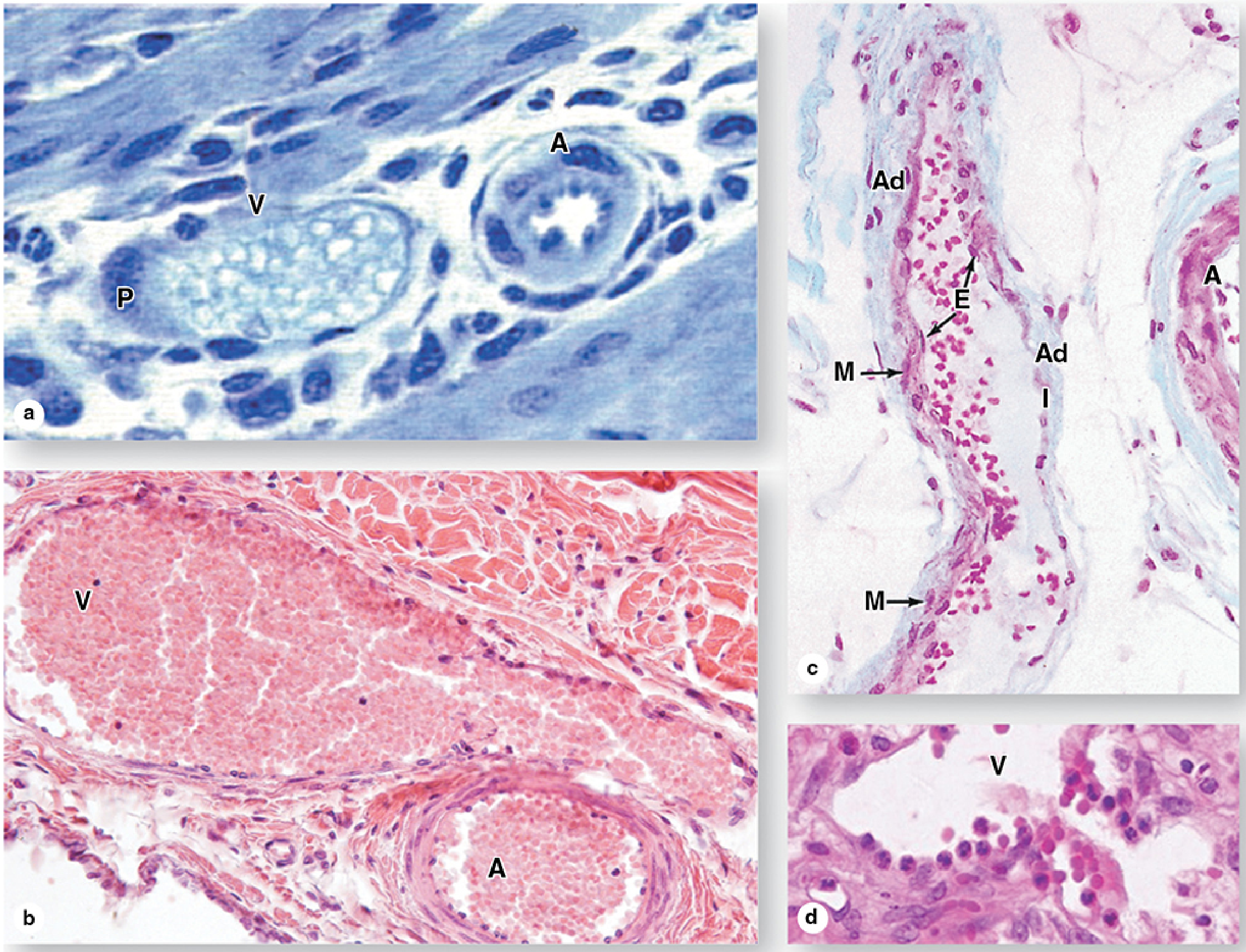
Sources: Junqueira's Basic Histology - Text and Atlas, 17e, pp. 579-580; Histology: A Text and Atlas with Correlated Cell and Molecular Biology, pp. 1111-1112.
Histology of human heart in detail with question and answers
Reading File
A detailed study of human heart histology includes:
-
Layers of the heart wall
- Endocardium
- Myocardium
- Epicardium and pericardium
-
Cardiac muscle tissue
- Cardiac myocyte structure
- Striations, branching, central nuclei
- Intercalated discs
- T-tubules and sarcoplasmic reticulum
-
Endocardium and valves
- Endothelium and subendothelial connective tissue
- Subendocardial layer and Purkinje fibers
- Histology of valve leaflets
-
Conducting system
- SA node, AV node, bundle of His, bundle branches, Purkinje fibers
-
Coronary vessels and cardiac connective tissue
-
Microscopic slide identification and comparisons
- Cardiac vs skeletal vs smooth muscle
- Atria vs ventricles
- Purkinje fibers vs ordinary cardiac muscle
High-level overview
The heart wall has three layers. The endocardium is the thin inner lining continuous with the vascular endothelium. The myocardium is the thick contractile middle layer formed by branching cardiac muscle cells linked through intercalated discs. The epicardium is the outer mesothelial layer, containing connective tissue, fat, nerves, and coronary vessels.
The ventricular myocardium is much thicker than the atrial myocardium, and the left ventricular wall is thickest because it pumps against systemic arterial pressure. Specialized conducting cells, particularly Purkinje fibers, lie in the subendocardial region and rapidly distribute the cardiac impulse.
Which section would you like in detailed notes with examination-style questions and answers?
- Heart wall layers
- Cardiac muscle and intercalated discs
- Conducting system and Purkinje fibers
- Heart valves and endocardium
- Slide identification and viva Q&A
- All sections, one topic at a time
With refrence of pictures and diagram
Reading File
I will include labeled histology pictures and simplified diagrams alongside the explanations and Q&A.
Asking for Preferences
Which section should I prepare first with pictures/diagrams and questions with answers?
Venules are small blood vessels that carry blood from capillary beds toward progressively larger veins.
Types and structure
-
Postcapillary venules
- First vessels after capillaries.
- About 15-20 μm in diameter.
- Thin wall: endothelium, basal lamina, and pericytes, without a true smooth-muscle tunica media.
-
Collecting venules
- Formed by convergence of postcapillary venules.
- Larger lumen and increasing numbers of contractile cells.
-
Muscular venules
- Up to approximately 0.1 mm in diameter.
- Have a thin tunica adventitia and one or two layers of smooth muscle forming a tunica media.
Key functions
- Drain blood from capillaries into veins.
- Serve as the principal site of leukocyte adhesion and transmigration into tissues, especially during infection or inflammation.
- Inflammatory mediators such as histamine increase permeability of postcapillary venules, causing fluid leakage and edema.
- In lymph nodes and other secondary lymphoid organs, specialized postcapillary venules called high endothelial venules (HEVs) enable lymphocytes to enter lymphoid tissue from the bloodstream.
A distinguishing histologic feature is a large lumen with a wall that is thin relative to its diameter.
Sources: Junqueira's Basic Histology - Text and Atlas, 17e, pp. 579-580; Histology: A Text and Atlas with Correlated Cell and Molecular Biology, pp. 1111-1112.A detailed study of human heart histology includes:
-
Layers of the heart wall
- Endocardium
- Myocardium
- Epicardium and pericardium
-
Cardiac muscle tissue
- Cardiac myocyte structure
- Striations, branching, central nuclei
- Intercalated discs
- T-tubules and sarcoplasmic reticulum
-
Endocardium and valves
- Endothelium and subendothelial connective tissue
- Subendocardial layer and Purkinje fibers
- Histology of valve leaflets
-
Conducting system
- SA node, AV node, bundle of His, bundle branches, Purkinje fibers
-
Coronary vessels and cardiac connective tissue
-
Microscopic slide identification and comparisons
- Cardiac vs skeletal vs smooth muscle
- Atria vs ventricles
- Purkinje fibers vs ordinary cardiac muscle
High-level overview
The heart wall has three layers. The endocardium is the thin inner lining continuous with the vascular endothelium. The myocardium is the thick contractile middle layer formed by branching cardiac muscle cells linked through intercalated discs. The epicardium is the outer mesothelial layer, containing connective tissue, fat, nerves, and coronary vessels.
The ventricular myocardium is much thicker than the atrial myocardium, and the left ventricular wall is thickest because it pumps against systemic arterial pressure. Specialized conducting cells, particularly Purkinje fibers, lie in the subendocardial region and rapidly distribute the cardiac impulse.
Which section would you like in detailed notes with examination-style questions and answers?
-
Heart wall layers
-
Cardiac muscle and intercalated discs
-
Conducting system and Purkinje fibers
-
Heart valves and endocardium
-
Slide identification and viva Q&A
-
All sections, one topic at a timeI will include labeled histology pictures and simplified diagrams alongside the explanations and Q&A.Please choose the section you want first. I will provide labeled pictures/diagrams, detailed notes, and exam-style questions with answers.
-
Heart wall layers
-
Cardiac muscle cells and intercalated discs
-
Conducting system and Purkinje fibers
-
Endocardium and heart valves
-
Slide identification and viva questions
This is a shared conversation. Sign in to Orris to start your own chat.