
Vagus nerve
vagus nerve anatomy diagram cranial nerve X
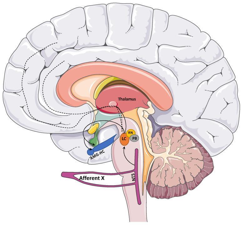
This medical anatomical diagram illustrates the central afferent pathways of the vagus nerve (Cranial Nerve X) within a sagittal section of the human brain. The pathway begins with the 'Afferent X' projecting to the Nucleus Tractus Solitarius (NTS) in the medulla. From the NTS, the diagram maps secondary projections to brainstem nuclei including the Locus Coeruleus (LC), Raphe Nuclei (RN), and Parabrachial Nucleus (PB). Further rostral connections are shown extending to limbic structures, specifically the Amygdala-Hippocampus complex (AMG-HC), and the Thalamus. Dotted lines indicate tertiary projections from the Thalamus and LC toward the cerebral cortex, representing the neuroanatomical basis for Vagus Nerve Stimulation (VNS) effects on cortical excitability and mood regulation. This schematic is designed for educational use in neurology and neurosurgery to demonstrate the integration of visceral sensory information into higher-order brain centers.
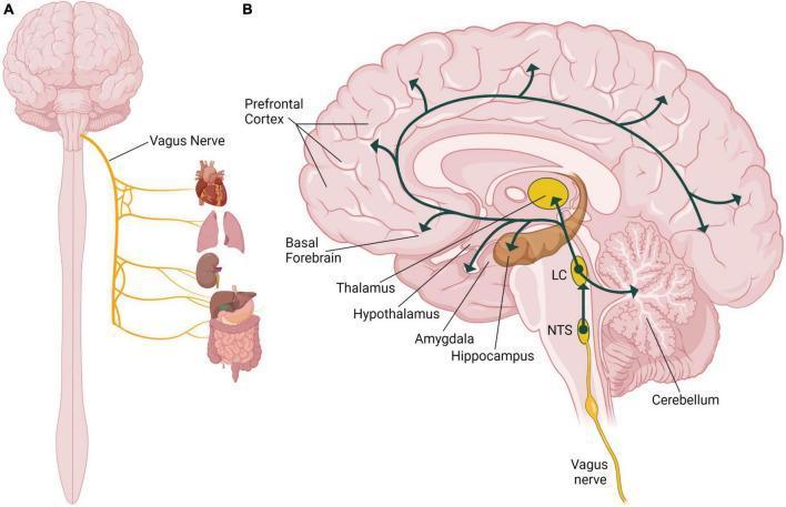
Anatomical diagram and pathophysiology schematic illustrating the systemic distribution and neuroanatomical pathways of the human vagus nerve (Cranial Nerve X). Section A depicts the peripheral innervation, showing the vagus nerve descending from the brainstem to supply parasympathetic fibers to the heart, lungs, kidneys, and gastrointestinal tract (liver, stomach, and intestines). Section B presents a sagittal view of the brain detailing the vagal afferent pathway. It illustrates the nerve projecting to the Nucleus of the Solitary Tract (NTS) in the medulla, which then connects to the Locus Coeruleus (LC). From the LC, widespread noradrenergic projections are shown reaching key functional regions including the prefrontal cortex, basal forebrain, thalamus, hypothalamus, amygdala, hippocampus, and cerebellum. This illustration highlights the structural basis for how vagus nerve stimulation (VNS) can modulate cognitive and emotional centers related to memory and homeostasis.
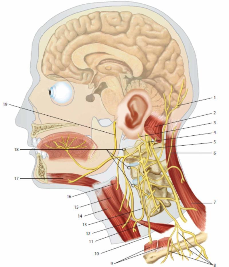
This medical anatomical diagram provides a lateral view of the human head and neck, focusing on the cervical plexus and its integration with cranial nerves. Key neural structures include the lesser occipital nerve (1), greater auricular nerve (3), and transverse cutaneous nerve of the neck (6), demonstrating the sensory distribution of the cervical plexus. The phrenic nerve (10) is shown descending inferiorly anterior to the prevertebral muscles (18). Cranial nerves X (vagus, 4), XI (accessory, 5), and XII (hypoglossal, 19) are illustrated to show their spatial relationships with cervical spinal branches. Muscular landmarks include the sternocleidomastoid (2, 9), trapezius (7), and infrahyoid muscles such as the omohyoid (11, 15) and thyrohyoid (16). The ansa cervicalis (13) is depicted innervating the sub-hyoid musculature. The diagram also illustrates the sagittal section of the brain and oral cavity, highlighting the innervation of the tongue and the relationship between the cervical spine vertebrae and emerging nerve roots. This resource is intended for medical education regarding the peripheral nervous system and regional anatomy of the neck.
Vagus Nerve (Cranial Nerve X)
Fiber Types
| Fiber type | Function |
|---|---|
| General visceral efferent (GVE) | Parasympathetic motor to thoracic and abdominal viscera (heart, lungs, GI tract to mid-transverse colon) |
| Special visceral efferent (SVE) | Voluntary motor to pharyngeal and laryngeal striated muscles (via nucleus ambiguus) |
| General visceral afferent (GVA) | Sensory from thoracic/abdominal organs - more than half of vagal fibers are sensory; cell bodies in the nodose (inferior) ganglion |
| General somatic afferent (GSA) | Sensory from external ear/tympanic membrane (Arnold's nerve); cell bodies in the superior (jugular) ganglion |
Brainstem Nuclei
| Nucleus | Role |
|---|---|
| Dorsal motor nucleus of CN X | Preganglionic parasympathetic efferents to thoracic/abdominal viscera |
| Nucleus ambiguus | Special visceral efferents; motor to pharynx, larynx, upper esophagus |
| Nucleus tractus solitarius (NTS) | Receives all visceral afferents; integrates signals for cardiovascular, respiratory, and GI reflex arcs |
Course and Key Branches
Exit from skull
- Leaves the brainstem as multiple rootlets from the lateral medulla (dorsolateral sulcus)
- Exits the skull through the middle compartment of the jugular foramen, alongside CN IX and XI
- Two ganglia lie near the foramen: the superior (jugular) ganglion (somatic afferent cell bodies) and the inferior (nodose) ganglion (visceral afferent cell bodies)
In the Neck
- Auricular branch (Arnold's nerve) - passes between mastoid and tympanic plate; sensory to tympanic membrane, posterior pinna, posterior ear canal. Stimulation can trigger the "vagal cough reflex" and referred otalgia.
- Carotid body branches - supplements CN IX supply to carotid body chemoreceptors
- Pharyngeal branch - carries spinal accessory fibers; motor to pharyngeal constrictors and soft palate via the pharyngeal plexus
- Superior laryngeal nerve (SLN) - divides at hyoid level into:
- Internal laryngeal nerve: sensory to laryngeal mucosa above the glottis and piriform fossa; penetrates thyrohyoid membrane
- External laryngeal nerve: motor to cricothyroid muscle (the only intrinsic laryngeal muscle NOT supplied by the recurrent laryngeal nerve)
- Cardiac branches - descend to the cardiac plexus
Right vs. Left in the Thorax
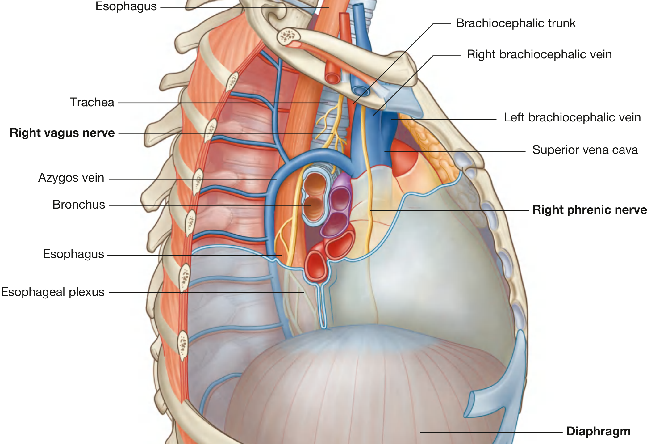
- Enters the superior mediastinum between the right brachiocephalic vein and the brachiocephalic trunk
- Crossed by the arch of the azygos vein
- Right recurrent laryngeal nerve hooks around the right subclavian artery in the root of the neck
- Enters posterior to the left brachiocephalic vein, between the left common carotid and subclavian arteries
- Crosses the arch of the aorta (medial to mediastinal pleura)
- Left recurrent laryngeal nerve hooks under the arch of the aorta just lateral to the ligamentum arteriosum, then ascends in the tracheoesophageal groove
Recurrent Laryngeal Nerve (RLN)
In the Abdomen
- Anterior vagal trunk (mostly left vagus) - enters abdomen through esophageal hiatus
- Posterior vagal trunk (mostly right vagus) - enters the same hiatus
Physiological Roles
Cardiovascular
- Slows heart rate (negative chronotropy) via M2 muscarinic receptors at the SA node
- Baroreceptor reflex: afferents from the carotid sinus and aortic arch travel via CN IX and X to the NTS, which modulates the dorsal motor nucleus to regulate heart rate
Respiratory
- Hering-Breuer reflex: pulmonary stretch receptors signal via vagal afferents to stop inhalation
Gastrointestinal
- Controls gastric acid secretion, gut motility, and the lower esophageal sphincter
- Vagal afferents carry satiety signals - cholecystokinin (CCK) released post-meal acts on vagal afferents in the gut via the CCK-A receptor to reduce meal size and initiate satiation
- More than half of all vagal fibers are sensory, relaying real-time information from the GI tract (up to the right colon) to the NTS
Metabolic/Appetite Regulation
Central Afferent Projections (NTS Onward)
- Locus coeruleus (LC) - noradrenergic arousal and attention
- Raphe nuclei - serotonergic modulation
- Parabrachial nucleus
- Thalamus - relay to cortex
- Amygdala and hippocampus - emotional and memory processing
Clinical Pearls
| Finding | Mechanism |
|---|---|
| Hoarseness (unilateral) | RLN palsy - most often left side (longer course); causes include lung/mediastinal tumors, aortic aneurysm, thyroid surgery |
| Dysphagia | Pharyngeal branch / nucleus ambiguus lesion |
| Absent gag reflex | Loss of efferent arc (CN X) or afferent arc (CN IX) |
| Referred otalgia | Arnold's nerve carries pain from deep structures to the external ear |
| Vasovagal syncope | Exaggerated vagal slowing of heart rate + vasodilation |
| Carotid sinus hypersensitivity | Excess vagal tone triggered by carotid baroreceptors |
| Hiccups (intractable) | Vagal/phrenic nerve irritation - sometimes treated with vagal maneuvers |