
1.Describe TETANUS****-Etiology,MOI,C/F,Management and Complications [15/6marks] 2. Cellulitis ** [3marks] 3.ABCESS A)pyogenic Abcess*** B)COLD ABCESS**** C)Antibioma *****.[3marks] D) Hiltons method of opening an Abcess. 4. CARBUNCLE******-Etiopathology,C/F,Treatment. 5. Erypsipelas [3Marks] 6.Write short note on NECROTISING FASCITIS***. [6/3Marks] 7.Add a note on SURGICAL SITE INFECTION**[PIRO classification ]. 8. GAS GANGRENE***- Define, Etiology,types, pathogenesis, C/F,Diagnosis&treatment. 9.Acute and Chronic Paronychia***[3Marks] 10.FELON****[3Marks] 11.Write About MYCETOMA FOOT(madura foot)**** 12.Onychocryptosis. 13.Universal precautions during surgery with HIV patients. -3M 14.FOURNIER'S GANGRENE **** 15. Direct and Indirect Traumatic gangrene. 16. Hydradenitis suppurativa. 17. MRSA [Marks]
SURGICAL INFECTIONS - Complete Study Guide
1. TETANUS ⭐⭐⭐⭐
Etiology
- Caused by Clostridium tetani - a Gram-positive, anaerobic, spore-forming bacillus
- Spores found in soil, road dirt, house dust, human and animal faeces
- Spores resist prolonged boiling and are extremely hardy
- Produces two exotoxins: Tetanospasmin (neurotoxin - causes clinical disease) and Tetanolysin (haemolysin)
Mechanism of Injury (MOI) / Pathogenesis
- Spores germinate into vegetative forms under anaerobic conditions (devitalized tissue, ischaemia, foreign bodies, or co-infection with other organisms)
- Deep puncture wounds involving muscle are most prone - wounds on farms, gardens, playing fields, roads, garages, sewers
- Tetanospasmin is absorbed by motor nerve endings and travels retrograde along axons to anterior horn cells
- At anterior horn: toxin blocks release of inhibitory neurotransmitters (GABA, glycine) from Renshaw cells
- Loss of inhibition → uncontrolled motor neuron firing → sustained muscle contraction (spasm)
- Toxin also affects the sympathetic nervous system → autonomic dysfunction
Clinical Features
- Trismus (lockjaw) - tonic spasm of masseter muscles; earliest sign
- Risus sardonicus - characteristic grinning expression due to facial muscle spasm
- Opisthotonus - severe arching of back due to spasm of paraspinal muscles
- Dysphagia (pharyngeal spasm)
- Neck stiffness
- Abdominal rigidity ("board-like abdomen")
- Reflex spasms - triggered by any stimulus (touch, sound, light) - can cause laryngospasm and asphyxia
- Autonomic instability: sweating, hyperthermia, labile BP, tachycardia
- Neonatal tetanus (tetanus neonatorum) - inability to suck, trismus
- Grade I: Mild - trismus only, no spasms
- Grade II: Moderate - trismus, dysphagia, mild spasms
- Grade III: Severe - reflex spasms, autonomic disturbance
- Grade IV: Very severe - autonomic storms
Management
- Proper wound care: thorough cleansing, debridement of all devitalized tissue, removal of foreign material, wound closure without tension
- Active immunity: Tetanus Toxoid (TT) 0.5 ml IM - 3 doses (0, 6-8 weeks, 6-12 months); booster every 10 years
- Passive immunity: Human Tetanus Immunoglobulin (HTIG) 250 units IM - for non-immunized patients post-wound
| Step | Action |
|---|---|
| Wound | Surgical debridement of wound; HTIG infiltration around wound |
| Neutralize toxin | HTIG 3000-6000 units IM (free toxin only; once fixed, cannot be neutralized) |
| Control spasms | Diazepam (first-line); Magnesium sulphate; Baclofen (intrathecal) |
| Airway | Early tracheostomy/intubation if laryngospasm risk |
| Antibiotics | Metronidazole (preferred) or Penicillin G IV - to kill vegetative organisms |
| Supportive | ICU nursing, quiet dark room (minimize stimuli), IV fluids, nutrition |
| Autonomic control | Labetalol, morphine for sympathetic storms |
Complications
- Laryngospasm → asphyxia and death
- Aspiration pneumonia
- Urinary retention, constipation
- Vertebral fractures (due to severe spasms)
- Autonomic cardiovascular crises
- Respiratory failure (most common cause of death)
- DVT, pressure sores
- Rhabdomyolysis
2. CELLULITIS ⭐⭐
- Definition: Acute spreading infection of the dermis and subcutaneous tissue
- Organisms: Most common - Streptococcus pyogenes (Group A), Staphylococcus aureus
- Predisposing factors: Skin breach (wound, tinea pedis), lymphoedema, diabetes, obesity, venous insufficiency
- C/F: Spreading erythema, warmth, oedema, tenderness; no sharp margin (distinguishes from erysipelas); fever, lymphangitis, lymphadenopathy may be present
- Investigations: Blood culture (often negative), raised WBC, CRP
- Treatment:
- Elevation of limb
- Oral flucloxacillin/amoxiclav for mild cases
- IV benzylpenicillin + flucloxacillin for severe cases
- MRSA: IV vancomycin
- Mark the border with a skin marker to monitor progression
- Treat underlying cause (e.g., tinea pedis)
3. ABSCESS
A) Pyogenic Abscess ⭐⭐⭐
- Definition: A localized collection of pus in a cavity formed by the disintegration of tissue
- Organisms: Staphylococcus aureus (most common), streptococci, coliforms, anaerobes
- Pathogenesis:
- Bacteria enter → acute inflammation → PMN infiltration → tissue necrosis → pus forms
- Surrounding wall of granulation/fibrous tissue = pyogenic membrane
- C/F:
- Cardinal signs of inflammation: rubor, calor, dolor, tumor
- Fluctuation (pathognomonic) - pus movement felt between two fingers
- Pointing - skin becomes thin, shiny, and tense at the site of impending rupture
- Fever, rigors
- Treatment:
- "Ubi pus, ibi evacua" - Where there is pus, drain it
- Incision and drainage (I&D)
- Send pus for C&S
- Antibiotics for surrounding cellulitis or systemic sepsis
- Daily wound dressings
B) Cold Abscess ⭐⭐⭐⭐
- Definition: An abscess without the usual signs of acute inflammation (no heat, no redness) - hence "cold"
- Etiology: Mycobacterium tuberculosis (most common); also atypical mycobacteria
- Pathogenesis: Caseous necrosis in a TB focus liquefies → pus tracks along fascial planes → presents at a site distant from the primary focus (hence also called "wandering abscess")
- Common sites:
- Cervical lymph node TB → collar-stud abscess
- Spinal TB (Pott's disease) → psoas abscess pointing to inner thigh/groin
- Rib TB → intercostal cold abscess
- C/F:
- Soft, fluctuant swelling
- No heat, no redness, no tenderness (hence "cold")
- Transillumination positive (clear fluid - unlike pyogenic)
- Collar-stud abscess: Deep abscess communicates through a defect in deep fascia → superficial component, with dumbbell/collar-stud shape
- Investigations: Aspirate for AFB, ZN staining, Lowenstein-Jensen culture; Mantoux test; CXR; FNAC
- Treatment:
- Aspiration (not incision - avoid sinus formation)
- Full course of anti-tubercular therapy (ATT)
- Surgical excision in selected cases
- Do NOT incise - leads to chronic discharging sinus
C) Antibioma ⭐⭐⭐⭐⭐
- Definition: A chronic indurated abscess that results from inadequate or incomplete antibiotic treatment of a pyogenic abscess
- Pathogenesis:
- Antibiotics given for an abscess that has already formed → bacteria killed but pus not drained
- Pus becomes inspissated (thickened), surrounded by dense fibrosis
- Results in a hard, firm, non-fluctuant lump - clinically mimics a tumour/carcinoma
- C/F:
- Hard, indurated, non-tender mass
- No fluctuation, no heat
- History of previous pain/swelling treated with antibiotics without drainage
- May be mistaken for malignancy (especially breast antibioma)
- Investigations: FNAC/biopsy to exclude malignancy; USS shows echogenic material
- Treatment:
- Surgical excision of the entire mass including the fibrous wall
- Send specimen for histopathology
D) Hilton's Method of Opening an Abscess
- Give local/general anaesthesia
- Make a small stab incision through the skin and superficial fascia only with a pointed scalpel (no. 11 blade)
- Insert closed sinus forceps (haemostat) through the incision
- Open the forceps widely in the direction of the important structure (blunt dissection) - this avoids cutting nerves and blood vessels
- Allow pus to escape; insert gloved finger to break all loculi
- Insert a corrugated rubber drain or pack with gauze
- Leave wound open for secondary healing
4. CARBUNCLE ⭐⭐⭐⭐⭐⭐
Etiopathology
- Definition: A carbuncle is an infective gangrene of the skin and subcutaneous tissue involving multiple adjacent hair follicles and their sebaceous glands
- Compared to a furuncle (involves one follicle), a carbuncle involves multiple follicles
- Organism: Staphylococcus aureus (usually MRSA in serious cases)
- Predisposing factors: Diabetes mellitus (most common), malnutrition, immunosuppression, obesity, poor hygiene, steroid use
- Pathogenesis:
- Staphylococci infect multiple adjacent hair follicles
- Infection spreads laterally through the deep dermis and subcutaneous fat
- Multiple pockets of pus with septa form the characteristic "riddled with holes" appearance
- Central area undergoes necrosis → sloughing → crater-like ulcer
- Intense fibrosis surrounds the lesion
Clinical Features
- Typically on the back of neck (nape), back, buttocks, thighs
- Begins as a hard, red, painful, indurated plaque
- Multiple pustules/follicular openings visible on surface - "sieve-like" or "watering can" appearance
- Central area becomes necrotic and sloughs, leaving a deep, ragged, undermined ulcer
- Significant surrounding cellulitis and oedema
- Marked systemic toxicity: high fever, rigors, malaise (more than furuncle)
- Regional lymphadenopathy
- Complications: Septicaemia, metastatic abscesses, cavernous sinus thrombosis (if on face)
Treatment
- Treat underlying diabetes - blood sugar control is essential
- Antibiotics: Flucloxacillin IV; clindamycin if MRSA; guided by culture and sensitivity
- Surgical treatment:
- Cruciate incision (cross-shaped) through the entire carbuncle
- All necrotic tissue and septa excised
- Wound packed with gauze (heals by secondary intention)
- Alternatively: wide excision with split-thickness skin grafting
- Nutritional support and correction of immunodeficiency
- Topical honey/povidone-iodine dressings post-op
5. ERYSIPELAS ⭐⭐ (3 Marks)
- Definition: Acute superficial infection of the upper dermis and superficial lymphatics with a sharply demarcated edge
- Organism: Almost exclusively Group A beta-haemolytic Streptococcus (Streptococcus pyogenes)
- Entry point: Skin fissures, ulcers, minor abrasions, tinea pedis
- Common sites: Face (butterfly distribution), lower limbs
Clinical Features
- Fiery red, shiny, oedematous plaque with a sharply raised, well-defined advancing border - the hallmark that distinguishes it from cellulitis
- Warm, tender, tense skin
- "Peau d'orange" appearance (orange peel skin) in some cases
- High fever, rigors, malaise preceding the rash
- Lymphangitis and lymphadenopathy
- Vesicles and bullae may develop in severe cases
- Facial erysipelas: "Butterfly" distribution across cheeks and nose
Treatment
- Penicillin V oral (mild) or IV benzylpenicillin (severe) - drug of choice
- Erythromycin if penicillin-allergic
- Elevation of affected part
- Cool saline compresses
- Recurrent erysipelas: monthly penicillin prophylaxis
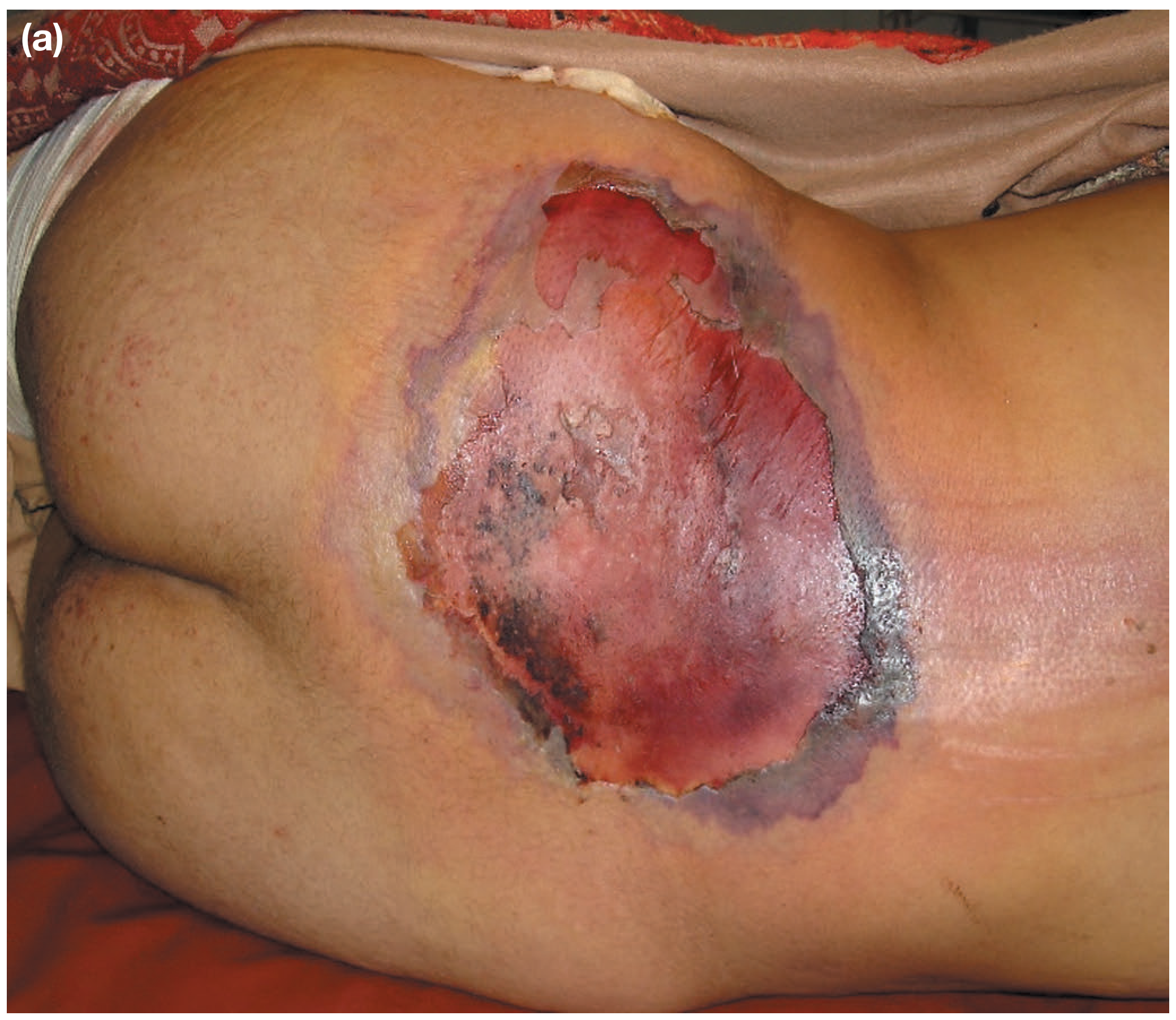
6. NECROTISING FASCIITIS ⭐⭐⭐ (6/3 Marks)
Definition
Etiology
- Type I (Polymicrobial): Most common. Mixed aerobic + anaerobic organisms: Streptococcus, Staphylococcus aureus, E. coli, Pseudomonas, Clostridium, Bacteroides, Klebsiella
- Type II (Monomicrobial): Group A Streptococcus pyogenes (flesh-eating bacteria) - most virulent; younger patients
- Type III: Marine organisms (Vibrio vulnificus) - after seafood exposure
Predisposing Factors
Pathogenesis
Clinical Features (Summary Box)
- Unusual, disproportionate pain (pain out of proportion to appearance - early sign)
- Erythema, oedema, warmth
- Crepitus (gas in tissues)
- Blisters, bullae
- Grey/black discolouration - skin turns dusky blue-black
- "Dishwater pus" (grey, thin fluid) from wound
- Cutaneous anaesthesia (necrosis of nerves)
- Fixed skin staining
- High fever, tachycardia, tachypnoea
- Septic shock
- Coagulopathy (DIC)
- Multiorgan failure
- Renal failure (hypovolaemia + septic shock)
Specific Variants
- Fournier's gangrene: NF involving perineum, scrotum, and perianal region
- Meleney's gangrene: NF of abdominal wall
Diagnosis
- Primarily clinical - do not delay treatment for investigations
- Elevated creatine kinase (enormous elevation)
- CT scan: gas tracking along fascial planes (confirmatory when positive)
- Biopsy of fascia: fascial necrosis + acute inflammatory infiltrate
- LRINEC score (Laboratory Risk Indicator for Necrotising Fasciitis): uses CRP, WBC, Hb, Na, creatinine, glucose - score ≥6 is high risk
Treatment
- ICU admission - fluid resuscitation, haemodynamic monitoring
- IV antibiotics: High-dose Penicillin G + 3rd generation cephalosporin + metronidazole (empiric broad-spectrum cover); adjust per culture
- Add clindamycin (anti-toxin effect against Streptococcus)
- MRSA: add vancomycin
- Urgent radical surgical debridement - cornerstone of management
- Wide excision of ALL necrotic tissue going beyond the zone of induration
- Wound packed and dressed
- Second-look operation in 24-48 hours - multiple debridements often required
- Hyperbaric oxygen - adjunct (where available)
- Reconstructive surgery after infection controlled (skin grafting)
7. SURGICAL SITE INFECTION (SSI) ⭐⭐ - PIRO Classification
Definition (CDC/NHSN)
Classification of SSI
- Superficial incisional SSI: Involves only skin and subcutaneous tissue
- Deep incisional SSI: Involves deep soft tissue (fascia, muscle)
- Organ/space SSI: Involves any organ or space opened during the procedure (e.g., anastomotic leak, intra-abdominal abscess)
PIRO Classification (Predisposition, Insult/Injury, Response, Organ dysfunction)
| Component | Meaning | Examples |
|---|---|---|
| P - Predisposition | Patient host factors | Diabetes, obesity, malnutrition, immunosuppression, smoking, old age, ASA score, MRSA colonisation |
| I - Insult/Injury | Nature and severity of the surgical insult | Contaminated/dirty wound, emergency surgery, prolonged operative time (>2 hrs), bowel perforation, ischaemic tissue |
| R - Response | Host inflammatory response | Fever, raised WBC, raised CRP, procalcitonin - SIRS criteria |
| O - Organ dysfunction | End-organ failure as consequence | AKI, respiratory failure, shock, multiorgan dysfunction |
Prevention of SSI
- Pre-op: Treat remote infections, glycaemic control, smoking cessation, MRSA screening and decolonisation (mupirocin nasal + chlorhexidine baths), hair clipping (not shaving)
- Intra-op: Antibiotic prophylaxis (within 60 min of incision), maintain normothermia, maintain normovolaemia, minimise dead space, minimize operative time
- Post-op: Proper wound dressings, early removal of drains, glucose control
8. GAS GANGRENE ⭐⭐⭐
Definition
Etiology
- Primary organism: Clostridium perfringens (welchii) - 80-90% of cases
- Others: Clostridium oedematiens (novyi), Clostridium septicum, Clostridium histolyticum
- Gram-positive, anaerobic, spore-forming rods
- Found in soil, road dirt, human and animal faeces
Types
- Clostridial myonecrosis (true gas gangrene) - involves muscle
- Clostridial cellulitis - involves subcutaneous tissue, not muscle (less severe)
- Anaerobic streptococcal myositis - caused by anaerobic streptococci
Pathogenesis
- Spores enter devitalized tissue (trauma, ischaemia, surgery)
- Germinate under anaerobic conditions → vegetative organisms proliferate
- Produce exotoxins:
- Alpha toxin (lecithinase) - most important - lyses cell membranes, destroys muscle, causes haemolysis
- Hyaluronidase, collagenase, protease, DNase - spread infection through tissue planes
- Gas (CO2, H2) produced by fermentation → crepitus in tissues
- Toxins enter bloodstream → systemic toxaemia → cardiovascular collapse
- Alpha toxin produces intravascular haemolysis → haemoglobinuria → renal failure
Clinical Features
- Bursting/throbbing pain at wound site - out of proportion, often the first sign
- Wound appears tense, indurated; skin is red, shiny, tense
- Sero-sanguinous, thin, watery discharge with characteristic sickly sweet smell ("mousy" odour)
- Crepitus on palpation (late sign) - gas crackling in tissues
- High fever, rapid thready pulse (rising pulse rate with falling BP)
- Bronze discolouration of overlying skin → blue-black gangrenous change
- Profound toxaemia, restlessness, confusion
- Haemolytic jaundice (haemoglobinuria - "port wine" urine)
- Septic shock, multiorgan failure, death (if untreated)
Diagnosis
- Clinical - characteristic smell, crepitus, wound appearance
- X-ray: Gas shadows in muscle planes (feathery pattern)
- Gram stain of wound discharge: Large Gram-positive rods, absence of PMNs (toxin kills WBCs)
- Wound swab for C&S
- Blood culture
- CT scan: Gas in muscle compartments
Treatment
- Surgical - the cornerstone:
- Wide, radical opening of wound
- Excision of all necrotic/doubtful muscle (muscle that does not bleed or contract when cut is dead)
- Wound left open, lightly packed with fluffed gauze
- Amputation may be necessary to save life (sacrificial surgery)
- Antibiotics:
- IV Penicillin G (high dose) - all Clostridia sensitive
- Add metronidazole and clindamycin (anti-toxin)
- Clindamycin preferred adjunct - inhibits toxin production
- Supportive: IV fluids, blood transfusion for haemolysis, vasopressors
- Hyperbaric oxygen (HBO): Valuable adjunct - raises tissue O2 tension, inhibits Clostridial growth; available in limited centres
- Antitoxin: Of doubtful value - not routinely used
9. PARONYCHIA ⭐⭐⭐ (3 Marks)
Acute Paronychia
- Definition: Acute infection of the nail fold (paronychium) - the skin fold surrounding the nail
- Organism: Staphylococcus aureus (most common)
- Causes: Hangnail, manicure injury, nail biting, splinter
- C/F: Throbbing pain, redness, swelling of nail fold; fluctuation; pus visible under nail fold
- Treatment:
- Early: warm soaks + oral antibiotics (flucloxacillin)
- Pus present: Incision and drainage - longitudinal incision along nail fold under ring block anaesthesia; if pus under nail - remove portion of nail; Hilton's method not suitable here
- Severe: Remove entire nail if subungual abscess
Chronic Paronychia
- Definition: Chronic infection/inflammation of the nail fold lasting >6 weeks
- Organism: Candida albicans most common (+ bacteria e.g. Pseudomonas)
- Causes: Repeated wet work, chemical exposure, diabetes, immunosuppression
- C/F: Boggy, swollen, tender nail fold; nail becomes discoloured, ridged, thickened; pus present intermittently; loss of cuticle (earliest sign)
- Treatment:
- Keep hands dry, avoid wet work
- Topical antifungals (clotrimazole, miconazole)
- Systemic antifungals (itraconazole, fluconazole) for persistent cases
- Surgical: Eponychial marsupialisation (Keystone flap) for refractory cases
10. FELON ⭐⭐⭐⭐ (3 Marks)
- Definition: An abscess of the pulp space (distal closed fibrous compartment) of the fingertip
- Most common digit: Index finger and thumb
- Organism: Staphylococcus aureus
- Cause: Penetrating injury (needle prick, thorn, splinter), diabetic finger prick
Anatomy (Crucial to understand)
Clinical Features
- Intense throbbing pain - severe, worsens at night
- Tense, shiny, red swelling of the volar (palmar) aspect of the fingertip
- Dorsum of fingertip is NOT involved (distinguishes from herpetic whitlow initially)
- Tenderness on deep palpation of pulp
- Complications if untreated:
- Osteomyelitis of distal phalanx (bone is adherent to the septa - pus strips periosteum)
- Septic arthritis of DIP joint
- Flexor tenosynovitis
- Ischaemic necrosis of distal phalanx (septa compress digital vessels)
Treatment
- Early (cellulitis only): Antibiotics + splint + elevation
- Pus present - Surgical drainage (mandatory):
- Preferred: Longitudinal volar incision (midline/unilateral longitudinal) - avoids scar on gripping surface
- Alternatively: "Fish mouth" incision (no longer recommended - causes unstable fingertip)
- Break all septa inside the pulp space
- Pack wound open, leave to heal by secondary intention
- X-ray to exclude osteomyelitis
- Antibiotics: Flucloxacillin IV post-op
11. MYCETOMA FOOT (Madura Foot) ⭐⭐⭐⭐
Definition
Etiology
- Madurella mycetomatis (most common worldwide, black grains)
- Scedosporium apiospermum (white grains)
- Nocardia brasiliensis (pale yellow grains)
- Actinomadura madurae (white/yellow grains)
- Streptomyces somaliensis (yellow grains)
Epidemiology
- Endemic in tropical and subtropical regions - India (Tamil Nadu, Gujarat), Africa, Latin America
- "Madura foot" - named after Madurai, South India
- Predominantly affects males, agricultural workers (barefoot)
Pathogenesis
- Inoculation of organism via penetrating trauma (thorn, splinter) in barefoot walkers
- Chronic granulomatous inflammation
- Spread through fascial planes → invades muscle and bone
- Sulfur granules = compact masses of fungal hyphae/actinomycetes in a polysaccharide matrix, surrounded by PMNs
Clinical Features
-
Classic triad:
- Swelling - painless, progressively enlarging
- Sinuses - multiple discharging sinuses on the surface
- Grains - coloured granules discharged from sinuses (colour varies by organism - diagnostic)
-
Foot most commonly affected (dorsum); also hand, leg, back
-
Overlying skin: indurated, violaceous, peppered with sinuses
-
Bone involvement: moth-eaten appearance on X-ray, pathological fractures
-
No lymph node involvement (distinguishes from other infections)
-
No systemic symptoms (chronic, indolent)
Investigations
- Discharge for KOH mount: hyphae in granules
- Gram stain, ZN stain (for actinomycetes)
- Culture: Sabouraud's medium (fungi); blood agar (actinomycetes)
- X-ray foot: Bone destruction, periosteal reaction, "soap bubble" appearance, new bone formation
- MRI: Most sensitive for extent of tissue involvement; shows "dot in circle" sign
- FNAC/biopsy for histology
Treatment
- Co-trimoxazole + amikacin (Domingos-Abreu regimen) - first-line
- Add rifampicin for resistant cases
- Long treatment (6-12 months)
- Itraconazole (most effective) or voriconazole
- Very prolonged treatment (1-2 years)
- Surgical debulking to reduce fungal load
- Wide excision for localised disease
- Amputation for extensive bone involvement - last resort
12. ONYCHOCRYPTOSIS (Ingrowing Toenail)
- Definition: Ingrowth of the nail edge into the adjacent lateral nail fold (paronychium)
- Most common site: Great toe (hallux), lateral edge
Etiology
- Incorrect nail cutting (cutting too short at sides, curved cut)
- Tight/ill-fitting footwear
- Excessive sweating
- Trauma
- Genetic predisposition (curved nail plate)
Stages (Heifetz Classification)
- Stage I: Pain, erythema, swelling of nail fold - no discharge
- Stage II: Infected with discharge (pus/granulation tissue)
- Stage III: Chronic granulation tissue, recurrent episodes
Treatment
- Conservative (Stage I): Warm soaks, antibiotics, proper nail cutting, cotton wool under nail edge, wider footwear
- Surgical (Stage II/III):
- Wedge resection (Partial nail avulsion): Under ring block; tourniquet; remove the offending 1/4 of nail plate + germinal matrix ablation (phenol 80% applied for 3 min = chemical matricectomy - gold standard) or surgical excision of matrix
- Zadik's procedure (Total nail ablation): Complete removal of nail + entire germinal matrix - reserved for severe/recurrent cases or onychogryphosis
- Winograd procedure: Excision of the nail edge, nail fold, and underlying nail bed in a wedge
13. UNIVERSAL PRECAUTIONS DURING SURGERY IN HIV PATIENTS (3 Marks)
Standard Universal Precautions:
- Double gloving - reduces needlestick injury risk by 87%
- Waterproof gown/apron
- Face shield or goggles (eye protection)
- N95 mask (for aerosol-generating procedures)
- Shoe covers
- No hand-to-hand passing of sharps - use a neutral zone/kidney dish
- Never recap needles by hand
- Blunt-tip suture needles wherever possible
- Announce before passing sharp instruments ("sharp coming")
- Use stapler/harmonic scalpel/electrocautery to reduce sharp use
- Dispose in puncture-proof sharps containers immediately
- Minimise number of staff in OT
- One-person rule - only one sharp instrument at a time
- Avoid finger-guided suturing (use retractors/instruments, not fingers)
- Use closed-system transfers for blood/fluids
- Instruments-first technique for locking blades
- Electrocautery preferred over sharp dissection
- Immediately wash with soap and water for 5 minutes; squeeze out blood
- Report to occupational health immediately
- Test donor patient (HIV status, Hep B, Hep C) with consent
- Test exposed worker (baseline)
- Start Post-Exposure Prophylaxis (PEP) within 2 hours (max 72 hours): TDF + FTC + raltegravir for 28 days
- Follow-up testing at 6 weeks, 3 months, 6 months
- All blood-soaked materials in yellow biohazard bags
- Sharps in puncture-proof containers
- Incineration of waste
14. FOURNIER'S GANGRENE ⭐⭐⭐⭐
Definition
Etiology / Risk Factors
- Diabetes mellitus (most common - 40-60%)
- Perirectal/perianal abscess (most common source)
- Perineal trauma
- Urinary tract infection, urethral stricture, phimosis
- Colorectal pathology (Crohn's, colorectal carcinoma)
- Obesity, alcoholism, immunosuppression, malignancy
Organisms
Pathogenesis
- Source of infection: perineal skin, anal canal, urethra, or haematogenous
- Bacteria spread along Colles' fascia (superficial perineal fascia), Dartos fascia (scrotum), Scarpa's fascia (anterior abdominal wall)
- Endarteritis obliterans → thrombosis of subcutaneous vessels → ischaemia → necrosis
- Deep scrotal/penile fascia (Buck's fascia) and testes are usually spared (independent blood supply - internal spermatic artery)
Clinical Features
- Severe perineal pain, swelling
- Crepitus in scrotum/perineum (pathognomonic)
- Skin erythema → dusky/black discolouration → gangrene
- Odour - foul-smelling discharge
- Systemic sepsis: fever, tachycardia, hypotension
- Testes preserved (unlike scrotal skin)
- Mortality: 20-40% (up to 67% in some series)
Fournier's Gangrene Severity Index (FGSI)
Treatment
- ICU resuscitation - aggressive fluid replacement
- Broad-spectrum IV antibiotics:
- Piperacillin-tazobactam + metronidazole + gentamicin
- MRSA: add vancomycin
- Urgent surgical debridement - wide excision of ALL necrotic tissue
- Testes/penis usually preserved (not involved)
- Wound left open; daily dressings
- Faecal diversion (colostomy) if perianal source or extensive contamination
- Urinary diversion (suprapubic catheter) if urethral involvement
- Second-look surgery at 24-48 hours; multiple debridements common
- Hyperbaric oxygen - adjunct
- Reconstruction after infection control: skin grafting, orchiectomy if testicular necrosis
15. DIRECT AND INDIRECT TRAUMATIC GANGRENE
Traumatic Gangrene
Direct Traumatic Gangrene
- Definition: Gangrene resulting from direct, immediate vascular injury at the site of trauma
- Mechanism:
- Crush injuries - direct destruction of blood vessels + tissue
- Avulsion injuries - tearing of vessels
- Gunshot wounds - primary cavitation destroys vessels
- Severe burns - direct thermal destruction of vessels and tissue
- Ischaemia is immediate at the site of trauma
- Gangrenous tissue is at the zone of injury
- Examples: Degloving injury, traumatic amputation, blast injury
Indirect Traumatic Gangrene
- Definition: Gangrene occurring at a site remote from or consequent to the primary trauma, due to indirect disruption of blood supply
- Mechanisms:
- Arterial injury/thrombosis from blunt trauma (intimal tear, spasm) → distal gangrene
- Compartment syndrome → increased compartment pressure → venous then arterial obstruction → distal ischaemia
- Prolonged tourniquet application → distal limb gangrene
- Thromboembolism following trauma → distal gangrene
- Fractures causing arterial compression/laceration → distal limb gangrene
- Gangrenous tissue is distal to the site of injury
- Classic example: Supracondylar fracture of humerus in children → brachial artery injury → forearm/hand gangrene (Volkmann's ischaemia progressing to gangrene)
16. HIDRADENITIS SUPPURATIVA (Hydradenitis Suppurativa)
Definition
Pathogenesis
- Not a primary infection - primarily a follicular occlusion disease
- Follicular hyperkeratosis → follicular plugging → follicular rupture → secondary inflammation and infection
- Apocrine glands secondarily involved
- Organisms: Mixed flora - S. aureus, coagulase-negative staphylococci, anaerobes (secondary)
- Chronic suppuration leads to fistulae and scar formation
Risk Factors
- Obesity, smoking (strongest modifiable risk factors)
- Family history (autosomal dominant)
- Female gender (3:1 ratio), onset post-puberty
- Diabetes, hyperandrogenism
Sites (Apocrine-bearing areas)
Clinical Features
- Stage I: Solitary/multiple abscesses, no sinus tracts, no scarring
- Stage II: Recurrent abscesses, single/multiple widely separated sinus tracts and scars
- Stage III: Multiple interconnected sinus tracts and abscesses over an entire region; diffuse involvement
Treatment
- Topical clindamycin 1% (mild, Stage I)
- Oral tetracycline/doxycycline - first-line systemic
- Oral clindamycin + rifampicin (300 mg each BD x 10 weeks) - most effective medical regimen
- Biological therapy: Adalimumab (anti-TNF-alpha) - only FDA-approved biologic; for Stage II/III
- Weight loss, smoking cessation
- Intralesional triamcinolone for acute flares
- I&D: Temporary relief only - high recurrence
- Deroofing/unroofing: Sinus tracts laid open - good local control
- Wide local excision (Stage III) - definitive; wound heals by secondary intention or skin grafting
- Laser hair removal: Nd:YAG laser - adjunct
17. MRSA (Methicillin-Resistant Staphylococcus Aureus)
Definition
Mechanism of Resistance
- mecA gene on a mobile genetic element (SCCmec - Staphylococcal Cassette Chromosome mec)
- mecA encodes PBP2a (Penicillin-Binding Protein 2a) - modified PBP with low affinity for all beta-lactams
- Beta-lactams cannot inhibit PBP2a → bacteria continue cell wall synthesis → resistance
Types
- HA-MRSA (Healthcare-Associated MRSA): Hospitals, nursing homes; typically multi-drug resistant
- CA-MRSA (Community-Associated MRSA): Community setting, no healthcare contact; Panton-Valentine Leucocidin (PVL) toxin positive - causes severe skin/soft tissue infections and necrotising pneumonia; usually more susceptible to non-beta-lactam antibiotics
- LA-MRSA (Livestock-Associated MRSA): Animal contact (ST398)
Clinical Significance
- SSI, wound infections, bacteraemia, pneumonia, osteomyelitis, septic arthritis, endocarditis
Screening and Decolonisation (Pre-op)
- Nasal swab (most common colonisation site) + groin/perineum/axilla swab
- Decolonisation: Mupirocin 2% nasal ointment TDS x 5 days + Chlorhexidine 4% full-body washes x 5 days
- Repeat swab after decolonisation
Treatment of MRSA Infection
| Severity | Drug of Choice |
|---|---|
| Mild/moderate skin & soft tissue | Oral co-trimoxazole (TMP-SMX) or doxycycline or clindamycin |
| Severe/systemic/bacteraemia | IV Vancomycin (trough 15-20 mg/L) |
| Vancomycin failure/VISA | IV Daptomycin (not for pneumonia) |
| Pneumonia | IV Linezolid (penetrates lung well) |
| Alternative systemic | Teicoplanin, ceftaroline (5th-gen ceph), tigecycline |
Prevention
- Contact precautions: Gloves, apron; cohort nursing; single rooms
- Hand hygiene (alcohol gel between patients)
- Antibiotic stewardship
- Pre-op MRSA screening and decolonisation
- Environmental cleaning with chlorhexidine/hydrogen peroxide vapour
(SAQS & VSAQS 1.Suture Materials. 2.Incision & Drainage Of Abcess*** [ BREAST abcess**]. 3.CIRCUMCISION*** 4.VASECTOMY** 5.Tracheostomy-indications -3M 6.Mention the Indications for Amputations***-3m 7.APPENDICECTOMY****. 8. Bassini's Herniorrhaphy. 9.Staplers in Surgery. 10.LAPAROSCOPIC SURGERY**** 11. MESH REPAIR*** • 12. Bipolar Electro cautery. 13. Harmonic Scalpel. 14. ROBOTIC SURGERY. Answer all the questions
SURGICAL PROCEDURES & INSTRUMENTS - Complete Study Guide
1. SUTURE MATERIALS
Definition
Classification
A. Based on Absorption
| Suture | Type | Material | Absorption | Tensile Strength | Uses |
|---|---|---|---|---|---|
| Plain Catgut | Natural | Collagen from sheep/cattle submucosa | 7-10 days | Lost within 7-10 days | Superficial vessel ligation, subcutaneous tissue |
| Chromic Catgut | Natural | Catgut tanned with chromium salts | 90 days | Lost in 21-28 days | As plain catgut, slower degradation |
| Polyglactin (Vicryl®) | Synthetic | Copolymer of lactide + glycolide (90:10) | Hydrolysis - complete at 60-90 days | 60% at 2 weeks; 30% at 3 weeks | Workhorse suture - gut anastomosis, vascular ligation, subcuticular closure |
| Polydioxanone (PDS®) | Synthetic | Polyester polymer | Complete at 180 days | 70% at 2 wks; 50% at 4 wks | Where longer support required; fascial closure |
| Polyglycaprone (Monocryl®) | Synthetic | Glycolite + caprolactone | 90-120 days | 21 days max | Subcuticular skin, GI, muscle |
| Suture | Tissue Reaction | Tensile Strength | Common Uses |
|---|---|---|---|
| Silk | Moderate-High | Loses 80-100% by 6 months | Ligation, securing drains; NOT recommended for skin/vascular prostheses |
| Linen | Moderate | Stronger when wet; 30% at 2 years | GI surgery (historical) |
| Steel (Stainless) | Minimal | Infinite | Sternotomy closure, tendon repair |
| Nylon (Ethilon®) | Low | Degrades 15-20%/year | Skin closure, abdominal wall, hernia repair, neurosurgery |
| Polyester (Ethibond®) | Low | Infinite | Cardiovascular, ophthalmic, plastic surgery |
| Polypropylene (Prolene®) | Low | Infinite | Cardiovascular surgery, subcuticular closure, hernia mesh fixation |
B. Based on Number of Strands
- Monofilament: Single strand - less drag, less infection harbour (Prolene, PDS, Monocryl, Nylon)
- Multifilament (Braided/Twisted): Multiple strands twisted together - better handling, knot security, but more friction and infection risk (Silk, Vicryl, Ethibond)
C. Based on Origin
- Natural: Catgut (collagen), Silk (silkworm protein), Linen (flax fibres), Steel
- Synthetic: Nylon, Polypropylene, PDS, Vicryl, Monocryl, Polyester
Suture Sizes
- Measured in the USP system (United States Pharmacopeia): 0, 1, 2 (thicker) → 2-0, 3-0, 4-0... 10-0 (thinner, finest)
- Also metric gauge system (diameter in mm x 10)
Suture Needles
- Cutting needle: Triangular cross-section; 3 cutting edges; for tough tissues (skin, fascia)
- Reverse cutting: Cutting edge on the outside of the curve; stronger; less risk of tearing
- Round/Taper needle: Splits tissue without cutting; for bowel, blood vessels, peritoneum
- Blunt needle: Reduced needlestick injury risk; for fascial closure
Properties of an Ideal Suture
- High tensile strength
- Secure knot holding
- Minimal tissue reaction
- No electrolytic or carcinogenic potential
- Sterile, easily handled, inexpensive
- Predictable absorption
2. INCISION & DRAINAGE OF ABSCESS ⭐⭐⭐
Principle
General Technique of I&D
- Confirm fluctuation (pus present)
- Mark site of maximum fluctuation
- Consent; anaesthesia (local ring block / general)
- Skin preparation
- Incision: Make a bold, single incision through the centre of the abscess at the point of maximum fluctuation - large enough for adequate drainage
- Direction: Langer's lines (tension lines of skin) when possible for cosmesis
- Hilton's method near neurovascular structures: stab incision + blunt sinus forceps opening
- Drain pus: Allow pus to flow; send sample for C&S
- Finger exploration: Break all loculi with a gloved finger
- Wash out: Irrigate cavity with saline/hydrogen peroxide
- Do NOT close primarily - leave open for secondary healing
- Pack/drain: Insert corrugated rubber drain or pack lightly with ribbon gauze
- Dressings: Change daily; allow granulation from base upwards
- Oral antibiotics for surrounding cellulitis
- Daily dressings
- Review culture result and adjust antibiotics
BREAST ABSCESS ⭐⭐
Types
- Lactational (Puerperal) Breast Abscess - During breastfeeding
- Non-lactational Breast Abscess - Periareolar (young women, smokers) or peripheral
Etiology
- Lactational: Staphylococcus aureus (most common); also streptococci, coliforms
- Non-lactational/periareolar: Mixed anaerobes, Bacteroides, enterococci (associated with duct ectasia)
C/F
- Painful, tender, fluctuant swelling in breast
- Overlying skin: red, oedematous, shiny
- Fever, malaise
- Nipple discharge (sometimes)
Management
- Aspiration under USS guidance (preferred for <3 cm) - can be repeated; preserves cosmesis; allows continued breastfeeding
- Incision and Drainage (I&D): For larger/multilocular abscesses
- Incision placement:
- Circumareolar (periareolar) incision for central/subareolar abscesses (best cosmetic result)
- Radial incision (along Langer's lines, from areola outward) for peripheral abscesses - avoids cutting lactiferous ducts
- Inframammary incision for large/lower pole abscesses
- Make generous incision; break loculi; irrigate; pack or drain
- Breastfeeding can continue from the contralateral breast; discourage or pump from affected side
- Incision placement:
- Antibiotics: Flucloxacillin (anti-staphylococcal); clindamycin if MRSA
- Often recurrent
- I&D + antibiotics (metronidazole for anaerobes)
- Hadfield's operation (duct excision) for recurrent cases
- Advise smoking cessation
3. CIRCUMCISION ⭐⭐⭐
Definition
Indications
- Phimosis - tight prepuce that cannot be retracted over the glans (congenital or acquired/scarring BXO)
- Paraphimosis - retracted prepuce trapped behind glans causing venous engorgement/gangrene (after acute reduction)
- Recurrent balanoposthitis (repeated infection of glans + prepuce)
- Balanitis xerotica obliterans (BXO/Lichen sclerosus) - chronic inflammatory condition causing phimosis
- Penile carcinoma (partial or total as part of treatment)
- Religious/ritual (Judaism, Islam) - most common worldwide indication
- Prevention of UTI in infants with recurrent UTI
- Prevention of STIs (HIV, HPV) - evidence supports modest protection
- Hygiene
Pre-operative preparation
- Consent; anaesthesia (local penile block or GA in children)
- Dorsal penile nerve block or caudal block
Technique (Sleeve Resection / Dorsal Slit method)
- Retract prepuce (if possible); mark circumferential lines on inner and outer prepuce
- Mark inner incision line: 5-8 mm proximal to coronal sulcus (preserves enough inner prepuce)
- Mark outer incision line: corresponding mark on outer prepuce
- Incise along both marks; excise the sleeve of skin
- Achieve haemostasis (diathermy/ligatures for frenular artery)
- Approximate inner and outer skin edges with interrupted absorbable sutures (Vicryl 3-0)
- Dressing: Vaseline gauze wrap
- Single incision through dorsal prepuce to relieve constriction; formal circumcision later
Complications
- Early: Bleeding (most common), haematoma, infection, urinary retention, meatal injury
- Late: Meatal stenosis, inadequate skin removal (recurrence of phimosis), excess skin removal, buried penis, unsatisfactory cosmesis, loss of sensation, inclusion dermoid cyst
4. VASECTOMY ⭐⭐
Definition
Pre-operative Counselling
- Permanent procedure; reversal (vasovasostomy) possible but success not guaranteed
- Not immediately effective - residual sperm persist for ~20 ejaculations or 3 months
- Does NOT affect testosterone, libido, or erectile function
- Written informed consent
Technique
- Anaesthesia: Local (1% lignocaine bilaterally into scrotal skin + perivasal tissue)
- Palpate and isolate vas deferens at the neck of the scrotum
- Skin incision: Two separate small (1 cm) lateral scrotal incisions or single midline incision
- Deliver vas through incision; strip perivasal tissue (bare a 2-3 cm segment)
- Apply two haemostats 2-3 cm apart
- Excise a 1-2 cm segment of vas between haemostats
- Ligate both cut ends with non-absorbable sutures (Prolene 2-0)
- Fascial interposition (Uchida/modified): fold back the fascial sheath to separate the two cut ends (reduces failure rate)
- Send excised segment for histopathology (confirm vas)
- Skin closure with absorbable suture (Vicryl 3-0)
- Uses a vas ring clamp + sharp dissecting forceps (Li forceps)
- Punctures scrotal skin instead of incision; stretches the puncture hole
- Lower complication rates than conventional method
- Faster; less haematoma, less infection
Post-operative
- Scrotal support for 48 hours
- Ice pack to reduce swelling
- Abstain from sexual intercourse for 1 week
- Use alternative contraception until semen analysis at 12 weeks confirms azoospermia (or after 20 ejaculations)
- Semen analysis: two consecutive samples showing zero sperm = confirmed sterility
Complications
- Early: Haematoma (most common - 1-3%), wound infection, pain
- Late: Sperm granuloma (tender nodule at cut end), recanalization (failure ~1:2000), post-vasectomy pain syndrome, epididymo-orchitis
- Failure: Recanalization; operation on the wrong structure (lipoma of cord, artery)
Reversal (Vasovasostomy)
- Success (pregnancy) rates: 75% if reversed within 3 years; falls to 30% if >15 years
- Microscopic technique
5. TRACHEOSTOMY - INDICATIONS (3 Marks)
Definition
Indications
- Laryngeal carcinoma
- Foreign body in larynx/upper trachea (cannot be removed)
- Severe oropharyngeal/laryngeal oedema (trauma, anaphylaxis, Ludwig's angina)
- Bilateral vocal cord paralysis
- Tracheal/subglottic stenosis
- Severe head and neck burns
- Long-term mechanical ventilation (avoids complications of prolonged endotracheal intubation - subglottic stenosis, sinusitis)
- Severe neurological injuries (TBI, GBS, high spinal cord injuries)
- Post-operative head/neck surgery
- Copious secretions the patient cannot clear
- Unconscious patients with retained secretions
- Post-thoracic/cardiac surgery
- Maxillofacial trauma making oral/nasal intubation impossible
- Laryngotracheal trauma
- Base of skull fractures (contraindicate nasal intubation)
- Before major head and neck operations where post-op airway swelling is anticipated
Types
- Surgical tracheostomy: Standard open procedure
- Percutaneous dilatational tracheostomy (PDT): Seldinger technique; bedside ICU procedure
- Mini-tracheostomy: For secretion clearance only (4 mm tube)
6. INDICATIONS FOR AMPUTATION ⭐⭐⭐ (3 Marks)
Mnemonic: "DIVT CAMP"
Indications:
- Critical limb ischaemia not amenable to revascularization
- Diabetic gangrene with extensive tissue loss
- Traumatic amputation (mangled extremity - MESS score >7)
- Crush injury with non-viable limb
- Blast/gunshot injury - irreparable vascular/skeletal injury
- Degloving with loss of vital structures
- Gas gangrene threatening life
- Necrotising fasciitis not responding to debridement
- Chronic osteomyelitis with non-functional limb
- Fournier's gangrene with extensive penile/scrotal involvement
- Bone tumours (osteosarcoma, Ewing's sarcoma) - when limb salvage is not possible
- Soft tissue sarcomas - inadequate margins otherwise
- Fungating recurrent tumours
- Non-functional congenital limb anomalies where prosthetics would be superior
- Severe post-injury paralysis with non-functional, insensate limb prone to pressure sores
- Full-thickness circumferential burns with established gangrene
7. APPENDICECTOMY ⭐⭐⭐⭐
Definition
Indications
- Acute appendicitis (principal indication)
- Perforated appendicitis with peritonitis
- Appendicular mass/abscess (interval appendicectomy after 6-8 weeks)
- Appendicular tumours (carcinoid, mucocele)
- Incidentally during other surgeries (controversial)
Pre-operative Preparation
- IV fluids, IV antibiotics (cefuroxime + metronidazole before incision)
- Consent; NPO; catheterization
- Mark the site (RIF)
OPEN APPENDICECTOMY
- Gridiron (McBurney's) incision: Most common - oblique, centered over McBurney's point (junction of lateral 1/3 and medial 2/3 of line joining ASIS to umbilicus), perpendicular to this line
- Lanz incision: Transverse incision at McBurney's point - better cosmesis, preferred
- Rutherford Morrison incision: If appendix or perforation extends further (lateral extension)
- Lower midline/Pfannenstiel: If diagnosis uncertain (gynaecological pathology possible)
- Incision through skin and subcutaneous fat
- Grid-iron dissection: External oblique split in direction of fibres → internal oblique + transversus split perpendicular → peritoneum opened
- Deliver caecum and follow taenia coli to base of appendix
- Mesoappendix divided: Between haemostats; appendicular artery ligated with 2-0 Vicryl
- Appendix base: Crush the base with a haemostat; reapply haemostat 5 mm distal; tie a Vicryl 1 ligature at crush mark; divide between ligature and haemostat
- Stump: Cauterise mucosa; (invaginate into caecum with purse-string suture is optional/historical)
- Peritoneal lavage with warm saline if peritonitis/perforation
- Close in layers: Peritoneum (Vicryl 1), muscle layers (Vicryl 1), fascia, subcutaneous, skin
- In perforation: Drain may be left; skin may be left open for delayed primary closure
- 10 mm umbilical port (camera)
- 5 mm suprapubic port
- 5 mm left iliac fossa (LIF) port
- Pneumoperitoneum with CO2 (12-14 mmHg)
- 30-degree laparoscope; identify appendix
- Mesoappendix divided with Harmonic scalpel/LigaSure/endoloop
- Appendix base: apply 2 endoloops (Roeder loops) proximally, 1 distally; divide
- Retrieve appendix in endobag through umbilical port (avoids contamination)
- Irrigate; deflate; close ports
- Better visualisation of entire abdomen (diagnostic advantage - especially in women)
- Less post-operative pain
- Shorter hospital stay
- Faster return to activity
- Lower wound infection rate
- Better cosmesis
Complications of Appendicectomy
- Early: Wound infection (most common), haematoma, ileus, intra-abdominal abscess (pelvic, subphrenic), urinary retention
- Late: Adhesive intestinal obstruction, stump appendicitis (if stump too long), incisional hernia, faecal fistula (stump blowout)
- Specific: Injury to ileum, right ureter, or right spermatic cord
8. BASSINI'S HERNIORRHAPHY
History
Principle
Anatomy Revised
- Inguinal canal: 4 cm long; runs from deep ring (midpoint of inguinal ligament) to superficial ring (above pubic tubercle)
- Posterior wall (inguinal floor): Transversalis fascia (weak area in hernia)
- Conjoint tendon: Fusion of transversus abdominis + internal oblique aponeuroses
Steps of Bassini's Operation
- Incision: Oblique incision in the skin crease above and parallel to the medial 2/3 of inguinal ligament
- External oblique aponeurosis divided in the direction of its fibres, from the superficial ring outward
- Spermatic cord is isolated and held on a sling; ilioinguinal nerve protected
- Hernial sac identified, dissected free, transfixed at neck, and ligated → redundant sac excised
- Triple layer reconstruction (the key step):
- The transversus abdominis aponeurosis + transversalis fascia (conjoined together = "triple layer") are sutured to the shelving edge (undersurface) of the inguinal ligament using interrupted non-absorbable sutures (Prolene 1-0 or nylon)
- Sutures placed from pubic tubercle medially to the deep inguinal ring laterally
- At the deep ring, the repair is made snug around the cord ("new deep ring")
- External oblique aponeurosis closed over the cord to reconstruct the inguinal canal
- Skin closure
Limitations of Bassini's Repair
- Tension repair - suturing structures not naturally in apposition
- Recurrence rate: 10-15% (higher than mesh repairs)
- Shouldice repair (modification with 4 rows of running sutures) has lower recurrence (~1-2%)
- Now largely replaced by tension-free mesh repairs (Lichtenstein, TEP, TAPP)
9. STAPLERS IN SURGERY
Principle
Types of Surgical Staplers
- Applies 2 or 4 parallel rows of staples WITHOUT cutting
- Used to close (not divide) structures: bronchus, bowel stumps
- Available in lengths: 30, 45, 60, 90 mm
- Applies 4 rows of staples AND cuts between the middle two rows simultaneously
- Creates a side-to-side (functional end-to-end) anastomosis
- Used in: small bowel anastomosis, bowel resection, gastric bypass, lung resection
- Available in lengths: 60, 80, 100 mm; with multiple staple heights (for different tissue thickness)
- Circular array of staples; anvil placed in proximal bowel + cartridge in distal
- Creates an end-to-end circular anastomosis by firing and simultaneously cutting a circular "donut" of tissue
- Used for: colorectal anastomosis (especially low anterior resection), oesophago-gastric, gastrojejunostomy
- Available in diameters: 21, 25, 28, 31, 33 mm
- Donuts (two rings of tissue cut out) must be checked for completeness after firing
- Applies stainless steel staples to approximate skin edges
- Rapid; particularly for long incisions
- Articulating linear cutting staplers for endoscopic use
- Used in: laparoscopic appendicectomy (base of appendix), sleeve gastrectomy, VATS lung resection
Advantages
- Faster than hand-sewn anastomosis
- Consistent, reproducible anastomosis
- Reduced ischaemia (faster)
- Particularly useful in difficult-to-access areas (low pelvis, thorax)
- Reduced risk of contamination
Disadvantages
- Expensive
- Anastomotic leak still possible (≥3%)
- Staple line bleeding
- Require specific tissue thickness for proper firing
- Learning curve
10. LAPAROSCOPIC SURGERY ⭐⭐⭐⭐
Definition
Basic Principles and Equipment
- Laparoscope - 0° (straight) or 30° angled telescope; rod-lens system
- Camera + monitor - high-definition/4K display
- CO2 insufflator - maintains pneumoperitoneum at 12-14 mmHg
- Light source - xenon or LED
- Trocars/ports - 5 mm, 10 mm, 12 mm
- Instruments: Graspers, dissectors, scissors, clip appliers, suction-irrigation
- Energy devices: Monopolar, bipolar, harmonic scalpel, LigaSure
Creating Pneumoperitoneum
- Veress needle technique (closed): Blind puncture (usually umbilicus); safer in virgin abdomen
- Hasson (open) technique (preferred): Mini-laparotomy at umbilicus; direct port insertion; safest; used in re-do cases, pregnant patients, obese patients
Advantages Over Open Surgery
| Parameter | Laparoscopic | Open |
|---|---|---|
| Pain | Less | More |
| Hospital stay | 1-2 days | 5-7 days |
| Recovery | Faster | Slower |
| Wound infection | Lower | Higher |
| Cosmesis | Better | Scar |
| Visualisation | Magnified (x10) | Direct |
| Blood loss | Less | More |
Disadvantages / Limitations
- 2D vision (loss of depth perception - 3D cameras/robotic systems address this)
- Reduced tactile feedback (haptics)
- Long learning curve
- Equipment-dependent; expensive
- Difficult in previous abdominal surgery (adhesions)
- CO2 pneumoperitoneum effects: diaphragm elevation, CO2 absorption (hypercarbia), port-site hernias
- Not suitable for all procedures (e.g., complex vascular)
Complications Specific to Laparoscopy
- Pneumoperitoneum-related: Gas embolism (rare), subcutaneous emphysema, pneumothorax, diaphragmatic injury
- Port insertion: Injury to bowel, major vessels (iliac vessels), bladder
- Trocar-site hernia (esp. 10 mm+ ports - fascial closure mandatory)
- Port-site metastasis (in oncological surgery)
- Conversion to open: Not a complication - a decision
Common Laparoscopic Procedures
11. MESH REPAIR ⭐⭐⭐
Rationale
Types of Mesh
- Polypropylene (Prolene mesh): Most commonly used; permanent, monofilament; promotes good fibroblast ingrowth
- Polyester (Mersilene): Multifilament; good tensile strength
- PTFE (Gore-Tex): Expanded polytetrafluoroethylene; smooth surface; less adhesion (used for intra-peritoneal placement)
- Composite mesh: Polypropylene on parietal side + anti-adhesion coating on visceral side (for intraperitoneal use - IPOM)
- Biological mesh: Derived from human/porcine/bovine dermis or intestinal submucosa; absorbable; for contaminated fields
- Lightweight mesh (large pore, >1 mm): Less foreign body reaction; better patient comfort; preferred now
- Heavyweight mesh (small pore, <1 mm): Stronger; more reaction/pain; older generation
Open Mesh Repair - Lichtenstein Tension-Free Repair (Gold Standard for Open Inguinal Hernia)
- Standard inguinal incision; open external oblique; isolate spermatic cord
- Sac reduced/ligated
- Polypropylene mesh (approx 8x16 cm) tailored to cover the posterior inguinal wall
- Mesh sutured medially to pubic tubercle; inferiorly to inguinal ligament (continuous Prolene suture)
- Superiorly to conjoint tendon/internal oblique
- Keyhole created at the lateral edge to accommodate the cord (creating a new deep ring)
- Tails of mesh overlapped and sutured; excess trimmed
- External oblique closed over the mesh
Laparoscopic Mesh Repairs
- Mesh placed in the preperitoneal space without entering the peritoneal cavity
- Approach via umbilical port, balloon dissection of preperitoneal space
- Mesh covers entire myopectineal orifice (inguinal + femoral spaces)
- Advantage: No intraperitoneal entry; less adhesion risk; earlier recovery
- Peritoneal cavity entered; peritoneum flap raised; mesh placed in preperitoneal space; peritoneum closed over mesh
- Allows visualisation of entire abdomen
Complications of Mesh Repair
- Short-term: Seroma (most common), haematoma, wound infection, urinary retention
- Long-term: Chronic groin pain (most significant), mesh migration, mesh erosion (rare), mesh infection (requires mesh removal), testicular atrophy (spermatic cord compression), recurrence
12. BIPOLAR ELECTROCAUTERY
Principle
Mechanism
Key Difference from Monopolar
| Feature | Monopolar | Bipolar |
|---|---|---|
| Circuit | Current flows through entire patient (from active electrode to ground plate) | Current flows only between two forceps tips |
| Tissue effect | Cutting and coagulation | Coagulation only (no cutting) |
| Power required | Higher | Lower (4-5W sufficient) |
| Spread of heat | Wider; risk of collateral damage | Minimal spread; precise |
| Interference with pacemaker | Yes - dangerous | No - safe in pacemaker patients |
| Use near nerves | Risky | Safer |
| Safe for neurosurgery | No | Yes |
Indications for Bipolar
- Neurosurgery (coagulating small vessels near brain/nerve tissue)
- Laparoscopy (fallopian tube sterilization, bleeding control)
- Ophthalmic surgery
- Patients with cardiac pacemakers/implantable defibrillators (ICD)
- Microsurgery
- Coagulating vessels near vital structures (ureter, vas deferens)
Limitations
- Cannot cut tissue
- Cannot be used in saline irrigation (disperses current)
- Requires dry field for effective coagulation
13. HARMONIC SCALPEL
Principle
Mechanism
- Ultrasonic vibration → hydrogen bond disruption in protein molecules → protein coagulum forms → seals vessels
- Simultaneously: mechanical cutting action from blade movement
Key Features
| Feature | Harmonic Scalpel | Electrocautery |
|---|---|---|
| Energy type | Ultrasonic mechanical | Electrical (thermal) |
| Working temperature | 50-100°C | 150-400°C |
| Lateral thermal spread | Minimal (1-3 mm) | Wider (5-7 mm) |
| Smoke/vapour | Minimal | More (can obscure view) |
| Cutting + coagulation | Both simultaneously | Monopolar: both; Bipolar: coagulation only |
| Vessel sealing | Up to 7 mm vessels | 3-5 mm (bipolar) |
| Interference with implants | None (no electrical current) | Pacemaker risk (monopolar) |
| Charring | Minimal | Yes |
Advantages
- Precise dissection with minimal thermal spread - safe near nerves, ureters, bowel
- Cuts and coagulates simultaneously - faster surgery
- Seals vessels up to 7 mm
- No electrical current through patient - safe for pacemaker patients
- Minimal smoke - better laparoscopic view
- Less dessication, less tissue sticking to instrument
Common Uses
- Laparoscopic cholecystectomy (dissection in Calot's triangle)
- Laparoscopic colectomy, thyroid surgery, bariatric surgery
- Open oncological resections requiring precise dissection near nerve structures
- Liver resection
Trade names: Harmonic Ace® (Ethicon), SonoSurg® (Olympus)
14. ROBOTIC SURGERY
Definition
The da Vinci Robotic System (Intuitive Surgical)
- Market leader - over 11 million robotic surgeries performed worldwide as of 2022
- Components:
- Surgeon's Console: Where surgeon sits, views 3D image, controls instruments via finger controls and foot pedals
- Patient-side Cart (Robotic unit): 3-4 robotic arms attached to ports in the patient; holds camera + instruments
- Vision Cart: High-definition 3D camera system, image processing
Advantages over Conventional Laparoscopy
| Feature | Robotic | Laparoscopic |
|---|---|---|
| Vision | 3D high-definition | 2D (mostly) |
| Degrees of freedom | 7 (wristed instruments) | 4 |
| Tremor | Filtered out | Surgeon's tremor transmitted |
| Ergonomics | Surgeon seated at console - comfortable | Standing, less ergonomic |
| Depth perception | Excellent | Reduced |
| Haptic feedback | Absent (tactile) | Reduced |
| Learning curve | Shorter (for complex tasks) | Longer |
Disadvantages
- No haptic/tactile feedback (surgeon cannot feel tissue resistance)
- Very expensive (system costs $2-3 million; maintenance + disposable instruments costly)
- Bulky: Takes up significant operating room space; difficult to reposition
- Setup time: Docking the robot takes additional time
- Only one patient-side operator possible; no assistant at table
- Proprietary system (Intuitive Surgical monopoly historically)
Common Applications
- Urology: Robotic-assisted radical prostatectomy (RARP) - most common robotic procedure worldwide; radical cystectomy; nephrectomy; pyeloplasty
- Colorectal: Low anterior resection (narrow pelvis), rectopexy
- Gynaecology: Hysterectomy, myomectomy
- Cardiac: Mitral valve repair
- General: Nissen fundoplication, Heller myotomy, Whipple's procedure
- Head & Neck: TORS (transoral robotic surgery) for oropharyngeal tumours
Newer Robotic Systems
- Hugo RAS (Medtronic)
- Versius (CMR Surgical)
- SPORT Surgical System (Titan Medical)
- Single-port robotic systems
SAQS & VSAQS 1. HypoNatremia***-Causes, C/F, Management. 2.A) Hypokalemia.***[3marks] B) Hyperkalemia. 3.Write short note on Total Parenteral Nutrition [TPN]**** 4.A)METABOLIC ACIDOSIS**** B Metabolic Alkalosis. 5. Feeding gastrostomy vs jejunostomy 6. Role of Electrolyte balance in Surgical practice.**** Miscellaneous SAQS 1. Write short note on SURGICAL AUDIT***:Cycle & steps. 2. Pre-op care, intra-op care, Post-op care**** 3. Analgesics in pain management. 4. Segregation of BIOMEDICAL WASTE. 5.a)Asepsis & Antisepsis b)Sterilization & Disinfection. 6. Routes of administration of nutrition. A)Enteral {ryle's tube, feeding Gastrostomy, Feeding jejunostomy)*** B) Parenteral. 7. DAY CARE SURGERY-Define,Indications,Types, Criteria, Assessment, Mx & Discharge. 8.TRIAGE**** 9. Write short note on Minimally Invasive General Surgery.*** 10.TISSUE TYPING/ Human Leukocyte Antigen(HLA)****. Answer all the questions
FLUID & ELECTROLYTES + SURGICAL MISCELLANEOUS - Complete Study Guide
PART A: FLUID & ELECTROLYTE BALANCE
1. HYPONATRAEMIA ⭐⭐⭐
Definition
Pathophysiology
Classification & Causes (by Volume Status)
A. Hypovolaemic Hyponatraemia (Low TBNa, Low TBW - but Na loss > water loss)
| Urine Na < 10 mEq/L (Renal Na conservation) | Urine Na > 20 mEq/L (Renal Na loss) |
|---|---|
| Vomiting, diarrhoea | Thiazide diuretics (most common drug cause) |
| Burns | Loop diuretics (less common) |
| Third space sequestration (peritonitis, pancreatitis, ileus) | Salt-wasting nephropathy |
| Haemorrhage | Addison's disease (adrenal insufficiency) |
B. Euvolaemic Hyponatraemia (Normal TBNa, slightly increased TBW)
- SIADH (Syndrome of Inappropriate ADH secretion) - most common cause of euvolaemic hyponatraemia
- CNS: meningitis, SAH, stroke, head injury
- Pulmonary: pneumonia, TB, SARS
- Drugs: SSRIs, carbamazepine, chlorpropamide, cyclophosphamide, opioids
- Malignancies: small cell lung carcinoma (ectopic ADH)
- Post-operative state
- Hypothyroidism
- Glucocorticoid deficiency
- Psychogenic polydipsia (excess water intake)
C. Hypervolaemic Hyponatraemia (High TBNa, even higher TBW)
- Congestive cardiac failure (CCF)
- Liver cirrhosis (ascites)
- Nephrotic syndrome
- Acute and chronic renal failure
D. Pseudohyponatraemia (measured Na low, actual Na normal)
- Severe hyperlipidaemia, severe hyperproteinaemia (displaces water in lab sample)
- Severe hyperglycaemia (osmotic shift of water into ECF diluting Na)
Clinical Features
- Nausea, vomiting
- Headache, lethargy
- Muscle cramps, weakness
- Altered cognition
- Confusion, disorientation
- Drowsiness progressing to stupor and coma
- Seizures (major risk)
- Respiratory arrest
- Cerebral oedema → cerebral herniation → death
Management
Step 1: Identify the cause and type
- Clinical assessment of volume status
- Urine sodium, urine osmolality, serum osmolality
- TFTs, cortisol, blood glucose
Step 2: Correct based on type
- Faster correction → Osmotic Demyelination Syndrome (ODS) / Central Pontine Myelinolysis (CPM) - irreversible neurological damage
- Correct volume deficit first with isotonic saline (0.9% NaCl)
- Treat the underlying cause
- Fluid restriction (800-1000 mL/day) - cornerstone
- Demeclocycline (tetracycline that causes nephrogenic DI) - for chronic SIADH
- Vaptans (Tolvaptan, Conivaptan): V2-receptor antagonists; block ADH at collecting duct → free water excretion without Na loss ("aquaretics"); for euvolaemic/hypervolaemic hyponatraemia
- Treat underlying cause
- Fluid restriction + salt restriction
- Treat underlying cause (diuretics for CCF, but use cautiously)
- Vaptans
- Hypertonic saline (3% NaCl) - IV infusion
- Raise Na by 1-2 mEq/L/hour until symptoms resolve or Na reaches 125 mEq/L
- Then switch to slower correction
- Do NOT exceed 10-12 mEq/L in 24 hours total
- ICU monitoring; neurological observation
2. POTASSIUM DISORDERS
A) Hypokaalaemia ⭐⭐⭐
Causes
- Inadequate intake: Starvation, prolonged IV therapy with K+-free fluids
- Excessive GI loss:
- Vomiting (loss of HCl → metabolic alkalosis → renal K+ loss)
- Diarrhoea, fistulas, ileostomy
- High nasogastric output
- Excessive renal loss:
- Diuretics (loop + thiazide most common)
- Hyperaldosteronism (Conn's syndrome)
- Cushing's syndrome (cortisol has mineralocorticoid effect)
- Renal tubular acidosis (RTA)
- Drugs: amphotericin B, aminoglycosides, cisplatin (cause Mg2+ depletion → renal K+ wasting)
- Intracellular shift:
- Metabolic alkalosis (K+ shifts into cells; K falls 0.3 mEq/L per 0.1 rise in pH)
- Insulin therapy (especially in DKA correction)
- Beta-2 agonists (salbutamol)
- Refeeding syndrome
Clinical Features
- ECG: U waves (characteristic), T-wave flattening, ST depression, prolonged QU interval, arrhythmias
- Increased sensitivity to digoxin toxicity
- Ventricular fibrillation (severe) Renal: Polyuria, polydipsia (K+ depletion impairs tubular concentrating ability)
Management
- Mild (K 3.0-3.5): Oral K+ replacement (KCl tablets/syrup); correct underlying cause
- Moderate-severe (K < 3.0 or symptomatic):
- IV potassium chloride (KCl) - max 10-20 mEq/hour (never rapid bolus - cardiac arrest)
- Max concentration peripherally: 40 mEq/L (via central line: up to 80 mEq/L)
- Monitor ECG and serum K+ during infusion
- Correct hypomagnesaemia first (if Mg2+ low, K+ repletion is very difficult)
- Identify and treat the underlying cause
B) Hyperkalaemia
Causes
- Excess intake: Excess oral/IV K+ supplementation; massive blood transfusion (old blood releases K+)
- Decreased excretion:
- Acute/Chronic Renal Failure (most common cause)
- Addison's disease (aldosterone deficiency)
- Drugs: K+-sparing diuretics (spironolactone), ACE inhibitors, ARBs, NSAIDs
- Shift from ICF to ECF:
- Acidosis (H+ moves into cells, K+ moves out - opposite of alkalosis)
- Crush injury, rhabdomyolysis, haemolysis (massive)
- Rapid rise in extracellular osmolality (hyperglycaemia, mannitol)
- Burns, extensive tissue necrosis
Clinical Features
- Tall, peaked (tented) T waves - earliest ECG change
- Prolonged PR interval
- Flattened/absent P waves
- Widened QRS complex
- Sine wave pattern
- Ventricular fibrillation → cardiac arrest
Management (Emergency if K+ > 6.5 or ECG changes)
| Step | Drug | Mechanism | Onset | Duration |
|---|---|---|---|---|
| 1. Membrane stabilisation | 10% Calcium gluconate IV 10 mL over 2 min | Raises resting membrane potential; protects heart from arrhythmia (does NOT lower K+) | 1-2 min | 30-60 min |
| 2. Shift K+ into cells | 50% Dextrose 50 mL + Insulin 10 units IV | Insulin drives K+ into cells with glucose | 30 min | 4-6 hr |
| Salbutamol (albuterol) nebuliser | Beta-2 agonist promotes K+ cellular uptake | 30 min | 2 hr | |
| Sodium bicarbonate (if acidosis) | Corrects acidosis → K+ shifts into cells | 30 min | Variable | |
| 3. Remove K+ from body | Calcium resonium (Kayexalate) oral/rectal | Ion exchange resin binds K+ in gut | Hours | Hours |
| Furosemide IV | Renal K+ excretion (if adequate renal function) | 30 min | 2-3 hr | |
| Haemodialysis | Definitive removal; for renal failure or refractory cases | Immediate during dialysis | During session |
3. TOTAL PARENTERAL NUTRITION (TPN) ⭐⭐⭐⭐
Definition
Indications
- "If the gut works, use it" - TPN only when enteral feeding is not possible
- Prolonged intestinal failure: Short bowel syndrome, entero-cutaneous fistula (high output)
- Severe acute pancreatitis (when enteral route not tolerated)
- Prolonged ileus post major abdominal surgery
- Inflammatory bowel disease with severe acute flare
- Major trauma/burns with inaccessible gut
- Pre-operative nutritional support in severely malnourished patients (>7-10 days)
- Malabsorption syndromes
- Radiation enteritis
Contraindications
- Functioning GI tract (always prefer enteral)
- Short-term illness (< 7 days) in well-nourished patients
- Aggressive treatment not consistent with patient's wishes
Components of TPN
| Component | Example Solutions | Requirements |
|---|---|---|
| Carbohydrates | Dextrose 50%, 70% | 50-60% of non-protein calories; 3-4 g/kg/day |
| Proteins | Amino acid solutions (Aminosyn, Aminoplasmal) | 1-1.5 g/kg/day (normal); up to 2 g/kg (hypercatabolic) |
| Fats (Lipid emulsions) | Intralipid 10%, 20% | 30-40% of calories; 1-1.5 g/kg/day; soy-based |
| Electrolytes | Na, K, Mg, Ca, Phosphate, Cl | Per daily requirements and serum levels |
| Vitamins | Multivitamin preparations | Water + fat soluble vitamins daily |
| Trace elements | Zinc, selenium, copper, manganese, chromium | Daily |
| Water | As appropriate | 30-35 mL/kg/day |
Administration
- Central venous catheter (CVC) - mandatory for standard TPN (high osmolarity 1500-2000 mOsm/L causes peripheral vein thrombophlebitis)
- Subclavian vein, internal jugular, PICC line
- "All-in-one" (3-in-1 or AIO bags): all components premixed in single bag under aseptic conditions (pharmacy prepared)
- Infusion over 24 hours via infusion pump; or cyclic TPN (12-16 hours overnight) for long-term patients
Monitoring
- Daily: Blood glucose (6-hourly initially), fluid balance, weight, electrolytes
- Weekly: LFTs, TFTs, albumin, phosphate, Mg, Zn, CRP
- Catheter site inspection daily
Complications
- Pneumothorax, haemothorax, subclavian artery puncture (insertion)
- Catheter-related bloodstream infection (CRBSI) - most serious complication
- Catheter thrombosis, air embolism
- Hyperglycaemia (most common) - needs insulin
- Refeeding syndrome - sudden drop in phosphate, K+, Mg2+ when feeding starts after starvation; can cause cardiac arrhythmias, respiratory failure, neurological problems
- Prevent by starting TPN slowly and supplementing phosphate
- Hypoglycaemia (if TPN suddenly stopped)
- Electrolyte disturbances (hypo/hypernatraemia, hypokaalaemia)
- Hypertriglyceridaemia (excess fat)
- Metabolic acidosis (excess chloride in amino acid solutions)
- TPN-associated liver disease (fatty liver, cholestasis, hepatic steatosis)
- Biliary sludge and gallstones (biliary stasis from no enteral stimulation)
- Gut mucosal atrophy (no enteral stimulation → bacterial translocation)
- Increased infection risk
4. ACID-BASE DISORDERS
Normal Values
- pH: 7.35-7.45
- PaCO2: 35-45 mmHg
- HCO3-: 22-26 mEq/L
- Base Excess (BE): -2 to +2
A) Metabolic Acidosis ⭐⭐⭐⭐
Expected PaCO2 = (1.5 × HCO3-) + 8 ± 2
Causes - Using ANION GAP
| Cause | Mechanism |
|---|---|
| M - Methanol | Formic acid accumulation |
| U - Uraemia (renal failure) | Retention of sulphates, phosphates |
| D - Diabetic ketoacidosis (DKA) | Ketoacids (acetoacetate, beta-hydroxybutyrate) |
| P - Propylene glycol / Paracetamol | Toxic metabolites |
| I - Isoniazid / Iron | Lactic acid |
| L - Lactic acidosis | Shock, hypoxia, metformin, sepsis |
| E - Ethylene glycol | Oxalic acid |
| S - Salicylates | Uncoupling oxidative phosphorylation |
- Diarrhoea (loss of HCO3-)
- Renal Tubular Acidosis (RTA)
- Ureterosigmoidostomy (colonic Cl-/HCO3- exchange)
- Pancreatic fistula, small bowel fistula
- Acetazolamide (carbonic anhydrase inhibitor)
Clinical Features
- Kussmaul breathing (deep, rapid breathing) - compensatory
- Fatigue, weakness, nausea, vomiting
- Confusion, drowsiness, coma (severe)
- Cardiac: dysrhythmias, decreased contractility, vasodilation → hypotension
- Peripheral vasodilation with warm peripheries (paradoxically)
Management
- Treat the underlying cause - primary treatment
- DKA: insulin + fluids
- Lactic acidosis: treat shock/sepsis
- Renal failure: dialysis
- Sodium bicarbonate (NaHCO3): Only for severe acidosis (pH < 7.1 or HCO3- < 10)
- Formula: HCO3- deficit (mEq) = 0.5 × body weight (kg) × (desired HCO3- - measured HCO3-)
- Give half the calculated dose; reassess
- Risks of NaHCO3: paradoxical CNS acidosis, fluid overload, hypokalaemia, overshoot alkalosis
- Haemofiltration/dialysis for renal failure or refractory cases
B) Metabolic Alkalosis
Causes
- Vomiting / NG aspiration (most common surgical cause - loss of HCl)
- Diuretics (thiazide/loop) - loss of Cl- and H+
- Hyperaldosteronism (Conn's/Cushing's) - renal H+ loss
- Bartter's/Gitelman's syndrome
- Volume depletion (avid Na+ reabsorption coupled with H+ secretion)
- Chloride depletion (urine Cl- < 15 mEq/L = "saline-responsive")
- Hypokaalaemia (drives H+ into cells, K+ out)
Classification
- Saline-responsive (Urine Cl- < 15 mEq/L): Vomiting, diuretics, post-hypercapnia - treat with IV normal saline + KCl
- Saline-resistant (Urine Cl- > 25 mEq/L): Hyperaldosteronism, severe K+ depletion - treat underlying cause + potassium
Clinical Features
- Nausea, vomiting
- Paraesthesias, muscle cramps, tetany (low ionised Ca2+)
- Hypoventilation (compensatory)
- Confusion, cardiac arrhythmias
Management
- Saline-responsive (most cases): Isotonic saline (0.9% NaCl) IV + KCl replacement
- Treat underlying cause: Stop diuretics; treat hyperaldosteronism
- Acetazolamide: Causes renal HCO3- excretion (for severe/resistant cases)
- Dilute HCl infusion (0.1 N HCl via central line) - only for severe, refractory, life-threatening cases
5. FEEDING GASTROSTOMY vs JEJUNOSTOMY
| Parameter | Feeding Gastrostomy | Feeding Jejunostomy |
|---|---|---|
| Definition | Feeding tube placed directly into stomach via abdominal wall | Feeding tube placed into proximal jejunum via abdominal wall |
| Indication | Long-term feeding when oropharynx/oesophagus inaccessible (head/neck cancers, oesophageal obstruction) | Post major upper GI surgery; severe gastroparesis; acute pancreatitis; high aspiration risk |
| Aspiration risk | Higher (stomach = reservoir) | Lower (bypasses stomach) |
| Type | PEG (Percutaneous Endoscopic Gastrostomy) - preferred; Surgical gastrostomy (Stamm, Witzel) | Needle catheter jejunostomy; PEJ (Percutaneous Endoscopic Jejunostomy); Surgical |
| Formula used | Standard enteral formulae | Elemental/semi-elemental formulas initially |
| Bolus feeding possible? | Yes (stomach accommodates bolus) | No - must use continuous infusion pump (small bowel cannot accommodate bolus → dumping) |
| When started post-op | 24-48 hours | 6-12 hours post-op (earlier start) |
| Contraindications | Gastroparesis, high aspiration risk, previous gastric surgery | Severe IBD, radiation enteritis |
| Complications | Leakage, tube dislodgement, buried bumper syndrome (PEG), aspiration, peristomal infection | Tube kinking/blockage, diarrhoea, jejunal necrosis (rare), dumping |
6. ROLE OF ELECTROLYTE BALANCE IN SURGICAL PRACTICE ⭐⭐⭐⭐
- Hyponatraemia can cause cerebral oedema, seizures, and cardiovascular instability during anaesthesia induction
- Hypokalaemia (< 3.0 mEq/L) increases the risk of intraoperative cardiac arrhythmias (especially in patients on digoxin) and should be corrected before elective surgery
- Hyperkalaemia (> 5.5 mEq/L) can cause fatal arrhythmias; surgery best deferred until corrected
- Hypocalcaemia and hypomagnesaemia impair neuromuscular function
- Fluid shifts during major surgery (third spacing, blood loss) cause acute electrolyte disturbances
- Massive transfusion causes hyperkalaemia, hypocalcaemia (citrate binds Ca2+), acidosis
- Prolonged surgery with incorrect IV fluids (excess 0.9% saline) causes hyperchloraemic metabolic acidosis
- Balanced crystalloids (Hartmann's/Plasmalyte) are preferred to 0.9% saline for this reason
- SIADH is common post-operatively (pain, opioids, stress all stimulate ADH) → hyponatraemia
- Intestinal losses from NG tubes, drains, fistulas → hypokaalaemia, hyponatraemia
- Refeeding syndrome after prolonged starvation and TPN initiation
- Monitoring post-thyroidectomy for hypocalcaemia (hypoparathyroidism)
- Post-adrenalectomy: Addisonian crisis (hyponatraemia, hyperkalaemia)
PART B: MISCELLANEOUS SAQS
1. SURGICAL AUDIT ⭐⭐⭐
Definition
Types of Audit
- Structure audit: Evaluates the setting (equipment, facilities, staffing)
- Process audit: Evaluates what is done (adherence to guidelines, waiting times)
- Outcome audit: Evaluates results (mortality, morbidity, complication rates, readmission)
THE AUDIT CYCLE (Most important concept)
1. Identify a problem/set a standard
        ↓
2. Observe current practice (data collection)
        ↓
3. Compare against the standard
        ↓
4. Implement change
        ↓
5. Re-audit (close the loop)
        ↓ (back to Step 1)
Steps of a Surgical Audit
- Areas with known problems, high volume procedures, or high-risk outcomes
- Examples: SSI rates, anastomotic leak rates, 30-day mortality, time to surgery for hip fracture
- Based on evidence-based guidelines (NICE, SIGN, Royal College of Surgeons)
- Example standard: "100% of patients undergoing elective colectomy should receive pre-operative antibiotic prophylaxis within 60 minutes of incision"
- Retrospective (case notes review) or prospective
- Tools: Operative registers, discharge summaries, complications register, patient surveys
- Databases: CEPOD, NCEPOD, NSQIP (National Surgical Quality Improvement Program)
- Calculate compliance rate
- Identify outliers and areas of non-compliance
- Statistical analysis
- Feedback results to team (mortality & morbidity - M&M meetings)
- Change protocol/guidelines
- Education and training
- Procurement of resources/equipment
- Confirm change has occurred
- Assess whether standard is now being met
- If not: further investigation and action needed
Importance in Surgical Practice
- Identifies variation in care and substandard practice
- Drives continuous quality improvement
- Medico-legal protection (documented standards)
- Peer accountability (M&M meetings)
- Required for surgical training (FRCS/MCh programmes)
2. PRE-OPERATIVE, INTRA-OPERATIVE & POST-OPERATIVE CARE ⭐⭐⭐⭐
A) Pre-operative Care
- Full surgical, medical, drug, allergy, family, social history
- Systems examination; airway assessment (Mallampati classification)
- Assessment of fitness for anaesthesia: ASA (American Society of Anaesthesiologists) grading
- Routine: FBC, U&E, coagulation, blood group & save; ECG (>40 yrs or cardiac risk); CXR (if indicated)
- Specific to procedure
- Correct anaemia, electrolyte imbalances, coagulopathy
- Glycaemic control (HbA1c < 8.5% for elective surgery)
- Stop anticoagulants/antiplatelets (warfarin - stop 5 days; DOACs - 24-48 hours; aspirin - as per guidelines)
- Treat respiratory infections; optimise COPD/asthma
- Nutritional optimization (malnourished patients)
- MRSA screening and decolonisation
- Written informed consent: nature of procedure, intended benefit, material risks, alternatives
- Never obtained under duress
- Pre-anaesthetic assessment with anaesthetist
- DVT prophylaxis: LMWH (low molecular weight heparin) + TED stockings
- Antibiotic prophylaxis: Given within 60 min of incision
- Fasting: Solids 6 hours, clear fluids 2 hours pre-operatively (ERAS guidelines)
- Pre-medication if indicated (anxiolytics, antacids)
- Mark the operative site
- Shave vs clip: Electric clipping preferred over razor shaving (less SSI)
- Bowel preparation (if indicated for colorectal surgery)
- Urinary catheterisation as needed
B) Intra-operative Care
- General / regional / local anaesthesia as appropriate
- Continuous monitoring: ECG, SpO2, ETCO2, BP, temperature, urine output
- Appropriate for procedure; padding of pressure points; protect nerves (brachial plexus, ulnar, lateral popliteal)
- Prevent DVT: calf compressors (IPC - intermittent pneumatic compression)
- Strict aseptic technique: sterile gowns, gloves, drapes
- Gentle tissue handling (Halsted's principles)
- Careful haemostasis
- Avoid dead space; minimal use of diathermy
- Temperature maintenance: warming blanket/warm IV fluids (hypothermia increases infection, coagulopathy)
- Maintain normovolaemia; goal-directed fluid therapy
- Antibiotic redosing if procedure > 3 hours
- Operation note, anaesthetic record, swab and instrument counts (count in/count out - sign off by scrub nurse)
C) Post-operative Care
- ABC monitoring: airway, breathing, circulation
- SpO2, ECG, BP every 15 min
- Pain assessment and analgesia (WHO ladder)
- Nausea management (anti-emetics)
- Monitor surgical site for bleeding
- Reversal of anaesthesia; assess consciousness
- IV fluids until oral intake established
- DVT prophylaxis: LMWH + TED stockings (restart after haemostasis secured)
- Early mobilisation (reduces DVT, pneumonia, ileus)
- Incentive spirometry; physiotherapy
- Wound care; drain management
- Regular vital signs; urine output monitoring
- Oral intake: ERAS protocols allow early oral feeding
- Stable vital signs, adequate pain control on oral analgesia
- Tolerating oral fluids/diet
- Passing flatus/stool (if bowel surgery)
- Wound satisfactory, no signs of infection
- Patient understands follow-up and warning signs
3. ANALGESICS IN PAIN MANAGEMENT
WHO Analgesic Ladder (originally for cancer pain; applied to all surgical pain)
Step 3: Severe pain
Strong opioids (Morphine, Oxycodone, Fentanyl) ± adjuvants

Step 2: Moderate pain
Weak opioids (Codeine, Tramadol) ± non-opioids ± adjuvants

Step 1: Mild pain
Non-opioids (Paracetamol, NSAIDs) ± adjuvants
Classes of Analgesics
| Drug | Mechanism | Dose | Notes |
|---|---|---|---|
| Paracetamol (Acetaminophen) | Inhibits COX centrally; descending serotonin pathway | 1 g QID | Safest; first-line; hepatotoxic in overdose |
| NSAIDs (Ibuprofen, Diclofenac, Ketorolac) | COX-1 + COX-2 inhibition → ↓PGs | Variable | Anti-inflammatory; avoid in renal impairment, peptic ulcer, post-op renal at-risk; Ketorolac - injectable NSAID |
| COX-2 Inhibitors (Celecoxib, Parecoxib) | Selective COX-2 inhibition | Variable | Less GI and platelet effects; avoid post-cardiac surgery |
| Drug | Type | Route | Notes |
|---|---|---|---|
| Morphine | Strong opioid | Oral, IV, IM, SC | Gold standard; euphoria, constipation, respiratory depression, nausea |
| Fentanyl | Strong opioid | IV, transdermal patch, intranasal | 100x potency vs morphine; rapid onset (IV); patch for chronic pain |
| Tramadol | Weak opioid + SNRI | Oral, IV | Dual mechanism; lower addiction potential; avoid in seizure disorder |
| Codeine | Weak opioid | Oral | Prodrug (converted to morphine by CYP2D6); variable efficacy |
| Pethidine (Meperidine) | Strong opioid | IM | Toxic metabolite (norpethidine) - avoid in elderly/renal failure; use in pancreatitis pain (traditional) |
- Gabapentin/Pregabalin: Neuropathic pain; reduces opioid requirements post-op
- Amitriptyline: Chronic neuropathic pain
- Ketamine (low dose): NMDA receptor antagonist; reduces opioid tolerance; useful for procedural pain and opioid-resistant pain
- Dexamethasone: Reduces post-op pain and nausea
- Local anaesthetics (Lignocaine, Bupivacaine, Ropivacaine)
- Epidural analgesia (gold standard for major abdominal/thoracic surgery)
- Spinal analgesia
- Nerve blocks (femoral, brachial plexus, TAP block)
- Local anaesthetic wound infiltration
4. SEGREGATION OF BIOMEDICAL WASTE
Legal Basis (India)
Colour-Coded Segregation System
| COLOUR | CATEGORY | TYPE OF WASTE | TREATMENT & DISPOSAL |
|---|---|---|---|
| YELLOW | Human anatomical waste | Tissues, organs, body parts, foetus below viability, soiled dressings, plaster casts, blood-soaked cotton | Incineration / plasma pyrolysis / deep burial |
| Discarded medicines | Expired antibiotics, cytotoxic drugs, glass/plastic ampoules | Incineration (cytotoxics >1200°C) or return to manufacturer | |
| Chemical waste | Used/discarded disinfectants | Incineration / encapsulation | |
| Microbiology lab waste | Blood bags, cultures, live/attenuated vaccines | Pre-sterilisation on-site then incineration | |
| RED | Contaminated recyclable waste | Disposables: IV sets, catheters, syringes (without needles), urine bags, gloves | Autoclaving/microwaving → shredding → recycling |
| WHITE (Translucent) | Sharps waste | Needles, syringes with needles, lancets, blades, scalpels | Autoclaving/dry heat sterilisation → shredding/mutilation → disposal in secure landfill |
| BLUE | Glassware | Broken/unbroken glass, metallic body implants | Autoclaving/dry heat → disposal in secure landfill / recycling |
Key Principles
- Waste must be segregated at the point of generation (bedside, OT, lab)
- No mixing of categories
- All bags/containers labelled with biohazard symbol + name of hospital + date
- Bags tied/sealed when 3/4 full
- Transport in sealed containers to central waste area
- Records maintained for all biomedical waste generated and disposed
- Sharp needles: never recap - straight into white sharps container
5. ASEPSIS, ANTISEPSIS, STERILIZATION & DISINFECTION
A) Asepsis vs Antisepsis
| Asepsis | Antisepsis | |
|---|---|---|
| Definition | Complete absence of pathogenic microorganisms | Use of chemical agents on living tissue to inhibit/kill microorganisms |
| Goal | Prevention of entry of organisms | Reduction of microbial load on living tissue |
| Agents | Sterile technique, sterile instruments, sterile drapes, filtered air | Antiseptic agents (chemicals applied to skin/wounds) |
| Examples | Surgical aseptic technique, laminar flow OT | Povidone-iodine, chlorhexidine, alcohol, hydrogen peroxide |
| Applied to | Instruments, environment, sterile fields | Skin, mucous membranes, wounds |
B) Sterilization vs Disinfection
| Sterilization | Disinfection | |
|---|---|---|
| Definition | Complete destruction of ALL forms of microbial life, including spores | Reduction of microbial load; kills most pathogens but NOT necessarily spores |
| Level | Absolute (100% kill) | High, intermediate, or low level |
| Methods | See below | Glutaraldehyde, formaldehyde, chlorine-based, alcohol, phenolics |
| Used for | Surgical instruments, implants, items entering sterile body cavities | Endoscopes (flexible), surfaces, non-critical items |
Methods of Sterilization
-
Autoclave (Steam under pressure) - Gold standard
- 121°C at 15 psi for 15 min (gravity cycle) OR 134°C at 30 psi for 3 min (pre-vacuum cycle)
- Kills all organisms including spores (latent heat of vaporisation)
- Used for: metals, glassware, textiles, rubber goods
- Not for: heat-sensitive materials
-
Dry Heat (Hot air oven)
- 160°C for 2 hours OR 180°C for 30 min
- Less efficient than steam (requires higher temp)
- For: oils, powders, glass syringes, metal instruments that rust in steam
-
Radiation:
- Gamma radiation (Co-60): Industrial sterilisation of single-use items (syringes, catheters, sutures, implants); penetrates sealed packaging; cold sterilization
- UV radiation: Air and surface sterilisation in OTs; limited penetration
- 37-63°C, 40-60% humidity; 2-6 hours + aeration time
- For heat-sensitive items: plastic, rubber, endoscopes, electronics
- Toxic/carcinogenic; requires dedicated equipment and aeration
-
Formaldehyde gas (formaldehyde cabinets)
-
Hydrogen peroxide plasma (Sterrad):
- Low temperature; rapid cycle (45-75 min)
- Ideal for heat/moisture-sensitive items; OR-compatible
- Not for liquids or long lumened instruments
- Bowie-Dick test (autoclave function)
- Autoclave tape (chemical indicator strips change colour)
- Biological indicators: Spore tests (Bacillus stearothermophilus for autoclave; Bacillus subtilis for ETO) - gold standard confirmation
6. ROUTES OF ADMINISTRATION OF NUTRITION
A) Enteral Nutrition
- Ryle's tube: Large bore (14-16 Fr); for gastric decompression AND feeding; short-term (< 4 weeks)
- Fine bore nasogastric tube (6-8 Fr): Dedicated feeding; more comfortable for longer use
- Insertion: Through nostril → nasopharynx → oesophagus → stomach
- Confirm position: Aspirate pH (< 5.5 confirms gastric); CXR (gold standard if doubt)
- Indications: Dysphagia, unconsciousness, post-head/neck surgery, neurological conditions
- Complications: Aspiration (head elevation 30° mandatory), tube displacement/migration, sinusitis, oesophageal erosion
- Direct feeding into stomach via abdominal wall
- PEG (Percutaneous Endoscopic Gastrostomy): Inserted endoscopically under sedation; does NOT require GA or surgery; preferred method
- Contraindications to PEG: Cannot transilluminate abdominal wall, ascites, previous upper GI surgery, peritoneal dialysis
- Surgical gastrostomy (Stamm's technique): Open or laparoscopic; used when PEG not feasible
- Indications: Long-term feeding (> 4 weeks) for oropharyngeal/oesophageal pathology
- Allows bolus feeding
- Complications: Tube leakage, infection at stoma site, buried bumper syndrome, aspiration
- Direct feeding into proximal jejunum via abdominal wall
- Needle Catheter Jejunostomy: Fine needle (14 G) through abdominal wall at laparotomy; placed at time of major upper GI surgery (oesophagectomy, gastrectomy, Whipple's)
- PEJ: Percutaneous endoscopic jejunostomy
- Indications: Post major upper GI surgery; gastroparesis; pancreatitis; aspiration risk
- Must use continuous pump infusion - no bolus feeding
- Advantages: Allows early post-op feeding (within 6-12 hours); bypasses stomach; lower aspiration risk
- Complications: Tube kinking, diarrhoea (if too fast), rare jejunal necrosis
B) Parenteral Nutrition
- Via peripheral vein (forearm/hand)
- Only for low-osmolarity solutions (< 900 mOsm/L) - partial supplementation
- Short-term (< 7-10 days); limited caloric delivery; thrombophlebitis risk
- Not suitable for full nutritional replacement
- All nutrition via central venous catheter (subclavian, internal jugular, PICC)
- Full nutritional replacement possible
- Long-term (weeks to months)
- See TPN section above for full details
7. DAY CARE SURGERY
Definition
Indications
- ASA I, II, or stable ASA III
- BMI < 40 (ideally)
- Responsible adult to accompany and care post-op
- Lives within 1 hour of the facility
- Has access to a telephone
- Inguinal hernia repair (mesh)
- Laparoscopic cholecystectomy (selected)
- Varicose vein surgery
- Circumcision, vasectomy
- Cystoscopy, prostatectomy (TURP short stay)
- Laparoscopic appendicectomy (uncomplicated)
- Breast lump excision, sentinel node biopsy
- Dental surgery
- ENT: Tonsillectomy, grommet insertion
- Arthroscopy (knee, shoulder)
- Cataracts, squint correction
- Skin lesion excision, skin graft
Types
- True day surgery: Admission and discharge same day (< 12 hours)
- 23-hour surgery (Extended day surgery): Overnight stay up to 23 hours; for slightly complex procedures
Pre-operative Assessment Criteria (Aldrete Criteria - used for suitability)
- Social suitability: responsible carer, transport, home environment
- Medical stability: no uncontrolled systemic disease
- Procedure suitability: not requiring prolonged post-op monitoring
Discharge Criteria (Post-Anaesthetic Discharge Scoring System - PADSS / Modified Aldrete)
| Parameter | 2 | 1 | 0 |
|---|---|---|---|
| Vital signs | Within 20% of pre-op | 20-40% change | >40% change |
| Activity | Steady gait, no dizziness | With assistance | Unable |
| Pain/N&V | Minimal, controlled | Moderate | Severe, persistent |
| Bleeding | Minimal | Moderate | Severe |
| Oral intake | Able to drink | Nauseated only | Nausea + vomiting |
Advantages
- Cost-effective (bed resources)
- Reduced hospital-acquired infection risk
- Psychological benefit (home environment)
- Faster recovery
- Reduced patient anxiety
Disadvantages / Complications
- Inadequate pain control at home
- Post-op nausea/vomiting (PONV) requiring admission
- Surgical complications (bleeding) detected later
- Not suitable for all patients or complex procedures
Discharge Instructions
- Written instructions: diet, activity restrictions, wound care
- Warning signs requiring emergency attendance (bleeding, severe pain, fever)
- Responsible adult supervision for 24 hours
- No driving for 24 hours post GA/sedation
- Follow-up appointment
8. TRIAGE ⭐⭐⭐⭐
Definition
Principles
- Not based on first-come-first-served
- Based on clinical need and likelihood of survival
- Dynamic - patient condition changes, re-triage frequently
Types
| Category | Colour | Waiting Time | Examples |
|---|---|---|---|
| Immediate | Red | 0 min | Cardiac arrest, respiratory failure, major haemorrhage |
| Very urgent | Orange | 10 min | Severe chest pain, moderate trauma, altered consciousness |
| Urgent | Yellow | 60 min | Moderate pain, stable fractures, fever in child |
| Semi-urgent (Standard) | Green | 120 min | Minor injury, mild pain, chronic symptoms |
| Non-urgent | Blue | 240 min | Minor complaints, routine follow-up |
| Colour | Category | Criteria | Action |
|---|---|---|---|
| RED | Immediate | Life-threatening but survivable; needs treatment NOW | Treat immediately |
| YELLOW | Delayed | Serious but stable; can wait 30-60 min | Treat next |
| GREEN | Minor (Walking wounded) | Minor injuries; can wait or self-help | Treat last |
| BLACK | Expectant/Dead | Dead OR unsurvivable injuries (overwhelming resources for one would deny many) | Comfort only |
- Can patient walk? → Yes = GREEN
- Respirations? → Absent after repositioning = BLACK; > 30/min = RED; < 30/min → next step
- Perfusion (radial pulse / capillary refill > 2s)? → Absent = RED; present → next step
- Mental status (obey commands)? → Can't = RED; Can = YELLOW
| Priority | Description | Examples |
|---|---|---|
| P1 - Immediate | Life or limb threatening, must operate within 1 hour | Ruptured AAA, massive haemorrhage, vascular trauma |
| P2 - Urgent | Significant risk if not operated within 1-6 hours | Appendicitis, strangulated hernia |
| P3 - Expedited | Can wait 6-24 hours | Uncomplicated obstructed hernia, incarcerated hernia |
| P4 - Elective | Can be deferred to scheduled list | Elective hernia repair, varicose veins |
9. MINIMALLY INVASIVE GENERAL SURGERY ⭐⭐⭐
Definition
Components / Approaches
- Camera + instruments through 5-12 mm ports (3-5 ports typically)
- CO2 pneumoperitoneum (12-14 mmHg)
- 2D or 3D vision via monitor
- Applications: cholecystectomy, appendicectomy, hernia repair (TEP/TAPP), colectomy, fundoplication, bariatric surgery
- (Detailed in previous session)
- Da Vinci system (7 degrees of freedom, 3D HD vision, tremor filtration)
- Surgeon at console, robot arms at patient
- Applications: Prostatectomy (RARP), colorectal, gynaecological, thoracic
- (Detailed in previous session)
- All instruments and camera through a single umbilical incision
- Better cosmesis (nearly invisible scar)
- Technically more challenging (instrument clashing)
- Used for: cholecystectomy, appendicectomy
- Access through natural orifices (mouth, vagina, rectum) into peritoneal cavity
- No external incisions
- Still largely experimental; most developed: POET (per-oral endoscopic myotomy) for achalasia
- Laparoscopy applied to thoracic cavity
- Applications: lobectomy, pneumothorax repair, pleural biopsy/drainage, oesophageal surgery, thymectomy
- Intravascular access via catheters; guidance by fluoroscopy/USS
- EVAR (Endovascular Aneurysm Repair), TEVAR, balloon angioplasty, stenting, embolization
- ERCP: Bile duct stone removal, stenting, sphincterotomy
- EUS (Endoscopic Ultrasound): Tissue sampling, drainage of pseudocysts
- Endoscopic Submucosal Dissection (ESD): En-bloc resection of superficial GI neoplasms
- Transanal minimally invasive surgery (TAMIS/TEM): For rectal polyps and early rectal cancers
Advantages of MIS vs Open Surgery
- Less pain → less analgesia
- Shorter hospital stay → faster return to work
- Less blood loss
- Reduced infection (smaller wounds)
- Reduced ileus
- Better cosmesis
- Lower incisional hernia rate
Limitations
- Expensive equipment
- Long learning curve
- Not suitable for all pathologies (e.g., advanced malignancy, complex anatomy, previous multiple surgeries)
- Risk of conversion to open
- CO2 pneumoperitoneum physiological effects
- Equipment failure dependency
10. TISSUE TYPING / HLA (Human Leukocyte Antigen) ⭐⭐⭐⭐
Definition
MHC and HLA System
- MHC (Major Histocompatibility Complex): A cluster of genes on chromosome 6p21 encoding highly polymorphic cell surface glycoproteins
- In humans, MHC is called the HLA system
- HLA antigens are expressed on virtually all nucleated cells
- Function: Present peptide antigens to T lymphocytes → direct immune response
HLA Classes
| Class | Loci | Expression | Function | Relevance to Transplant |
|---|---|---|---|---|
| Class I | HLA-A, HLA-B, HLA-C | All nucleated cells + platelets | Present intracellular peptides to CD8+ cytotoxic T cells | Key targets of allograft rejection |
| Class II | HLA-DP, HLA-DQ, HLA-DR | APCs (dendritic cells, macrophages, B cells, activated T cells) | Present extracellular peptides to CD4+ helper T cells | HLA-DR most important for renal transplant matching |
Why HLA Matters in Transplantation
- Mismatched HLA antigens on donor cells are recognised as "foreign" by recipient T cells → allograft rejection
- Better HLA matching → lower rejection risk → better graft survival
Tissue Typing Methods
- Lymphocytes from donor/recipient + specific HLA antisera + complement
- Cell lysis = antigen present
- Historical method; now largely replaced
- PCR-SSP (Sequence-Specific Primers): Quick, low-resolution
- PCR-SSO (Sequence-Specific Oligonucleotides): Intermediate resolution; Luminex-based
- NGS (Next Generation Sequencing): High resolution, most accurate; identifies even subtle differences
- Uses blood (buccal swab) sample
Crossmatch Testing
- Recipient serum + donor lymphocytes + complement
- Cell death = positive crossmatch = absolute contraindication to transplant (hyperacute rejection)
- More sensitive than CDC; detects low-level antibodies
- Positive = significant risk; further evaluation required
- Compare recipient's Panel Reactive Antibody (PRA) profile against donor's HLA typing
- No donor cells needed; used for deceased donor transplants
Panel Reactive Antibody (PRA)
- Percentage of HLA antigens in the population to which the patient has antibodies
- High PRA (> 80%): Highly sensitised patient; difficult to find compatible donor; higher risk of rejection
- Sensitisation from: previous transplant, blood transfusion, pregnancy
HLA Matching Priority
| Transplant | Most Important HLA Matching |
|---|---|
| Renal | HLA-DR > HLA-B > HLA-A |
| Bone marrow/Stem cell | ALL HLA loci (A, B, C, DR, DQ, DP) - 10/10 match ideal |
| Cornea | HLA matching NOT required (avascular, immune-privileged) |
| Liver | HLA matching less critical (immunologically tolerant) |
| Heart/Lung | ABO + CDC crossmatch; full HLA not always possible (time constraints) |
Rejection Types
| Type | Timing | Mechanism | Treatment |
|---|---|---|---|
| Hyperacute | Minutes to hours | Pre-formed antibodies (positive CDC crossmatch) | No treatment - graft removed immediately |
| Acute | Days to weeks | T-cell mediated (cellular) | High-dose steroids; anti-thymocyte globulin (ATG) |
| Chronic | Months to years | Both cellular + humoral; fibrosis; antibody-mediated | Modify immunosuppression; poor prognosis |
ABO Blood Group Compatibility
- Always required before solid organ transplant
- Incompatible ABO → hyperacute rejection (anti-A/anti-B antibodies)
- Exception: ABO-incompatible transplantation possible with pre-treatment (plasmapheresis + rituximab) in some centres
LAQS 1.A) Define & Classify Shock. B)HYPOVOLAEMIC shock ****-pathophysiology, C/F, investigations & Management. (Haemorrhagic shock ****) (OR) SEPTIC SHOCK****-Pathophysiology, C/F& management. SAQS & VSAQs 1. Describe shock syndrome. 2. Write short note on SEPTIC SHOCK****. 3.A)classify Haemorrhage. B)Haemorrhagic shock****-pathophysiology, Management. 4. Measurement of Central venous Pressure. [CVP] 5.A)Systemic Inflammatory Response Syndrome [SIRS]*** B Multiple Organ Dysfunction Syndrome. [MODS] 6.A)Reactionary Haemorrhage *** B)Secondary Haemorrhage*** [3marks] Blood Transfusion SAQs & VSAQs 1. BLOOD TRANSFUSION: B)INDICATIONS**** A)Principles )Complications[Acute Transfusion rxn]***-3m 2. Blood Products (Fresh Frozen Plasma***, Cryoprecipitate) 3.Write short note on Massive blood transfusion & It's complications.**** 5. Blood group typing (BGT) & cross matching. 6.Hyperbaric Oxygen. [3marks]
SHOCK, HAEMORRHAGE & BLOOD TRANSFUSION - Complete Study Guide
PART A: SHOCK
1A. DEFINITION & CLASSIFICATION OF SHOCK
Definition
Pathophysiology (Common Pathway)
Classification of Shock
| Type | Mechanism | CO | SVR | CVP/Filling |
|---|---|---|---|---|
| Hypovolaemic | Reduced preload (low circulating volume) | Low | High | Low |
| Cardiogenic | Primary pump failure | Low | High | High |
| Obstructive | Mechanical obstruction to cardiac filling | Low | High | High |
| Distributive | Vasodilatation / maldistribution of flow | High | Low | Low |
- Haemorrhagic: trauma, GI bleed, ruptured AAA, obstetric
- Non-haemorrhagic: vomiting, diarrhoea, burns, pancreatitis, bowel obstruction (third spacing)
- Myocardial infarction, arrhythmias, valvular disease, blunt myocardial injury, cardiomyopathy
- Cardiac tamponade, tension pneumothorax, massive pulmonary embolism, air embolism
- Septic shock (most common distributive)
- Anaphylactic shock (histamine-mediated vasodilatation)
- Neurogenic shock (spinal injury - loss of sympathetic tone)
- Addison's disease/adrenal insufficiency, hypothyroidism
1B. HYPOVOLAEMIC / HAEMORRHAGIC SHOCK ⭐⭐⭐⭐
Definition
Pathophysiology
- Catecholamine release (adrenaline + noradrenaline) from adrenal medulla
- ↑ Heart rate (tachycardia) - tries to maintain CO (CO = HR × SV)
- Peripheral vasoconstriction (↑SVR) - maintains BP, redistributes blood to heart/brain
- Venoconstriction - increases venous return (autotransfusion from venous capacitance)
- RAAS activation → aldosterone → Na+ + water retention → ↑blood volume
- ADH release → free water retention by kidneys → ↑ blood volume
- Cortisol release → mobilises glucose, supports catecholamines
- Capillary refill from interstitium (transcapillary refill)
- Blood redirected from skin, muscle, GI tract, kidney → to heart and brain
- "Centralization of circulation"
- Anaerobic metabolism in under-perfused tissues → lactic acid + metabolic acidosis
- Blood pressure falls (after 30-40% volume loss)
- Progressive acidosis impairs cardiac contractility
- Coagulation factors diluted/consumed → coagulopathy
- Hypothermia worsens coagulopathy
- "Lethal triad": Hypothermia + Acidosis + Coagulopathy → further bleeding → death
- Ischaemic cells release inflammatory mediators, reactive oxygen species, potassium, acids on reperfusion
- Complement + neutrophil activation → damage to distant organs (lung, kidney)
- SIRS → MODS (systemic inflammatory response → multiorgan dysfunction)
ATLS Classification of Haemorrhagic Shock
| Class | Blood Loss (mL) | Blood Loss (%) | HR | BP | RR | UO (mL/hr) | Mental Status |
|---|---|---|---|---|---|---|---|
| Class I | < 750 | < 15% | < 100 | Normal | 14-20 | > 30 | Slightly anxious |
| Class II | 750-1500 | 15-30% | 100-120 | Normal/Low PP | 20-30 | 20-30 | Mildly anxious |
| Class III | 1500-2000 | 30-40% | 120-140 | ↓BP, ↓PP | 30-40 | 5-15 | Anxious, confused |
| Class IV | > 2000 | > 40% | > 140 | Very low | > 35 | Negligible | Confused, lethargic |
Clinical Features
- Tachycardia (earliest reliable sign)
- Hypotension (late sign - appears after 30% loss)
- Narrowed pulse pressure (systolic - diastolic gap) - early sign (vasoconstriction raises diastolic before systolic falls)
- Weak, thready, rapid pulse
- Raised JVP (cardiogenic/obstructive) vs low JVP (hypovolaemic)
- Cool, pale, clammy skin (vasoconstriction + sweating)
- Prolonged capillary refill > 2 seconds
- Collapsed peripheral veins
- Tachypnoea (compensatory, driven by metabolic acidosis)
- Oliguria < 0.5 mL/kg/hour (early = renal compensation; late = renal hypoperfusion)
- Anxiety → confusion → drowsiness → coma (parallel to haemodynamic deterioration)
Investigations
- BP, HR, SpO2, RR, temperature
- Urine output (most sensitive continuous indicator of resuscitation)
- ECG
- Blood glucose (capillary)
- FBC: Hb/Haematocrit (may be normal initially - dilution occurs later); WBC
- U&E, Creatinine: Renal function, electrolytes
- Coagulation (PT, APTT, fibrinogen, platelets): Especially if massive haemorrhage
- ABG: pH, PaO2, PaCO2, lactate (most important marker of shock severity and resuscitation), base excess/deficit
- Lactate > 4 mmol/L = severe shock; lactate clearance tracks resuscitation success
- Blood Group & Cross-match (4-6 units pRBC urgently)
- Liver function tests
- D-dimer (if PE or DIC suspected)
- E-FAST (Extended Focused Assessment with Sonography in Trauma): Bedside USS for free fluid in abdomen, pericardium, pneumothorax
- CXR, pelvic X-ray (in trauma)
- CT (Trauma survey) - only when haemodynamically stable
- CECT abdomen (for intra-abdominal haemorrhage)
Management
- Direct pressure on external wounds
- Tourniquets (limb haemorrhage) - TCCC/MARCH protocol
- Pelvic binder (pelvic fractures)
- Haemostatic dressings (Combat gauze - kaolin-impregnated)
- FAST call surgeon for internal haemorrhage → emergency surgery
- High-flow oxygen 15 L/min (non-rebreather mask)
- Treat tension pneumothorax (needle decompression + chest drain)
- IV access: Two large-bore cannulae (14-16G) in antecubital fossae; alternatively intraosseous (IO) access if IV fails
- Blood samples taken simultaneously (FBC, crossmatch, coagulation, ABG, U&E)
- Fluid resuscitation:
| Phase | Agent | Volume | Rationale |
|---|---|---|---|
| Immediate (< 10 min) | Warm isotonic crystalloid (Hartmann's/N saline) | 1 litre bolus (adults); reassess | Temporary volume expansion pending blood |
| Blood (preferred) | Packed Red Blood Cells (pRBC) | As required in 1:1:1 ratio with FFP:Platelets | Restores O2 carrying capacity + coagulation |
| Permissive hypotension (penetrating trauma) | Target SBP 80-90 mmHg until surgical control | - | Prevents clot disruption before definitive haemostasis |
-
Damage Control Resuscitation:
- pRBC : FFP : Platelets = 1:1:1 (whole blood equivalent)
- Avoid excessive crystalloids (dilutional coagulopathy, hypothermia, oedema)
- Tranexamic acid (TXA) 1g IV over 10 min within 3 hours of injury (CRASH-2 trial) - antifibrinolytic; reduces mortality
- Maintain normothermia (warm fluids, warm blankets)
- Correct coagulopathy with cryoprecipitate (fibrinogen) + platelets
-
Vasopressors (if needed):
- Noradrenaline (norepinephrine) - first choice vasopressor while volume is being restored
- Note: vasopressors do NOT replace volume; they temporise
- Neurological assessment: GCS, pupils
- Capillary glucose
- Full exposure + log roll; keep warm (prevent hypothermia)
- MAP ≥ 65 mmHg (or SBP > 90 mmHg)
- Urine output ≥ 0.5 mL/kg/hour
- Lactate clearance (fall > 10% per hour)
- Base excess normalising (> -6)
- CVP 8-12 mmHg
- ScvO2 > 70%
- Temperature > 36°C
- Surgery (damage control laparotomy/thoracotomy for internal bleeding)
- Interventional radiology (angioembolization for pelvic, hepatic, splenic bleeding)
2. SEPTIC SHOCK ⭐⭐⭐⭐
Definitions (Sepsis-3, 2016)
- Infection: Microbial invasion with host inflammatory response
- Sepsis: Life-threatening organ dysfunction caused by a dysregulated host response to infection (organ dysfunction = SOFA score rise ≥ 2)
- Septic shock: Sepsis + vasopressor requirement to maintain MAP ≥ 65 mmHg + serum lactate > 2 mmol/L despite adequate fluid resuscitation
- qSOFA (quick SOFA - bedside screening): 2 of 3: RR ≥ 22/min, altered mentation, SBP ≤ 100 mmHg
Common Causative Organisms
- Gram-negative rods: E. coli, Klebsiella, Pseudomonas, Enterobacter (release endotoxin/LPS)
- Gram-positive cocci: Staphylococcus aureus, Streptococcus (release exotoxins)
- Fungi: Candida (in immunocompromised)
- Source: GI (peritonitis, anastomotic leak), urinary tract, lung, biliary, wound, CVC
Pathophysiology
- LPS (gram-negative endotoxin) or peptidoglycan/teichoic acid (gram-positive) binds Toll-like receptors (TLRs) on macrophages and monocytes
- Triggers massive release of pro-inflammatory cytokines: TNF-α, IL-1, IL-6, IL-8
- Cytokines activate complement, coagulation cascade, neutrophils, endothelium
- Massive endothelial activation → vasodilation (NO synthase induction → ↑ nitric oxide (NO))
- ↓ SVR (vasodilation) → ↓ MAP despite initially ↑ cardiac output
- Endothelial damage → capillary leak → third spacing of fluid → hypovolaemia
- Microthrombi form in capillaries (DIC - disseminated intravascular coagulation)
- Arteriovenous shunting → cells cannot extract oxygen despite adequate delivery
- Mitochondrial dysfunction - cells unable to utilise oxygen (cytopathic hypoxia)
- Anaerobic metabolism → hyperlactataemia even without low cardiac output
- Lactic acidosis
- Initial: hyperdynamic state (high CO, bounding pulse, warm peripheries) - "warm shock"
- Later: myocardial depression from TNF-α, IL-1, NO → falls CO → "cold shock"
- Relative adrenal insufficiency (cortisol resistance)
Clinical Features
- High fever (> 38°C) or hypothermia (< 36°C) - both possible
- Bounding pulse, tachycardia
- Warm, flushed peripheries (vasodilation - different from hypovolaemic shock)
- Wide pulse pressure
- Tachypnoea
- Confusion/agitation
- Becomes cold, clammy (like hypovolaemic shock - myocardial depression + hypovolaemia)
- Severe hypotension (refractory to fluids)
- Oliguria → anuria
- Jaundice (liver involvement)
- ARDS (acute respiratory distress syndrome) - lung failure
- DIC → bleeding from multiple sites, petechiae
- Multiorgan failure: AKI, hepatic failure, encephalopathy, ileus
Investigations
- Blood cultures (2 sets before antibiotics) + cultures from all potential sources
- FBC: WBC ↑↑ (or ↓ in severe sepsis - leucopenia), Left shift (immature neutrophils), Thrombocytopaenia (DIC)
- Lactate: Most important prognostic marker; > 2 = sepsis; > 4 = worse prognosis
- Procalcitonin (PCT): Elevated in bacterial sepsis; useful for monitoring
- CRP (elevated)
- ABG: metabolic acidosis, hypoxaemia
- U&E: AKI (↑creatinine)
- LFTs: hepatic dysfunction
- Coagulation: prolonged PT/APTT, ↑D-dimer, ↓fibrinogen (DIC)
- SOFA score calculation (respiratory, coagulation, liver, cardiovascular, CNS, renal)
- Imaging: CXR, USS, CT to identify source
Management - Surviving Sepsis Campaign Bundles (SSC 2018)
- Measure lactate; re-measure if initial > 2 mmol/L
- Blood cultures x2 before antibiotics (but do NOT delay antibiotics > 45 min)
- IV broad-spectrum antibiotics (within 1 hour of recognition)
- Crystalloid resuscitation: 30 mL/kg IV if MAP < 65 or lactate ≥ 4 mmol/L
- Vasopressors if MAP < 65 despite fluids: Noradrenaline first choice
- Balanced crystalloids (Plasmalyte/Hartmann's) preferred over 0.9% saline
- 30 mL/kg initial bolus (Sepsis-3 guideline); reassess with dynamic measures (pulse pressure variation, PLR - passive leg raise test)
- Avoid excessive fluids (ARDS risk, AKI)
- Noradrenaline (Norepinephrine) - first-line vasopressor; target MAP ≥ 65 mmHg
- Vasopressin 0.03 units/min add-on (spares noradrenaline dose)
- Dopamine (now less preferred - more arrhythmias)
- Adrenaline - add-on for refractory septic shock
- Broad-spectrum empiric: (depending on source)
- Unknown source: Piperacillin-tazobactam + gentamicin (or meropenem if severe)
- Add metronidazole if GI/abdominal source
- Add vancomycin if MRSA suspected
- Antifungals (fluconazole/caspofungin) if fungal sepsis suspected
- De-escalate at 48-72 hours based on culture results
- Duration: typically 7-10 days
- Drain abscesses, remove infected devices (CVCs, drains, prostheses)
- Surgical debridement if necrotising fasciitis
- Biliary drainage for cholangitis
- Source control within 12 hours of diagnosis
- ARDS → Lung-protective ventilation: Tidal volume 6 mL/kg IBW; PEEP; prone positioning
- AKI → CRRT (Continuous Renal Replacement Therapy) or HD if oliguria persists
- Glucose control: Target 7.8-10 mmol/L with insulin infusion
- Stress ulcer prophylaxis: PPI or H2 blocker
- DVT prophylaxis: LMWH + IPC
- Steroids (Hydrocortisone 200 mg/day): Only if refractory septic shock (vasopressor-dependent) - "vasopressor-dependent septic shock"; not routine
- MAP ≥ 65 mmHg
- Lactate clearance ≥ 10%/hour or normalisation
- Urine output ≥ 0.5 mL/kg/hr
- ScvO2 ≥ 70% (central venous O2 saturation)
- CVP 8-12 mmHg
3A. CLASSIFICATION OF HAEMORRHAGE
By Timing (Surgical)
- Primary haemorrhage: Occurs at the time of injury/surgery
- Reactionary haemorrhage: Within 24 hours of surgery - due to dislodgement of clot by resuscitation, rise in BP, vasodilatation, or slipping of a ligature
- Secondary haemorrhage: 7-14 days post-injury - due to sloughing of vessel wall from infection, pressure necrosis (drain), or malignancy
By Visibility
- Revealed (external): Visible bleeding (wound, haematemesis, haematuria)
- Concealed (internal): Within body cavities (haemothorax, haemoperitoneum, retroperitoneal, limb fractures); must be suspected and actively sought
By Origin
- Arterial: Bright red, pulsatile, rapid
- Venous: Dark red, steady flow
- Capillary: Ooze from small vessels; usually self-limiting
By Volume (ATLS Classification) - See table above (Class I-IV)
4. MEASUREMENT OF CENTRAL VENOUS PRESSURE (CVP)
Definition
Significance
- Low CVP (< 5 cmH2O): Hypovolaemia, vasodilation
- High CVP (> 12 cmH2O): Fluid overload, right heart failure, cardiac tamponade, tension pneumothorax, PE
- CVP trend over time (response to fluid challenge) is more useful than a single reading
Technique
- Central venous catheter (CVC)
- Manometer/electronic pressure transducer + monitor
- 3-way stopcock
- IV saline + IV administration set
- Internal jugular vein (IJV) - right side preferred; less risk of pneumothorax
- Subclavian vein - higher pneumothorax risk but easier nursing care
- Femoral vein - avoid if possible (infection risk, inaccurate for CVP)
- Patient positioned supine (or 30° head-up if respiratory compromise)
- Strict aseptic technique; skin prep, drape, gown, gloves, mask
- Local anaesthesia; ultrasound guidance strongly recommended (reduces complications)
- Seldinger technique: Needle → aspiration of dark venous blood → guidewire → dilator → CVC inserted → guidewire removed
- Confirm position: CXR (tip should be at SVC-RA junction, not in RA - arrhythmia risk)
- Zero the transducer at the phlebostatic axis (mid-axillary line, 4th intercostal space) - represents level of right atrium
- Read CVP at end-expiration (respiratory variation is normal)
- Pressure transducer connected to monitor
- Shows characteristic a, c, v waves on CVP waveform:
- a wave: Atrial contraction
- c wave: Tricuspid valve closure
- v wave: Venous return during isovolumetric contraction
Complications of CVC insertion
- Pneumothorax (most feared with subclavian access)
- Arterial puncture (carotid with IJV access)
- Air embolism
- Haematoma
- Line infection/CRBSI
- Arrhythmias (guidewire in right ventricle)
- Thoracic duct injury (left IJV/subclavian)
Limitations
- Does not reliably predict fluid responsiveness
- Affected by venous compliance, PEEP (ventilation), tricuspid valve disease, cardiac tamponade
- Preferred: Dynamic measures (pulse pressure variation, stroke volume variation, passive leg raise test) for fluid responsiveness assessment in ICU
5A. SIRS (Systemic Inflammatory Response Syndrome) ⭐⭐⭐
Definition
SIRS Criteria (Bone et al., 1992)
| Parameter | Criterion |
|---|---|
| Temperature | > 38°C (fever) OR < 36°C (hypothermia) |
| Heart rate | > 90 beats/min |
| Respiratory rate | > 20 breaths/min OR PaCO2 < 32 mmHg |
| WBC count | > 12,000/mm³ OR < 4,000/mm³ OR > 10% band forms (immature neutrophils) |
Causes of SIRS
- Infectious: Sepsis (bacterial, viral, fungal)
- Non-infectious: Major trauma/surgery, burns, pancreatitis, ischaemia-reperfusion injury, multiple transfusions, autoimmune conditions
Pathophysiology
Clinical Significance
- SIRS + confirmed infection = Sepsis (old definition; superseded by Sepsis-3 but still widely used)
- SIRS is a spectrum - severity correlates with outcome
- Monitoring SIRS criteria guides clinical decision-making
5B. MULTIPLE ORGAN DYSFUNCTION SYNDROME (MODS)
Definition
Causes / Precipitants
- Prolonged shock (hypovolaemic, septic) - most common
- Major trauma/burns
- Pancreatitis
- Ischaemia-reperfusion injury
- SIRS from any cause
Pathogenesis
Organs Affected (in order of typical involvement)
| Organ | Manifestation |
|---|---|
| Lungs (first) | ARDS - PaO2/FiO2 < 200; bilateral infiltrates; requires mechanical ventilation |
| Kidneys | AKI - oliguria, ↑ creatinine; requires CRRT/HD |
| Liver | ↑ Bilirubin, ↑ transaminases; coagulopathy (↓ clotting factors); hepatic encephalopathy |
| Cardiovascular | Hypotension, arrhythmias, myocardial depression; requires vasopressors/inotropes |
| GI tract | Ileus, mucosal ulceration, stress ulcers, GI bleeding, bacterial translocation |
| CNS | Encephalopathy, altered GCS, delirium |
| Coagulation | DIC - ↑ PT/APTT, ↓ fibrinogen, ↓ platelets, ↑ D-dimer; bleeding + thrombosis simultaneously |
Mortality
- 2 organs failed: ~40-50%
- 3 organs failed: ~60-70%
- 4+ organs failed: > 80% (often unsurvivable)
Management
- No specific treatment - supportive care of each failing organ
- Remove/treat the underlying trigger (source control)
- Nutritional support (early enteral nutrition)
- Prevention is key - aggressive early resuscitation of shock to limit the duration of hypoperfusion
6A. REACTIONARY HAEMORRHAGE ⭐⭐⭐
Definition
Causes
- Rise in blood pressure during recovery from anaesthesia/shock → dislodges a clot that had temporarily sealed a vessel
- Vasodilatation as patient warms up post-operatively
- Ligature slippage from a tied vessel
- Technical failure of haemostasis during surgery (inadequate diathermy/ligation)
- Inadequate wound closure under tension
Clinical Features
- Fresh bleeding from the wound, drain, stoma, or natural orifice within first 24 hours post-op
- Increasing drain output (bright red)
- Haematoma formation
- Tachycardia, falling BP
- Wound expanding with haematoma
Management
- Resuscitate: IV access, fluids, blood transfusion
- Identify source - check wound, drain output
- Surgical re-exploration - often necessary to identify and control the bleeding point (ligate vessel, re-apply clips, re-suture anastomosis)
- Never just "wait and see" with significant reactionary haemorrhage
6B. SECONDARY HAEMORRHAGE ⭐⭐⭐
Definition
Causes
- Infection/sepsis (most common) - bacterial enzymes digest the vessel wall
- Pressure necrosis - from a drain lying against a vessel
- Malignancy - tumour eroding into a vessel
- Radiotherapy - radiation endarteritis weakens vessel wall
- Anastomotic dehiscence - breakdown of bowel anastomosis erodes adjacent vessel
- Aortoenteric fistula - classically presents as "herald bleed" then massive haemorrhage
Clinical Features
- Sudden or insidious fresh bleeding from wound, drain, or natural orifice 7-14 days post-op
- Often preceded by signs of infection (fever, wound discharge, erythema)
- Can be catastrophic (profuse arterial haemorrhage - rare but life-threatening)
- Typically darker blood (from wound infection bed) unless major vessel involved
Management
- Immediate resuscitation (IV access, crossmatch, blood transfusion)
- Control active bleeding: Direct pressure; if arterial - immediate surgical exploration
- Treat the underlying infection aggressively (antibiotics, drainage)
- Surgical ligation of the bleeding vessel ± bypass grafting if major vessel
- Angioembolization (interventional radiology) for non-surgically accessible sites
- Remove/replace the drain (if it caused the pressure necrosis)
- Pack the wound if infection is the cause; allow healing by secondary intention
PART B: BLOOD TRANSFUSION
1. BLOOD TRANSFUSION
A) Principles of Blood Transfusion
- "First, do no harm" - transfuse only when benefit outweighs risk
- Correct indication - use restrictive transfusion strategy (transfuse when Hb < 7 g/dL in stable patients; Hb < 8-9 in cardiac/symptomatic patients)
- ABO and Rh compatibility - mandatory
- Full cross-match before elective transfusion
- Patient identification verification before hanging blood (fatal ABO reactions are mostly due to patient ID errors)
- Informed consent where possible
- Warm blood for massive transfusion (cold blood exacerbates coagulopathy)
- Monitoring during transfusion (first 15 min most important for acute reactions)
- Single unit policy (stable patients) - transfuse one unit, reassess Hb before next unit
B) Indications for Blood Transfusion ⭐⭐⭐⭐
- Active haemorrhage with haemodynamic compromise
- Target Hb ≥ 7-8 g/dL during resuscitation
- Restrictive triggers: Hb < 7 g/dL (in stable, not bleeding patients) - TRICC trial evidence
- Liberal triggers: Hb < 8-9 g/dL in:
- Acute coronary syndrome / cardiac disease
- Post-cardiac surgery
- Symptomatic patients (angina, dyspnoea at rest, tachycardia, hypotension)
- Hb < 8 g/dL before major surgery where blood loss expected
- Or when surgery cannot be delayed
- When Hb < 7 g/dL or symptomatic AND not responding to haematinic/erythropoietin therapy
- Thalassaemia, aplastic anaemia, MDS (maintain Hb 9-10)
- Active bleeding requiring > 10 units pRBC in 24 hours
C) Complications of Blood Transfusion ⭐⭐⭐ (Acute Transfusion Reactions)
ACUTE REACTIONS (within 24 hours):
- Cause: ABO incompatibility (most commonly due to clerical/identification error)
- Mechanism: Pre-formed antibodies (IgM) in recipient → complement-mediated intravascular haemolysis
- C/F: Sudden onset during infusion - fever, chills, loin/back pain, haemoglobinuria (red-brown urine), hypotension, tachycardia, flushing, burning at IV site, DIC, renal failure
- In anaesthetised patients: Unexplained hypotension, haemoglobinuria, oozing from wound (DIC)
- Management:
- STOP transfusion immediately
- Maintain IV access; change giving set
- IV fluids - 0.9% saline; maintain urine output ≥ 1 mL/kg/hr
- Furosemide if anuric (force diuresis)
- Send remaining blood + fresh blood sample to blood bank; recheck ID
- DAT (Direct Antiglobulin Test / Coombs test)
- FBC, coagulation, renal function, LFTs
- Treat DIC if develops (FFP, platelets, cryoprecipitate)
- Dialysis if AKI
- Document; incident reporting
- Cause: Leukocyte antibodies in recipient reacting to donor leukocytes; cytokines in stored blood
- C/F: Fever, chills, rigors (usually mild; no haemolysis); temperature rise ≥ 1°C during transfusion
- Now rare with leucodepletion of all blood components in the UK
- Management: Stop transfusion temporarily; paracetamol; restart slowly if no evidence of haemolysis
- Cause: Antibodies to plasma proteins in transfused product
- Range: Mild urticaria (IgE-mediated) → anaphylaxis
- C/F (mild): Urticaria, pruritus, flushing
- C/F (severe - anaphylaxis): Bronchospasm, stridor, hypotension, collapse
- Management:
- Mild: slow/stop transfusion + antihistamine (chlorphenamine 10 mg IV)
- Severe anaphylaxis: STOP transfusion; adrenaline 0.5 mg IM; IV fluids; steroids; antihistamines; O2
- Cause: Donor antibodies (anti-HLA, anti-neutrophil) react with recipient leukocytes → neutrophil activation → capillary leak in pulmonary microvasculature
- Particularly associated with FFP (multiple donor exposure)
- C/F: Acute onset (within 6 hours of transfusion) of bilateral pulmonary infiltrates, hypoxaemia, non-cardiogenic pulmonary oedema (resembles ARDS); fever, hypotension
- Management: Stop transfusion; supportive (oxygen, mechanical ventilation); usually resolves in 48-96 hours; no specific treatment; NOT diuretics (not cardiogenic)
- Cause: Rapid infusion of blood products → volume overload → left heart failure
- Risk: elderly, cardiac disease, renal failure, small body weight
- C/F: Dyspnoea, hypertension (unlike TRALI), pulmonary oedema, raised JVP, cardiogenic features
- Management: Stop transfusion; furosemide IV; O2; sit upright
- Distinguish from TRALI: TACO = hypertensive + respond to diuretics; TRALI = hypotensive + diuretics harmful
- Due to bacterial contamination of blood (usually during platelet storage - stored at room temperature)
- Organisms: Pseudomonas, Staphylococcus, Yersinia
- C/F: Rigors, high fever, hypotension shortly after transfusion
- Management: Stop; blood cultures from patient and bag; broad-spectrum antibiotics; resuscitation
- Bradykinin accumulation with bedside leucofiltration in patients on ACE inhibitors → hypotension
2. BLOOD PRODUCTS
A) Fresh Frozen Plasma (FFP) ⭐⭐⭐
- Massive haemorrhage (1:1:1 with pRBC and platelets)
- Documented coagulation factor deficiency with active bleeding (PT or APTT > 1.5x normal)
- Reversal of warfarin in active bleeding (with Vitamin K)
- DIC with active bleeding
- Liver disease with coagulopathy + active bleeding or before invasive procedures
- TRALI-like reactions from FFP (common source of HLA antibodies)
- Thrombotic Thrombocytopaenic Purpura (TTP) - plasmapheresis with FFP (provides ADAMTS13)
B) Cryoprecipitate
- Factor VIII (major component)
- Von Willebrand Factor (vWF)
- Fibrinogen (most important surgical use)
- Factor XIII
- Fibronectin
- Low fibrinogen (most important indication - fibrinogen < 1.5 g/L with bleeding) - especially massive transfusion
- Haemophilia A (Factor VIII deficiency) - when DDAVP or specific concentrates unavailable
- Von Willebrand disease - when specific vWF concentrates unavailable
- DIC (fibrinogen replacement)
- Uraemic bleeding (fibronectin helps)
3. MASSIVE BLOOD TRANSFUSION & COMPLICATIONS ⭐⭐⭐⭐
Definition
Indications
- Major trauma with haemorrhagic shock (Class III/IV)
- Ruptured AAA, ruptured ectopic pregnancy
- Massive upper GI haemorrhage
- Perioperative catastrophic haemorrhage
- Obstetric haemorrhage (PPH)
Massive Transfusion Protocol (MTP)
- 1:1:1 ratio of pRBC : FFP : Platelets (simulates whole blood)
- Tranexamic acid (TXA) 1g IV ASAP (then 1g over 8 hours) - antifibrinolytic
- Cryoprecipitate if fibrinogen < 1.5 g/L
- Warm all blood products (blood warmer)
- Avoid crystalloids
- Calcium (10 mL 10% calcium chloride) - citrate in blood binds Ca2+
"Lethal Triad" of Massive Transfusion
- Hypothermia (< 35°C)
- Coagulopathy (dilutional + consumption of clotting factors)
- Acidosis (metabolic, from shock + anaerobic metabolism) These three are mutually reinforcing and together predict very high mortality.
Complications of Massive Transfusion
- Dilutional coagulopathy - dilution of clotting factors and platelets with each unit of pRBC
- DIC (consumptive coagulopathy)
- Thrombocytopaenia (dilution + consumption; platelet nadir at 1.5x blood volume replaced)
- Hypothermia - stored blood at 4°C + large volumes
- Hypocalcaemia - citrate (anticoagulant in stored blood) chelates ionised Ca2+; corrected with IV calcium
- Hyperkalaemia - stored blood leaks K+ (K+ rises during storage); risk of arrhythmias
- Hypokalaemia - delayed (as cells recover after transfusion; K+ moves back in)
- Metabolic acidosis - stored blood pH is low (CO2 accumulation during storage); resolves as metabolism clears citrate
- Citrate toxicity (in hepatic failure - cannot metabolise citrate)
- Iron overload (chronic repeated transfusion - each unit contains ~250 mg elemental iron; treat with desferrioxamine)
- TRALI (Transfusion-Related Acute Lung Injury)
- TACO (Transfusion-Associated Circulatory Overload)
- Immunosuppression (transfusion-related immunomodulation - TRIM)
- Increased infection risk
- Air embolism (via pressure bags)
- Hypothermia (addresses systemic heat loss)
4. BLOOD GROUP TYPING & CROSS-MATCHING
ABO Blood Group System
| Blood Group | RBC Antigens | Serum Antibodies | Can Receive from | Can Donate to |
|---|---|---|---|---|
| A | A | Anti-B | A, O | A, AB |
| B | B | Anti-A | B, O | B, AB |
| AB | A and B | None | A, B, AB, O (Universal Recipient) | AB only |
| O | Neither | Anti-A + Anti-B | O only | All groups (Universal Donor) |
Rhesus (Rh) System
- Rh positive (Rh+): D antigen present on RBCs (85% of population)
- Rh negative (Rh-): D antigen absent
- Anti-D antibodies are NOT naturally occurring (require prior sensitisation)
- Clinical importance: Rh- women sensitised by Rh+ pregnancy/transfusion → anti-D antibodies → haemolytic disease of the newborn (HDN) in subsequent Rh+ pregnancies
Blood Group Typing (BGT)
Cross-Matching
- Major cross-match: Recipient serum + donor red cells + incubation + anti-human globulin (AHG/Coombs test)
- Positive (agglutination) = incompatible; transfusion contraindicated
- This is the critical step
- Minor cross-match: Donor serum + recipient red cells (now rarely performed in blood banks)
- Takes 45 minutes
- ABO + Rh type determined
- Serum screened for unexpected antibodies against a panel of RBCs
- If screen negative: electronic cross-match can be done in 2-5 minutes
- If screen positive: full serological cross-match required
- Type-specific blood (ABO + Rh typed only, no full X-match): Available in 10-15 min; acceptable safety
- O-negative (universal donor): For immediate, life-threatening emergency before any typing done
- O-negative pRBC to females of childbearing age
- O-positive pRBC to males and post-menopausal females
DAT (Direct Antiglobulin Test / Direct Coombs Test)
- Detects antibodies/complement already bound to patient's red cells
- Positive in: AIHA, haemolytic transfusion reactions, HDN, drug-induced haemolysis
5. HYPERBARIC OXYGEN (3 Marks)
Definition
Mechanism
- At 3 ATA with 100% O2: dissolved O2 in plasma rises from 0.3 mL/dL to ~6 mL/dL (sufficient for tissue requirements without haemoglobin)
- Markedly increases tissue partial pressure of O2 → reverses hypoxia
- Bactericidal: High PO2 directly kills anaerobic organisms (no tolerance of O2)
- Bacteriostatic: Impairs toxin production
- Enhances leukocyte killing (oxidative burst requires O2)
- Promotes angiogenesis and fibroblast proliferation → wound healing
- Reduces oedema (vasoconstriction at high PO2)
- Displaces CO from haemoglobin (CO poisoning)
Indications
- Gas gangrene (Clostridial myonecrosis) - inhibits Clostridial growth; adjunct to surgery and penicillin
- Necrotising fasciitis - adjunct to surgical debridement
- Chronic non-healing wounds (diabetic foot ulcers, radiation wounds) - promotes angiogenesis
- Compromised skin flaps and grafts - rescues borderline flaps
Contraindications
- Untreated pneumothorax (absolute)
- Uncontrolled high fever
- Certain medications (bleomycin, cisplatin, disulfiram)
Complications
- Oxygen toxicity seizures (rare at standard pressures)
- Barotrauma (ear, sinus pain)
- Claustrophobia
- Reversible myopia
- Fire risk (100% O2 environment)
Make a pdf with above all search for tomorrow exam
mkdir -p /tmp/workspace/surgery-exam && ls /tmp/workspace/surgery-exam/
Make a pdf with above all search for tomorrow exam
Make a pdf with above all search for tomorrow exam
mkdir -p /tmp/workspace/surgery-exam && echo "Directory ready"