
Ionised calcium mediates muscle contractions. Justify
Reading File
Now let me search the medical library for relevant content on calcium and muscle contraction.
Finding Sources
Finding Sources
Finding Sources
Finding Sources
Excellent results. Now let me read the key sections from Guyton & Hall, Harper's Biochemistry, and Histology textbooks for comprehensive content.
Reading File
Reading File
Reading File
Reading File
Reading File
Reading File
Reading File
Now let me read the critical section from Guyton covering the calcium activation mechanism in detail, and also get smooth muscle calcium-calmodulin content:
Reading File
Finding Sources
Reading File
Reading File
Now I have comprehensive information from multiple authoritative textbooks. Let me fetch the key diagram image showing the actin filament structure with troponin/tropomyosin and calcium:
Loading Image
I now have everything needed for a thorough, well-cited answer. Here is the complete justification:
Ionised Calcium (Ca²⁺) Mediates Muscle Contractions - Justification
Ionised calcium (Ca²⁺) is the universal second messenger that links electrical excitation of a muscle fiber to the mechanical event of contraction - a process called excitation-contraction coupling. The evidence spans skeletal, cardiac, and smooth muscle.
1. The Contractile Machinery: A Brief Overview
Muscle contraction depends on the sliding of thin filaments (actin) past thick filaments (myosin), driven by the cyclic formation and breaking of cross-bridges between myosin heads and actin. At rest, this interaction is physically blocked. Ca²⁺ is the switch that removes this block.
The actin thin filament is composed of:
- F-actin - a double-stranded helix of polymerised G-actin molecules, each bearing an active (myosin-binding) site
- Tropomyosin - a long molecule lying in the groove of the F-actin helix, physically covering the active sites at rest
- Troponin complex - three subunits attached intermittently along tropomyosin:
- Troponin I (TnI): anchors the complex to actin
- Troponin T (TnT): binds to tropomyosin
- Troponin C (TnC): the Ca²⁺-binding subunit (binds up to 4 Ca²⁺ ions)
(Guyton and Hall Textbook of Medical Physiology)
2. The Calcium Switch: Troponin-Tropomyosin System (Skeletal & Cardiac Muscle)
At rest (low Ca²⁺): Tropomyosin lies over the active sites of actin, sterically preventing myosin heads from attaching. No cross-bridges form. No contraction.
On activation (high Ca²⁺): When Ca²⁺ concentration in the sarcoplasm rises (from ~0.1 µM to ~1-10 µM), Ca²⁺ binds to TnC. This causes a conformational change in the entire troponin complex, which tugs on tropomyosin and moves it deeper into the groove between the two actin strands, uncovering the myosin-binding active sites on actin.
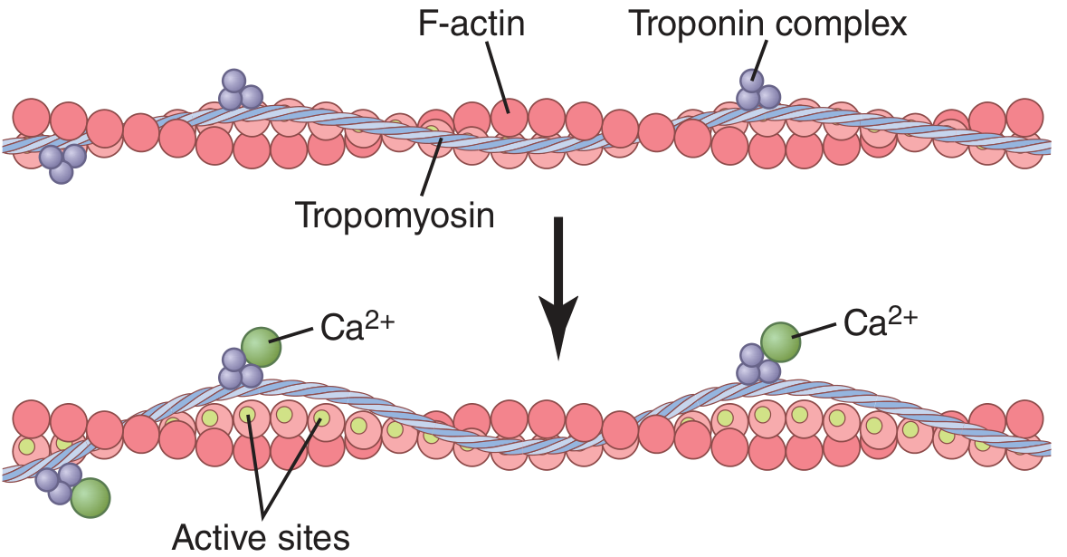
Figure: Top - at rest, tropomyosin covers active sites. Bottom - Ca²⁺ binds TnC, tropomyosin shifts, active sites (green) are exposed for myosin binding.
Once exposed, the myosin cross-bridge heads bind the actin active sites and undergo the power stroke (the "walk-along" or ratchet mechanism), pulling the actin filament toward the center of the sarcomere and shortening the muscle.
"When the troponin complex binds Ca²⁺, a conformation change occurs that tugs on the tropomyosin and exposes myosin binding sites on the actin, permitting myosin to bind to actin and allowing contraction."
- Guyton and Hall Textbook of Medical Physiology
3. Source of Activator Calcium: Excitation-Contraction Coupling
The sequence of events from nerve stimulus to Ca²⁺ release:
- Motor nerve fires → acetylcholine released at the neuromuscular junction → end-plate potential → action potential spreads along the sarcolemma
- Action potential travels down T tubules (transverse tubular invaginations of the plasma membrane), reaching the cell interior
- T tubule depolarisation activates dihydropyridine receptors (DHSRs) - voltage-sensing proteins in the T tubule membrane
- DHSR activation triggers opening of ryanodine receptors (RyR1) - gated Ca²⁺ release channels in the adjacent terminal cisternae of the sarcoplasmic reticulum (SR)
- Massive, rapid Ca²⁺ efflux from SR into the sarcoplasm (up to 20 mM stored in SR, bound to calsequestrin)
- Sarcoplasmic Ca²⁺ rises sharply → binds TnC → contraction begins
"The depolarization of the T-tubule membrane triggers the release of Ca²⁺ from the terminal cisternae to initiate muscle contraction by changes in the thin filaments."
- Histology: A Text and Atlas with Correlated Cell and Molecular Biology
Relaxation occurs when Ca²⁺-ATPase pumps in the SR membrane actively pump Ca²⁺ back into the SR. Cytosolic Ca²⁺ falls within ~30 ms, Ca²⁺ dissociates from TnC, tropomyosin returns to cover the active sites, cross-bridge cycling stops, and the muscle relaxes.
4. Smooth Muscle: Calcium Acts via Calmodulin (Different Mechanism)
Smooth muscle lacks the troponin complex, so Ca²⁺ uses a different pathway:
- Intracellular Ca²⁺ rises (from SR release or extracellular influx through voltage-gated Ca²⁺ channels)
- Ca²⁺ binds calmodulin (4 Ca²⁺ per calmodulin), causing a conformational change
- The Ca²⁺-calmodulin complex binds and activates myosin light chain kinase (MLCK)
- MLCK phosphorylates the regulatory myosin light chain (MLC₂₀) at serine-19
- Phosphorylated MLC₂₀ enables actin to activate myosin ATPase, initiating cross-bridge cycling and contraction
Relaxation requires a decrease in free Ca²⁺; calmodulin dissociates from MLCK, inactivating it. Myosin light chain phosphatase (MLCP) then dephosphorylates MLC₂₀, halting contraction.
"Smooth muscle contraction is regulated by intracellular free calcium (Ca²⁺) acting through calmodulin. Calcium-bound calmodulin undergoes a conformational change, increasing its affinity for myosin light chain kinase. MLC kinase is activated by binding of the calcium-calmodulin complex, leading to phosphorylation of the serine-19 residue of regulatory MLCm."
- Campbell Walsh Wein Urology
5. Cardiac Muscle: Calcium-Induced Calcium Release
In cardiac muscle, an additional mechanism operates - calcium-induced calcium release (CICR):
- Extracellular Ca²⁺ enters through L-type voltage-gated Ca²⁺ channels (unlike skeletal muscle, where DHSR is a purely mechanical trigger)
- This small Ca²⁺ influx triggers much larger Ca²⁺ release from SR via ryanodine receptors (RyR2)
- The resulting Ca²⁺ then binds TnC, activating the troponin-tropomyosin system as in skeletal muscle
"Ultimately, contraction results from the interaction of activator calcium (during systole) with the actin-troponin-tropomyosin system, thereby triggering the actin-myosin interaction."
- Katzung's Basic and Clinical Pharmacology, 16th Ed
Summary: Why Ca²⁺ Is Central
| Aspect | Skeletal Muscle | Cardiac Muscle | Smooth Muscle |
|---|---|---|---|
| Ca²⁺ source | SR (via RyR1) | SR + extracellular (CICR via RyR2) | SR + extracellular |
| Ca²⁺ sensor | Troponin C | Troponin C | Calmodulin |
| Effector | Tropomyosin shift | Tropomyosin shift | MLCK → MLC phosphorylation |
| Without Ca²⁺ | No contraction | No contraction | No contraction |
Ca²⁺ is not merely a modulator - it is the obligatory trigger for all types of muscle contraction. Without a rise in cytosolic ionised calcium, the molecular machinery of contraction cannot be activated in any muscle type.
References:
- Guyton and Hall Textbook of Medical Physiology
- Histology: A Text and Atlas with Correlated Cell and Molecular Biology
- Campbell Walsh Wein Urology
- Katzung's Basic and Clinical Pharmacology, 16th Ed
- Harper's Illustrated Biochemistry, 32nd Ed
This is a shared conversation. Sign in to Orris to start your own chat.