Now I have all the information needed. Let me compile the comprehensive response:
Chronic Liver Disease: Liver Cirrhosis and Complications
POINT 1: Etiology and Pathophysiology of Cirrhosis and Its Complications
Definition
Cirrhosis is defined as the diffuse transformation of the entire liver into regenerative parenchymal nodules surrounded by fibrous bands. It represents the morphologic endpoint of many chronic liver diseases and is technically a histological diagnosis, although most cases are diagnosed by a combination of imaging, clinical signs, and fibrosis markers.
— Robbins & Kumar Basic Pathology
Etiology
The causes of cirrhosis are categorized as follows:
| Category | Causes |
|---|
| Common | Chronic viral hepatitis B ± D, Chronic hepatitis C, Alcohol-related liver disease (ARLD), Non-alcoholic fatty liver disease (NAFLD)/MASH |
| Cholestatic | Primary biliary cholangitis (PBC), Primary sclerosing cholangitis (PSC), Post-surgical biliary strictures |
| Autoimmune | Autoimmune hepatitis |
| Metabolic/Genetic | Hereditary haemochromatosis, Wilson's disease, α1-antitrypsin deficiency, Cystic fibrosis, Galactosaemia, Glycogen storage disease |
| Vascular | Budd–Chiari syndrome, Sinusoidal obstruction syndrome (veno-occlusive disease), Cardiac hepatopathy |
| Drugs/Toxins | Methotrexate, amiodarone |
| Cryptogenic | No identifiable cause (~10–15%) |
— Goldman-Cecil Medicine; Harrison's Principles of Internal Medicine 22E
Worldwide, the three most common causes are chronic viral hepatitis (B and C), alcohol, and NAFLD.
Pathophysiology of Cirrhosis
The central pathophysiologic mechanism is hepatic stellate cell activation:
- Hepatocyte injury (from any cause) triggers Kupffer cells and damaged hepatocytes to release pro-inflammatory cytokines (TGF-β, TNF-α, IL-1).
- Hepatic stellate cells in the space of Disse are activated by these cytokines and transform into myofibroblast-like cells.
- Activated stellate cells produce:
- Collagen (types I and III) → fibrosis
- Pro-inflammatory cytokines → perpetuate hepatocyte damage
- Mediators promoting further tissue fibrosis
- Progressive fibrosis connects portal tracts to each other and to central veins, disrupting normal hepatic architecture.
- Regenerative nodule formation (ductular reactions increase with disease progression) replaces lost parenchyma.
Fibrosis Staging — Metavir Score:
- F0: No fibrosis
- F1: Portal fibrosis without septa
- F2: Portal fibrosis with few septa
- F3: Numerous septa without cirrhosis
- F4: Cirrhosis
Importantly, fibrosis can regress following disease remission or cure — scars become thinner, fragment, and adjacent nodules coalesce. However, even after regression, portal hypertension may persist due to irreversible shunts.
— Robbins & Kumar; Davidson's Principles
Pathophysiology of Major Complications
1. Portal Hypertension
The normal hepatic venous pressure gradient (HVPG) is 3–5 mmHg. Portal hypertension is defined as HVPG >5 mmHg; clinically significant portal hypertension = HVPG >10 mmHg; variceal bleeding risk = HVPG >12 mmHg.
Mechanism (sinusoidal type in cirrhosis):
- Increased resistance: Scarring, parenchymal nodules, and contraction of vascular smooth muscle/myofibroblasts obstruct sinusoidal blood flow.
- Increased portal inflow: Bacterial translocation → systemic inflammation → splanchnic arterial vasodilation (via nitric oxide and other vasodilators) → increased splanchnic blood flow → increased venous efflux into the portal system.
- This hyperdynamic circulation further perpetuates portal pressure.
2. Ascites
- Portal hypertension → increased hydrostatic pressure in hepatic sinusoids → fluid transudation into peritoneal cavity.
- Reduced hepatic synthesis of albumin → ↓ oncotic pressure.
- Splanchnic vasodilation → activation of RAAS and ADH → sodium and water retention.
- ~85% of all ascites is caused by portal hypertension from cirrhosis.
- Fluid is a transudate: protein <3 g/dL; serum-ascites albumin gradient (SAAG) ≥1.1 g/dL.
3. Esophageal Varices / Variceal Bleeding
- Sustained portal hypertension → dilation of portosystemic collateral vessels wherever the systemic and portal circulations share common capillary beds.
- Most dangerous: esophagogastric varices — arise in ~40% of advanced cirrhosis.
- One-third of patients with varices experience a bleed within the first year of diagnosis.
- Associated with coagulopathy → risk of massive, frequently fatal hematemesis.
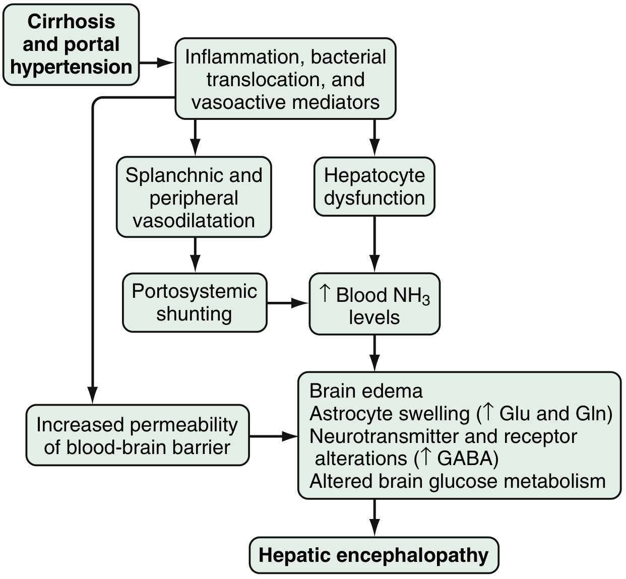
4. Hepatic Encephalopathy (HE)
Pathophysiology involves multiple converging mechanisms:
Fig. Proposed pathophysiology of hepatic encephalopathy. — Sleisenger & Fordtran's GI and Liver Disease
- Ammonia toxicity: Bacteria in the colon metabolize proteins → ammonia → enters portal circulation → normally cleared by hepatocytes, but in cirrhosis, reduced hepatocyte function + portosystemic shunting → hyperammonemia (present in up to 90% of HE patients). Ammonia causes astrocyte swelling, brain edema, and cytotoxic effects.
- GABA-benzodiazepine system upregulation: Enhanced sensitivity of astrocyte peripheral-type benzodiazepine receptors → neurosteroid production (allopregnanolone) → further GABA-A receptor activation → neuroinhibition.
- Other neurotoxins: serotonin, nitric oxide, manganese, circulating opioids.
- Gut–liver axis: Intestinal barrier dysfunction in cirrhosis → bacterial translocation → endotoxemia → Toll-like receptor activation → systemic inflammatory response.
- Increased blood-brain barrier permeability → enhanced cerebral extraction of ammonia and glutamine.
5. Hepatorenal Syndrome (HRS)
- Liver failure produces vasodilators (nitric oxide) → ↑ abdominal visceral blood flow → decreased renal perfusion pressure → ↓ GFR.
- Compensatory activation of renal sympathetic nervous system and RAAS → renal afferent arteriolar vasoconstriction → further ↓ renal perfusion — a vicious cycle.
- No intrinsic renal pathology — the kidney itself is structurally normal.
- Onset: ↓ urine output + rising BUN + rising creatinine (azotemia).
6. Spontaneous Bacterial Peritonitis (SBP)
- Impaired intestinal barrier + suppressed hepatic immune function (reduced Kupffer cell activity, ↓ complement) → bacterial translocation from gut to ascitic fluid.
- Most commonly caused by gram-negative enteric bacteria (E. coli, Klebsiella).
7. Hepatocellular Carcinoma (HCC)
- Most chronic liver diseases predispose to HCC, particularly hepatitis B, C, and alcohol-related cirrhosis.
- Persistent inflammation, regenerative nodule formation, and oxidative DNA damage promote oncogenic transformation.
POINT 2: Risk Factors and Clinical Manifestations
Risk Factors for Cirrhosis
| Category | Risk Factors |
|---|
| Viral | Chronic HBV, HCV, HDV co-infection |
| Alcohol | Prolonged excessive alcohol consumption (>14 units/week in women, >21 in men); duration and amount are most important |
| Metabolic | Obesity, type 2 diabetes, dyslipidemia, metabolic syndrome (NAFLD/MASH pathway) |
| Autoimmune | Female sex, HLA-DR3/DR4 (autoimmune hepatitis) |
| Genetic | Hereditary haemochromatosis (HFE gene), Wilson's disease (ATP7B), α1-AT deficiency (SERPINA1) |
| Biliary | Chronic biliary obstruction, PSC (associated with IBD) |
| Drugs | Long-term methotrexate, amiodarone, nitrofurantoin |
| Other | Ongoing drinking in established alcoholic cirrhosis (worst prognosis modifier) |
Clinical Manifestations
About 40% of patients with cirrhosis are asymptomatic until advanced stages. Early compensated cirrhosis may be an incidental finding.
General/Systemic Symptoms
- Anorexia, weight loss, fatigue, weakness (non-specific)
- Malnutrition/sarcopenia — affects up to 50% of decompensated patients
- Anaemia (multifactorial: chronic inflammation, bone marrow suppression, haemolysis, chronic blood loss)
Hepatic Signs
- Hepatomegaly (though liver may be small and firm in end-stage cirrhosis)
- Jaundice (impaired bilirubin conjugation and excretion)
- Pruritus — from bile salt accumulation in chronic cholestatic cirrhosis
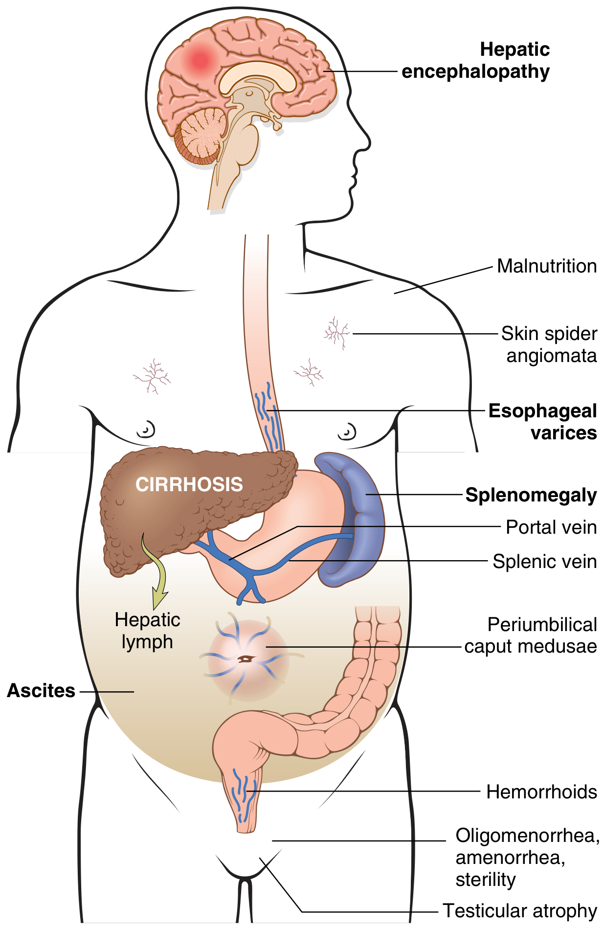
Signs of Portal Hypertension and Portosystemic Shunting
The following diagram illustrates the full spectrum of clinical consequences:
Fig. Major clinical consequences of portal hypertension in the setting of cirrhosis. — Robbins & Kumar Basic Pathology
- Ascites: Abdominal distension, shifting dullness, fluid thrill
- Splenomegaly → hypersplenism: thrombocytopenia, pancytopenia
- Esophagogastric varices → haematemesis, melaena
- Caput medusae: dilated periumbilical veins
- Haemorrhoids: from inferior mesenteric vein collaterals
Endocrine/Cutaneous Changes (from hyperestrogenemia in males due to impaired estrogen catabolism)
- Spider telangiectasia (spider naevi) — >5 is significant
- Palmar erythema
- Gynecomastia, testicular atrophy, loss of body hair (males)
- Oligomenorrhea/amenorrhea (females)
Synthetic Dysfunction Signs
- Coagulopathy: easy bruising, prolonged PT/INR (↓ clotting factor synthesis)
- Hypoalbuminaemia: peripheral oedema
- Hyponatraemia (defective free water clearance)
- Finger clubbing and leukonychia (Terry's nails)
- Dupuytren's contracture (in alcoholic cirrhosis)
- Parotid enlargement (alcoholic cirrhosis)
Signs of Decompensation (Box 24.30 features of chronic liver failure)
| Feature | Notes |
|---|
| Worsening synthetic function | ↑ PT, ↓ albumin |
| Jaundice | Progressive |
| Hepatic encephalopathy | Asterixis (flapping tremor), confusion, coma |
| Ascites | ± SBP |
| Variceal bleeding | Acute UGI haemorrhage |
| Hepatorenal failure | Oligo/anuria, rising creatinine |
Hepatic Encephalopathy — Clinical Grading (West Haven / SONIC Criteria)
| Grade | Intellectual Function | Neuromuscular Function | SONIC Classification |
|---|
| 0/Minimal | Normal exam; subtle work/driving changes | Minor psychometric abnormalities | Covert HE |
| 1 | Mild confusion, altered sleep-wake cycle, forgetfulness | Mild asterixis/tremor | Covert HE |
| 2 | Moderate confusion, disorientation (time) | Obvious asterixis, slurred speech | Overt HE |
| 3 | Marked confusion, somnolence | Asterixis, hyperreflexia, rigidity | Overt HE |
| 4 | Coma | No response to stimuli | Overt HE / Coma |
POINT 3: Management of Cirrhosis and Its Complications
General Principles
Management aims at: (1) treating the underlying cause, (2) maintaining nutrition, (3) monitoring for complications, (4) treating decompensating events, and (5) transplantation when indicated.
A. Treating the Underlying Cause
| Etiology | Specific Treatment |
|---|
| Chronic hepatitis B | Antiviral therapy (tenofovir, entecavir) |
| Chronic hepatitis C | Direct-acting antivirals (cure rates >95%) |
| Alcohol-related | Strict abstinence; nutritional support |
| NAFLD/MASH | Weight loss, metabolic control; emerging: resmetirom (THR-β agonist), obeticholic acid |
| Autoimmune hepatitis | Corticosteroids ± azathioprine |
| Haemochromatosis | Venesection |
| Wilson's disease | D-penicillamine, trientine, zinc |
| PBC | Ursodeoxycholic acid (UDCA) |
B. Nutritional Support
- Daily calorie intake: ≥35 kcal/kg body weight
- Protein: 1.2–1.5 g/kg/day (contrary to old teaching, protein restriction is harmful in most cases)
- Late evening snack to minimise overnight fasting and prevent catabolic state
- Handgrip strength to assess sarcopenia — predicts morbidity and mortality
- Enteral feeding if oral intake inadequate
- BMI unreliable alone — sarcopenia can coexist with obesity (NAFLD)
C. Screening and Surveillance
- 6-monthly liver ultrasound ± AFP for HCC screening (from time of diagnosis)
- Upper GI endoscopy at diagnosis to screen for varices; repeat every 6–12 months
- Child–Pugh score and MELD score for prognosis and transplant listing
Child–Pugh Classification:
| Score 1 | Score 2 | Score 3 | Parameter |
|---|
| None | Mild | Marked | Encephalopathy |
| <34 µmol/L | 34–50 | >50 | Bilirubin |
| >35 g/L | 28–35 | <28 | Albumin |
| <4 sec | 4–6 | >6 | PT prolongation |
| None | Mild | Moderate/Severe | Ascites |
- Child's A (≤6): 82% 1-year, 45% 5-year survival
- Child's B (7–9): 62% 1-year, 20% 5-year survival
- Child's C (≥10): 42% 1-year, 0% 10-year survival
MELD Score = 3.8×ln[bilirubin mg/dL] + 9.6×ln[creatinine mg/dL] + 11.2×ln[INR] + 6.4 → used for transplant prioritisation.
D. Management of Complications
1. Ascites
- Sodium restriction: <88 mmol/day (2 g/day)
- Diuretics (first-line): Spironolactone 100–400 mg/day (aldosterone antagonist) ± furosemide 40–160 mg/day; target weight loss 0.5–1 kg/day
- Large-volume paracentesis (LVP): for tense/refractory ascites; always with IV albumin (6–8 g per litre removed) to prevent post-paracentesis circulatory dysfunction
- TIPS (transjugular intrahepatic portosystemic shunt): for refractory ascites
- Spontaneous Bacterial Peritonitis (SBP): diagnosed by ascitic fluid PMN >250/mm³; treated with IV cefotaxime 2 g TDS or piperacillin-tazobactam; SBP prophylaxis with norfloxacin or ciprofloxacin (long-term)
2. Variceal Bleeding
Primary prevention (before first bleed):
- Non-selective beta-blockers (propranolol, nadolol, carvedilol) — reduce HVPG, reduce risk of bleeding and development of ascites/SBP
- Endoscopic variceal band ligation (EVL) — for medium to large varices
Active variceal bleeding:
- Resuscitation: IV access, blood transfusion (target Hb 70–80 g/L), FFP if coagulopathy
- Vasoactive drugs: Terlipressin (drug of choice in many guidelines), octreotide, or somatostatin infusion — reduce portal pressure
- Antibiotics: IV ceftriaxone (reduces risk of SBP and improves survival in acute variceal bleeding)
- Urgent endoscopy (within 12–24 hours): band ligation (first-line); sclerotherapy if ligation difficult
- Balloon tamponade (Sengstaken-Blakemore tube): temporary measure when endoscopy unavailable or fails; limit to 24 hours
- TIPS: for refractory or recurrent bleeding (effective in >90% of cases)
- Surgical shunts: reserved for cases where TIPS is not feasible
Secondary prevention:
- Combination of non-selective beta-blockers + EVL
3. Hepatic Encephalopathy
Identify and treat precipitating factors: GI bleeding, infection, electrolyte abnormalities (hyponatraemia, hypokalaemia), constipation, dehydration, medications (sedatives, opioids), renal failure.
Pharmacological treatment:
- Lactulose (first-line): 15–30 mL 2–3 times daily, titrated to 2–3 soft stools/day → acidifies the colon, reduces ammonia absorption
- Rifaximin (poorly absorbed antibiotic): 550 mg BD — reduces ammonia-producing gut bacteria with low resistance risk; significantly reduces recurrence of overt HE
- Zinc supplementation: corrects common deficiency in cirrhosis
- LOLA (L-ornithine L-aspartate): promotes hepatic and muscular ammonia detoxification
- Avoid benzodiazepines and sedatives
- Liver transplantation: generally reverses HE
4. Hepatorenal Syndrome (HRS)
- Precipitant identification and treatment: treat infection, stop NSAIDs/diuretics/nephrotoxins, treat hypovolaemia
- Terlipressin + IV albumin (1 g/kg on day 1, then 20–40 g/day): reverses splanchnic vasodilation and improves renal perfusion — first-line for HRS type 1
- Noradrenaline + albumin: alternative to terlipressin
- TIPS: may improve renal function in selected cases
- Renal replacement therapy: as bridge to transplantation
- Liver transplantation: only definitive treatment
5. Spontaneous Bacterial Peritonitis (SBP)
- Diagnosis: ascitic fluid neutrophil count ≥250/mm³ (even before culture result)
- Treatment: IV cefotaxime 2 g every 8 hours for 5 days + IV albumin (1.5 g/kg day 1, 1 g/kg day 3) — albumin prevents HRS development
- Primary prophylaxis: norfloxacin 400 mg/day if ascitic protein <10 g/L or Child's C; secondary prophylaxis lifelong after first SBP episode
6. Hepatocellular Carcinoma
- Surveillance: 6-monthly USS ± AFP
- Curative options: surgical resection (small tumours, compensated cirrhosis), liver transplantation (Milan criteria: single <5 cm or ≤3 nodules <3 cm each), radiofrequency/microwave ablation
- Palliative: TACE (transarterial chemoembolization), sorafenib/lenvatinib (systemic)
E. Liver Transplantation
Indications include:
- Decompensated cirrhosis (Child's C or MELD ≥15)
- Autoimmune hepatitis failing medical therapy
- Well-motivated patients with alcoholic liver disease (abstinent)
- Hereditary metabolic disorders (Wilson's, haemochromatosis, α1-AT deficiency)
- PSC, polycystic liver disease
- MASH cirrhosis (increasingly the leading indication globally)
- Recurrent HCV post-transplant (treated with DAAs)
MELD ≥49 in the UK (UKELD) is minimum score required for transplant listing for liver failure.
POINT 4: Clinical Approach to the Patient with Liver Cirrhosis and Its Complications
Step 1 — History
Establishing the cause:
- Alcohol history (CAGE questionnaire, quantity, duration)
- Risk factors for viral hepatitis (IVDU, blood transfusions, tattoos, sexual contacts)
- Family history (haemochromatosis, Wilson's disease, α1-AT deficiency)
- Drug history (methotrexate, amiodarone, nitrofurantoin)
- Metabolic history (obesity, T2DM, dyslipidaemia → NAFLD)
- Autoimmune symptoms (joint pain, rashes, thyroid disease → autoimmune hepatitis)
Assessing decompensation:
- Abdominal distension, ankle swelling → ascites/oedema
- Haematemesis, melaena → variceal bleeding
- Confusion, altered behaviour, sleep disturbance, driving difficulties → HE
- Oliguria, rising creatinine → HRS
- Fever, abdominal pain in a patient with ascites → SBP
- Weight loss, abdominal pain → HCC
Step 2 — Physical Examination
Systematic approach — look for stigmata of CLD:
| System | Signs |
|---|
| Hands | Palmar erythema, Dupuytren's contracture, leukonychia, finger clubbing, asterixis (flapping tremor) |
| Face | Jaundice (scleral icterus), parotid enlargement, facial telangiectasia |
| Chest/arms | Spider naevi (>5 pathological), gynaecomastia, pectoral/axillary hair loss |
| Abdomen | Hepatomegaly or small firm liver, splenomegaly, ascites (shifting dullness, fluid thrill), caput medusae, prominent superficial veins |
| Genitalia | Testicular atrophy |
| Neurological | Level of consciousness, asterixis, constructional apraxia, fetor hepaticus |
| Vitals | Blood pressure (hypotension in decompensation), temperature (infection/SBP) |
Step 3 — Investigations
Assessing severity of liver disease:
| Test | Significance |
|---|
| Serum albumin | Best indicator of synthetic function; <25 g/L = poor prognosis |
| Prothrombin time / INR | Reflects clotting factor synthesis; degree of prolongation ∝ severity |
| Serum bilirubin | Elevated in decompensation |
| Serum creatinine | >130 µmol/L = poor prognostic marker; HRS screening |
| Serum sodium | Hyponatraemia (<125 mmol/L) = severe disease |
| FBC | Thrombocytopenia (hypersplenism), anaemia, leukopenia |
| Liver enzymes (AST, ALT, ALP, GGT) | May be normal in compensated cirrhosis; deranged in decompensation |
| Serum AFP | >200 ng/mL suggestive of HCC |
| ELF test | <7.7 = none-mild fibrosis; ≥9.8 = severe fibrosis |
| Blood glucose | Hypoglycaemia in acute decompensation |
Identifying the cause:
- Hepatitis B surface antigen (HBsAg), anti-HCV antibodies, HCV RNA
- Serum ferritin, TIBC, transferrin saturation → haemochromatosis
- Serum copper, caeruloplasmin, 24-hour urine copper → Wilson's disease
- Serum α1-antitrypsin
- ANA, ASMA, AMA (anti-mitochondrial antibodies for PBC)
- In young patients: always measure copper and α1-AT
Imaging:
- Ultrasound abdomen (first-line): liver echogenicity, surface nodularity, portal vein patency/diameter, splenomegaly, ascites; excellent for HCC screening
- Transient elastography (FibroScan): liver stiffness — >25 kPa suggests cirrhosis and likely portal hypertension; not usable in ascites/obesity
- CT abdomen/pelvis: irregular liver contour, varices, ascites, spleen size, HCC characterisation
- MRI: superior for HCC characterisation, biliary anatomy (MRCP), vascular anatomy (MRA)
- Upper GI endoscopy: variceal size, stigmata of recent bleeding, portal hypertensive gastropathy
Liver biopsy (gold standard when aetiology/severity uncertain):
- Confirms diagnosis and grades/stages fibrosis
- Immunocytochemistry for viral antigens, oncogenic markers
- Chemical quantification of copper/iron
- Morphometric analysis with picrosirius red staining
- Limitations: sampling error in macronodular cirrhosis; not needed if non-invasive tests are diagnostic
Ascitic fluid analysis (diagnostic paracentesis):
| Test | Significance |
|---|
| Cell count (PMN) | ≥250/mm³ → diagnose SBP |
| Total protein | <25 g/L in portal hypertension ascites |
| SAAG | ≥1.1 g/dL confirms portal hypertension |
| Culture | Identify organism in SBP |
| Cytology | If malignant ascites suspected |
Step 4 — Prognostic Assessment
Calculate Child–Pugh and MELD score at every assessment to guide:
- Timing of referral for liver transplantation (MELD ≥15 = benefit from transplant)
- Intensity of follow-up
- Surgical risk stratification
- Prognosis discussion with patient
Poor prognostic indicators:
- Serum albumin <28 g/L; sodium <125 mmol/L
- PT >6 sec above normal; creatinine >130 µmol/L
- Persistent jaundice; unresponsive ascites
- Recurrent variceal haemorrhage with poor liver function
- Progressive neuropsychiatric complications
- Small liver on imaging
- Continued alcohol use in alcoholic cirrhosis
- Development of HCC
Step 5 — Management Plan (Individualised)
- Treat the underlying aetiology (as above)
- Nutritional optimisation
- Manage current complications (ascites, SBP, variceal bleeding, HE, HRS — as detailed in Point 3)
- Surveillance programme: 6-monthly USS (HCC), endoscopy (varices), regular MELD/Child–Pugh reassessment
- Avoid hepatotoxins: alcohol, NSAIDs (worsen renal function/ascites/GI bleeding), aminoglycosides, sedatives
- Vaccinations: Hepatitis A, Hepatitis B (if non-immune), pneumococcal, influenza
- Address Acute-on-Chronic Liver Failure (ACLF) if decompensation occurs:
- Identify precipitant (alcoholic hepatitis, infection, viral reactivation, GI bleed)
- Organ support as needed
- 28-day mortality: 23% (1 organ failure), 32% (2 organs), >75% (≥3 organs)
- Liver transplantation referral when appropriate
Summary Flow: Clinical Approach
Suspected CLD/Cirrhosis
         ↓
Thorough history (aetiology, decompensation, risk factors)
         ↓
Physical examination (stigmata of CLD, ascites, encephalopathy)
         ↓
Lab investigations (LFTs, FBC, coagulation, renal profile, aetiology screen)
         ↓
Imaging (USS ± FibroScan → CT/MRI if needed)
         ↓
Endoscopy (variceal screening) ± Liver biopsy (if needed)
         ↓
Prognostic scoring (Child–Pugh, MELD)
         ↓
Treat underlying cause + Manage complications + Surveillance
         ↓
Transplant assessment when MELD ≥15 or Child's C
Sources:
- Robbins & Kumar Basic Pathology (Elsevier)
- Goldman-Cecil Medicine, International Edition, 2-Volume Set (Elsevier)
- Harrison's Principles of Internal Medicine 22E, 2025 (McGraw-Hill)
- Davidson's Principles and Practice of Medicine (Elsevier)
- Sleisenger and Fordtran's Gastrointestinal and Liver Disease (Elsevier)
- Current Surgical Therapy 14e (Elsevier)
- Attached PDF: Liver Cirrhosis (clinical notes)This is a comprehensive synthesis covering all four points you requested, drawn from your uploaded PDF, Harrison's Principles, Robbins & Kumar, Davidson's, Goldman-Cecil, Sleisenger & Fordtran, and Current Surgical Therapy. Here is a summary of what was covered:
Quick Navigation Guide
Point 1 — Etiology & Pathophysiology
- Full etiology table (common, cholestatic, metabolic/genetic, vascular, drug causes)
- Stellate cell activation pathway → collagen deposition → fibrosis → nodule formation
- Metavir fibrosis staging (F0–F4)
- Pathophysiology of each major complication: portal hypertension (sinusoidal resistance + hyperdynamic circulation), ascites (SAAG ≥1.1), varices (HVPG >12 mmHg), hepatic encephalopathy (ammonia, GABA, gut-liver axis), hepatorenal syndrome (splanchnic vasodilation → renal vasoconstriction), SBP, and HCC
Point 2 — Risk Factors & Clinical Manifestations
- Risk factor table by category
- Full clinical picture from compensated (asymptomatic, early signs) to decompensated (ascites, variceal bleed, HE, HRS)
- West Haven/SONIC grading for encephalopathy
- Illustrated diagram of portal hypertension consequences
Point 3 — Management
- Cause-specific therapy (antivirals, abstinence, immunosuppression, venesection, UDCA)
- Nutrition (35 kcal/kg, 1.2–1.5 g protein/kg, late evening snack)
- Ascites: spironolactone, LVP + albumin, TIPS, SBP treatment/prophylaxis
- Variceal bleeding: terlipressin + antibiotics + EVL + TIPS
- HE: lactulose + rifaximin + precipitant removal
- HRS: terlipressin + albumin → transplant
- Child-Pugh and MELD scoring tables
- Liver transplant indications
Point 4 — Clinical Approach
- Structured history (aetiology + decompensation screening)
- Physical examination checklist (hands → face → chest → abdomen → genitalia → neurology)
- Full investigation grid (labs, imaging, endoscopy, liver biopsy, ascitic tap)
- Prognostic assessment and poor prognostic indicators
- Step-by-step management flow from diagnosis to transplant referral