
Medicine for allergic rhinitis
"allergic rhinitis"[MeSH Terms] AND treatment
allergic rhinitis treatment pharmacology drugs diagram
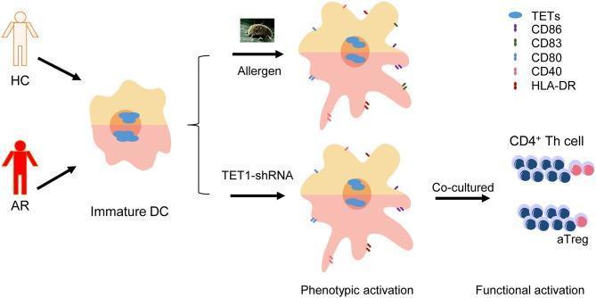
A pathophysiology diagram illustrating the signaling pathway and functional activation of dendritic cells (DCs) in Allergic Rhinitis (AR) compared to Healthy Controls (HC). The visual workflow begins with an immature DC, containing Ten-eleven translocation (TET) enzymes in the nucleus. Two experimental pathways for phenotypic activation are shown: allergen challenge (depicting a dust mite icon) and TET1-shRNA treatment. Both interventions result in the maturation of the DC, characterized by increased surface expression of costimulatory molecules including CD80, CD83, CD86, CD40, and HLA-DR. The final stage demonstrates functional activation through the co-culture of these activated DCs with CD4+ Th cells. The diagram highlights that these interactions lead to the differentiation of CD4+ T cell subsets, specifically showing the modulation of activated T regulatory cells (aTreg) and the persistent presence of TETs. This infographic summarizes how the loss or inhibition of TET1 in atopic DCs mimics allergen-induced maturation and influences subsequent T-cell mediated immune responses.
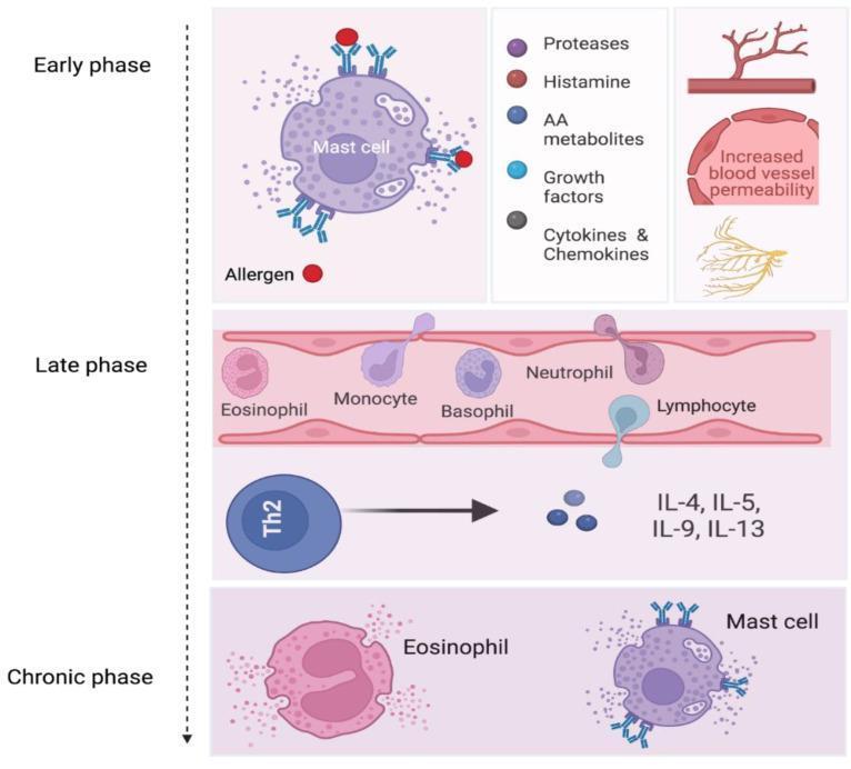
A pathophysiology diagram illustrating the three stages of allergic rhinitis: Early phase, Late phase, and Chronic phase. The 'Early phase' section depicts a mast cell activated by allergens binding to IgE on its surface receptors, triggering the release of inflammatory mediators including proteases, histamine, arachidonic acid (AA) metabolites, growth factors, and cytokines/chemokines. Associated vascular changes such as increased blood vessel permeability and nerve stimulation are shown. The 'Late phase' section demonstrates leukocyte recruitment and extravasation through a blood vessel wall, featuring an eosinophil, monocyte, basophil, neutrophil, and a migrating lymphocyte. It also shows a Th2 cell secreting interleukins IL-4, IL-5, IL-9, and IL-13. The 'Chronic phase' section illustrates the persistent presence and coexistence of degranulating eosinophils and mast cells in the inflamed tissue. This diagram provides a clinical overview of the immunological cascade and cellular interactions involved in allergic airway inflammation and tissue remodeling.
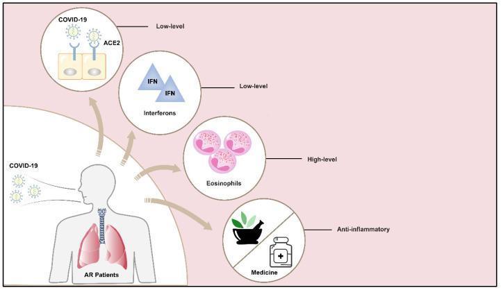
This medical infographic illustrates the pathophysiological landscape of Allergic Rhinitis (AR) in the context of COVID-19 susceptibility. A central human figure representing an AR patient is shown inhaling COVID-19 viral particles. Four circular callouts detail specific immunological and pharmacological factors influencing disease outcomes. The diagram highlights that AR patients exhibit 'low-level' ACE2 receptors on epithelial cells and 'low-level' Interferons (IFN), both of which are factors that may limit viral entry and replication. Conversely, the image depicts 'high-level' eosinophils, which are noted for their protective role in antiviral host defense. The final callout represents treatment strategies, labeled 'Anti-inflammatory Medicine,' featuring icons of a mortar and pestle with botanicals alongside a medical bottle, reflecting the use of both inhaled corticosteroids and traditional medicine. The illustration serves as an educational summary of why patients with allergic rhinitis might experience different susceptibility or severity levels when infected with SARS-CoV-2.
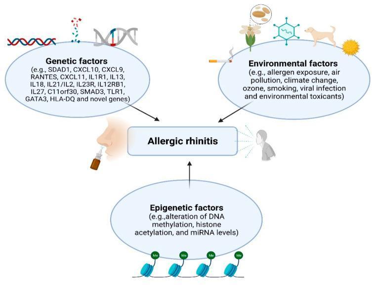
A pathophysiology diagram illustrating the multifactorial etiology of Allergic Rhinitis (AR). The graphic is organized into three major thematic bubbles converging on a central 'Allergic rhinitis' node, which is accompanied by icons of a nasal spray and an affected human profile. 1. Genetic Factors: Depicts a DNA double helix and lists specific susceptibility genes including SDAD1, Various Interleukins (IL1R1, IL13, IL18, IL21/IL2, IL23R, IL12RB1, IL27), Chemokines (CXCL9, CXCL10, CXCL11, RANTES), and others like SMAD3, GATA3, and HLA-DQ. 2. Environmental Factors: Features icons representing cigarette smoke (smoking), a dog (pet dander), a flower (pollen), a virus (infection), and the sun (climate change/ozone), alongside air pollution and toxicants. 3. Epigenetic Factors: Highlights mechanisms such as DNA methylation, histone acetylation, and miRNA level alterations, visually represented by chromatin structures with 'Me' (methylation) tags. This educational infographic summarizes the complex interplay between hereditary predisposition, external triggers, and gene-expression modifications in the pathogenesis of respiratory allergic disease.
Medicines for Allergic Rhinitis
ARIA Management Algorithm (ARIA 2008 Update)
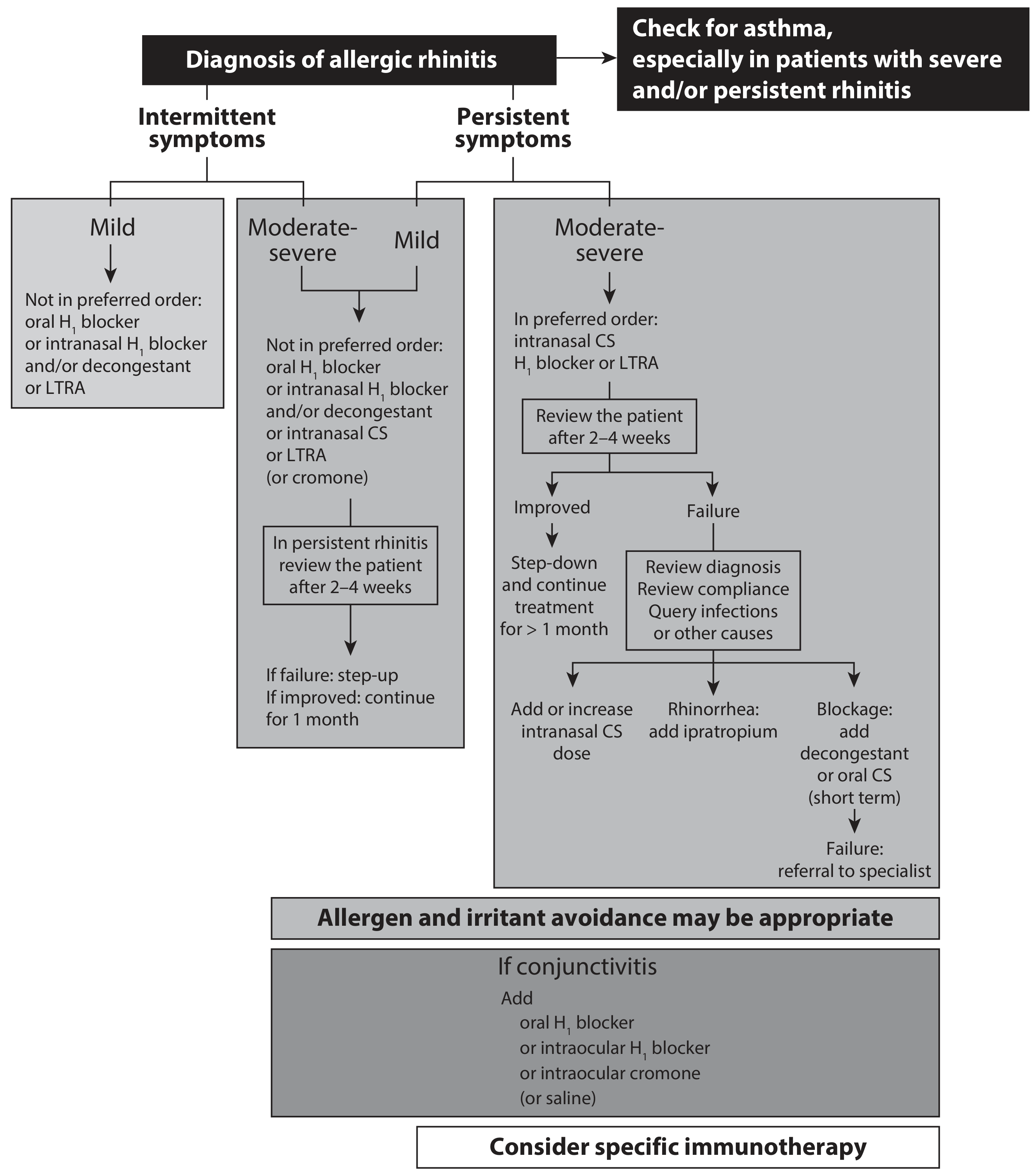
Drug Classes & Their Effects on Symptoms
| Drug | Itch/Sneezing | Discharge | Blockage | Impaired Smell |
|---|---|---|---|---|
| Oral antihistamines | +++ | ++ | +/- | - |
| Topical corticosteroids | +++ | +++ | ++ | + |
| Oral corticosteroids | +++ | +++ | +++ | ++ |
| Ipratropium bromide | - | +++ | - | - |
| Topical decongestants | - | - | +++ | - |
| Antileukotrienes | - | ++ | + | +/- |
| Sodium cromoglicate | + | + | +/- | - |
1. Antihistamines (H1 Blockers)
- Chlorphenamine (chlorpheniramine)
- Diphenhydramine
- Note: Ketotifen still has some use due to additional mast cell stabilizer activity
- Problem: Sedation
- Loratadine - once daily, non-sedating, safe for children
- Cetirizine - once daily, non-sedating, safe for long-term use
- Fexofenadine - non-sedating
- Onset: Usually < 1 hour; better when used regularly rather than as-needed
- Azelastine - rapid symptom control; can combine with intranasal steroid; downside is bitter taste and twice-daily dosing
- Azelastine eyedrops also useful for ocular symptoms
2. Intranasal Glucocorticosteroids (Most Effective Overall)
- Beclometasone dipropionate (older; mild growth retardation noted in children)
- Budesonide
- Fluticasone propionate - lower systemic bioavailability, preferred in children
- Mometasone furoate - very low systemic bioavailability
- Triamcinolone
3. Systemic Corticosteroids
- Prednisolone 20-40 mg/day is usually sufficient
- Keep treatment as short as possible to minimize systemic side effects
- Depot injectable steroids (e.g., triamcinolone acetonide IM): effective but NOT recommended - effects cannot be reversed once administered
4. Leukotriene Receptor Antagonists (LTRAs)
- Montelukast - as effective as loratadine for nasal symptoms; less effective than intranasal steroids
- Licensed for allergic rhinitis associated with asthma - dual benefit for both conditions
- Variable individual responsiveness; a monitored trial is reasonable
- Part of the ARIA treatment guidelines
5. Decongestants
- Xylometazoline
- Oxymetazoline
- Warning: Use for no more than 5-7 days - prolonged use causes rhinitis medicamentosa (rebound congestion/dependency)
- Pseudoephedrine (oral) - less rebound risk but systemic side effects (hypertension, palpitations, insomnia)
6. Sodium Cromoglicate (Mast Cell Stabilizer)
- Modest effect on rhinitis symptoms
- Must be used 4x daily - limits compliance
- No side effects; safe for young children
- Cromoglicate eyedrops are effective for ocular itching
- Also available as intranasal spray and eye drops
7. Ipratropium Bromide (Anticholinergic)
- Intranasal spray
- Specifically targets rhinorrhea (watery discharge) - very effective (+++)
- No effect on itching, sneezing, or blockage
- Added when persistent rhinorrhea remains after intranasal corticosteroids
8. Allergen-Specific Immunotherapy (AIT)
- Subcutaneous immunotherapy (SCIT): Injections of gradually increasing allergen doses; requires specialist referral
- Sublingual immunotherapy (SLIT): Under-tongue tablets or drops; better safety profile
- Considered for moderate-severe persistent rhinitis not controlled by pharmacotherapy
- Effective for: grass pollen, house dust mite, cat dander, tree pollen
Quick Treatment Summary by Dominant Symptom
| Dominant Symptom | First Choice |
|---|---|
| Sneezing + itching + runny nose | Oral or intranasal antihistamine |
| Nasal blockage | Intranasal corticosteroid |
| All symptoms (moderate-severe) | Intranasal corticosteroid +/- antihistamine |
| Watery discharge not responding | Add ipratropium bromide |
| Seasonal mild symptoms | Oral antihistamine or LTRA |
| Rhinitis + asthma | LTRA (montelukast) + intranasal steroid |
| Ocular symptoms | Antihistamine (oral or eye drops) or cromoglicate drops |
| Refractory disease | Specialist referral + consider immunotherapy |
Important Practical Points
- Medications control symptoms but do not cure allergy - they are needed for as long as allergen exposure persists
- For seasonal allergy (e.g., grass pollen), start treatment 1 month before pollen season begins
- Unified airway concept: Treat both nose and lungs together - nasal steroids reduce asthma symptoms and vice versa
- Recheck patients at 2-4 weeks to assess response and step up/down therapy
Anti histamine
antihistamine H1 receptor pharmacology classification generations
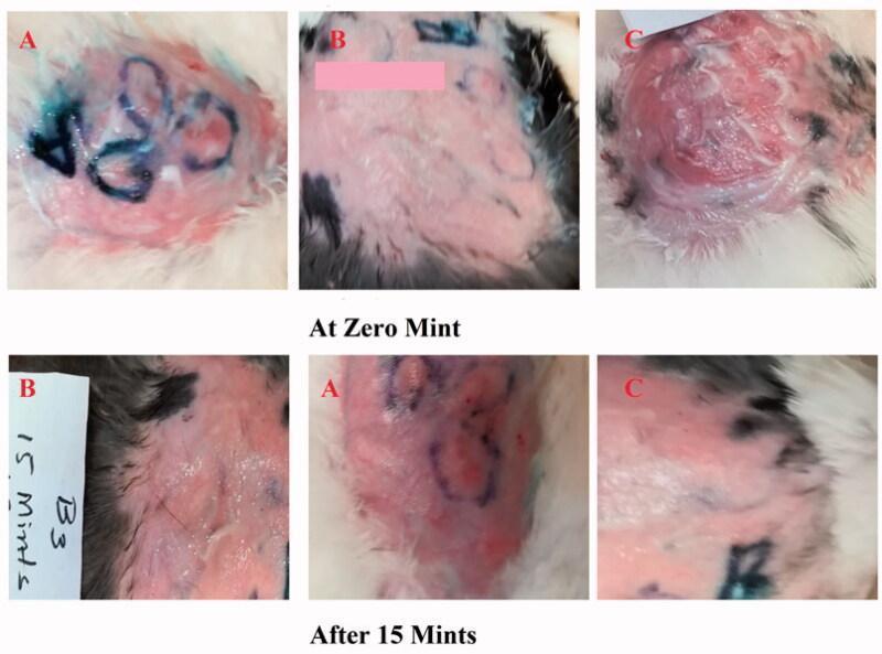
This comparison clinical photograph depicts the efficacy of topical antihistamine formulations on induced skin reactions in a rabbit model. The visual is organized into two rows representing time points: 'At Zero Mint' and 'After 15 Mints', subdivided into three experimental groups (A, B, and C). Group A evaluates a novel Ebastine emulgel, Group B serves as a positive control using a standard antihistamine (Benadryl), and Group C acts as the untreated negative control. At 0 minutes, all groups show histamine-induced erythema and wheal formation, with Group C exhibiting the most severe, raised, and inflamed lesion. Surface markings in blue and pink demarcate treatment zones. At 15 minutes, Group A and Group B demonstrate a visible reduction in the intensity and area of erythema, indicating effective drug absorption and inhibition of the H1-receptor-mediated allergic response. In contrast, Group C maintains significant, persistent inflammation and redness. The images illustrate the clinical progression of allergic skin lesions and the therapeutic effect of topical emulgel formulations on inflammatory symptoms like redness and swelling.
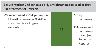
Summary : This figure presents a clinical recommendation regarding the use of modern 2nd generation H1-antihistamines as first-line treatment for urticaria, supported by strong consensus and evidence. flowchart: # Recommendation Statement : • "Should modern 2nd generation H1-antihistamines be used as first-line treatment of urticaria?" (header). • Recommendation: "We recommend a 2nd generation H1-antihistamine as first-line treatment for all types of urticaria." # Consensus and Evidence : • Strong consensus (indicated by "↑↑" and the phrase "Strong consensus¹"). • Evidence- and consensus-based (reference to an Evidence Report). # Visual Elements : • Central green vertical bar with two upward arrows ("↑↑") indicating strong recommendation. • Text boxes to the left (recommendation) and right (consensus/evidence) of the green bar. Analysis : • The figure visually and textually communicates a strong, evidence-based consensus to use modern 2nd generation H1-antihistamines as the first-line treatment for all types of urticaria. • The use of bold, boxed text and arrows emphasizes the strength and clarity of the recommendation.
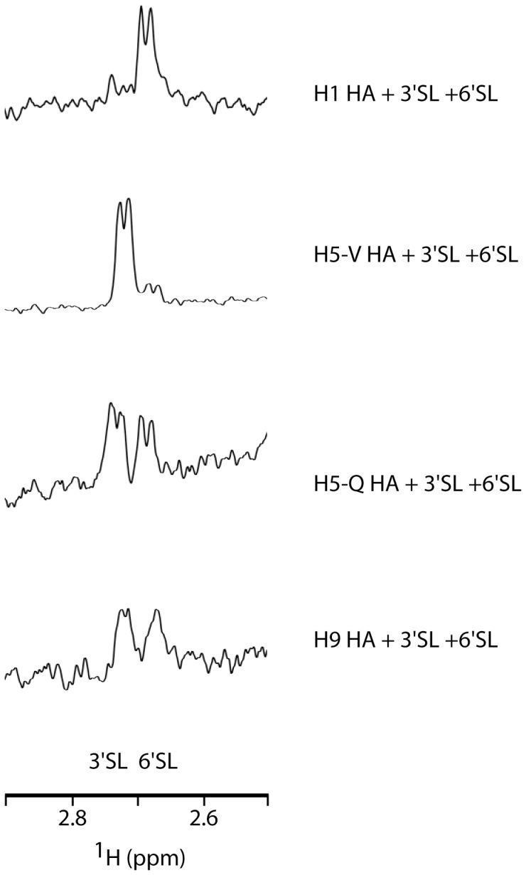
The image displays Saturation Transfer Difference (STD) NMR spectra illustrating competition assays between different influenza A hemagglutinin (HA) subtypes and receptor analogs. Four vertically stacked spectra compare the binding affinities of H1 HA, H5-V HA, H5-Q HA, and H9 HA toward two sialic acid analogs: 2,3-sialyllactose (3'SL, human-like) and 2,6-sialyllactose (6'SL, avian-like). The x-axis represents the 1H chemical shift in ppm, ranging from 2.6 to 2.8, specifically focusing on the H3eq resonance peaks. In the H1 HA spectrum, a dominant peak at 2.67 ppm signifies a higher binding preference for 6'SL. Conversely, the H5-V HA spectrum shows a prominent peak at 2.73 ppm, indicating a strong preference for 3'SL. The H5-Q and H9 HA spectra exhibit dual binding patterns with relatively equal peak intensities for both 3'SL and 6'SL, though with varying levels of signal resolution. This diagnostic visualization is used in virology and pharmacology to determine host-receptor specificity and pandemic potential of different influenza strains.
Antihistamines (H1-Receptor Blockers)
Mechanism of Action
Classification
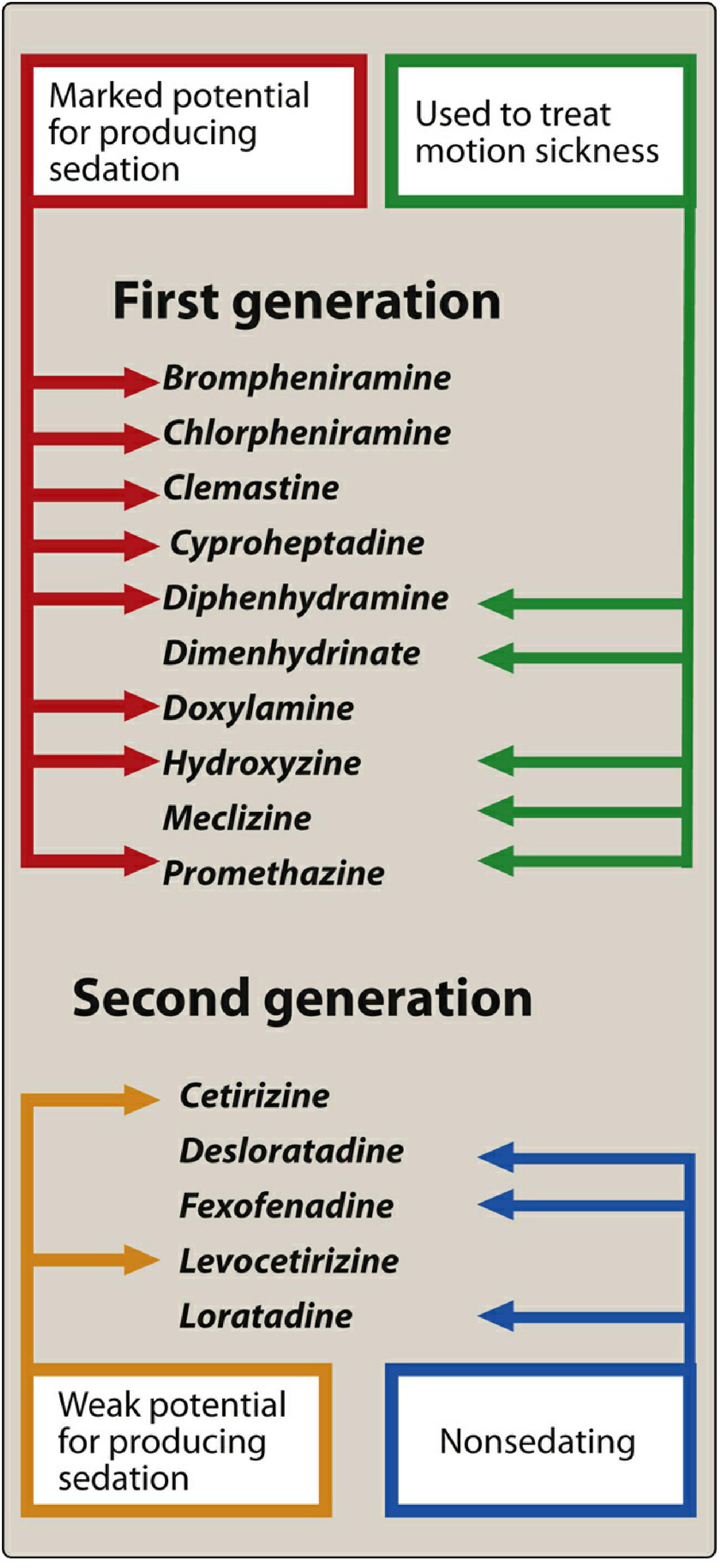
First-Generation Antihistamines
| Chemical Class | Drugs | Duration |
|---|---|---|
| Alkylamines | Chlorpheniramine, Brompheniramine | 24h / 4-6h |
| Ethanolamines | Diphenhydramine, Clemastine, Doxylamine | 12h |
| Piperazines | Hydroxyzine, Cyclizine, Meclizine | 6-24h |
| Phenothiazines | Promethazine | 4-6h |
| Piperidines | Cyproheptadine | 4-6h |
| Tricyclic | Doxepin | 6-24h |
Key Properties - First Generation:
- Sedation - marked CNS depression (cross BBB)
- Anticholinergic effects - dry mouth, blurred vision, urinary retention, constipation
- Antimuscarinic - explains antiemetic and antinausea uses
- Cyproheptadine - uniquely also blocks serotonin (5-HT2A) receptors; used for appetite stimulation and serotonin syndrome
- Ketotifen - has additional mast cell stabilizing activity
- Diphenhydramine, dimenhydrinate, meclizine, promethazine - effective for motion sickness (must be taken before symptoms start)
Second-Generation Antihistamines
| Drug | Sedation | Notes |
|---|---|---|
| Loratadine | Nonsedating | Active metabolite = desloratadine |
| Desloratadine | Nonsedating | Active metabolite of loratadine |
| Fexofenadine | Nonsedating | Does not penetrate CNS at all |
| Cetirizine | Weakly sedating | Carboxylated derivative of hydroxyzine |
| Levocetirizine | Weakly sedating | Active R-enantiomer of cetirizine |
| Azelastine | Minimal | Also has mast cell stabilizing action; available as nasal spray and eye drops |
| Olopatadine | Minimal | Ophthalmic formulation; used in allergic conjunctivitis |
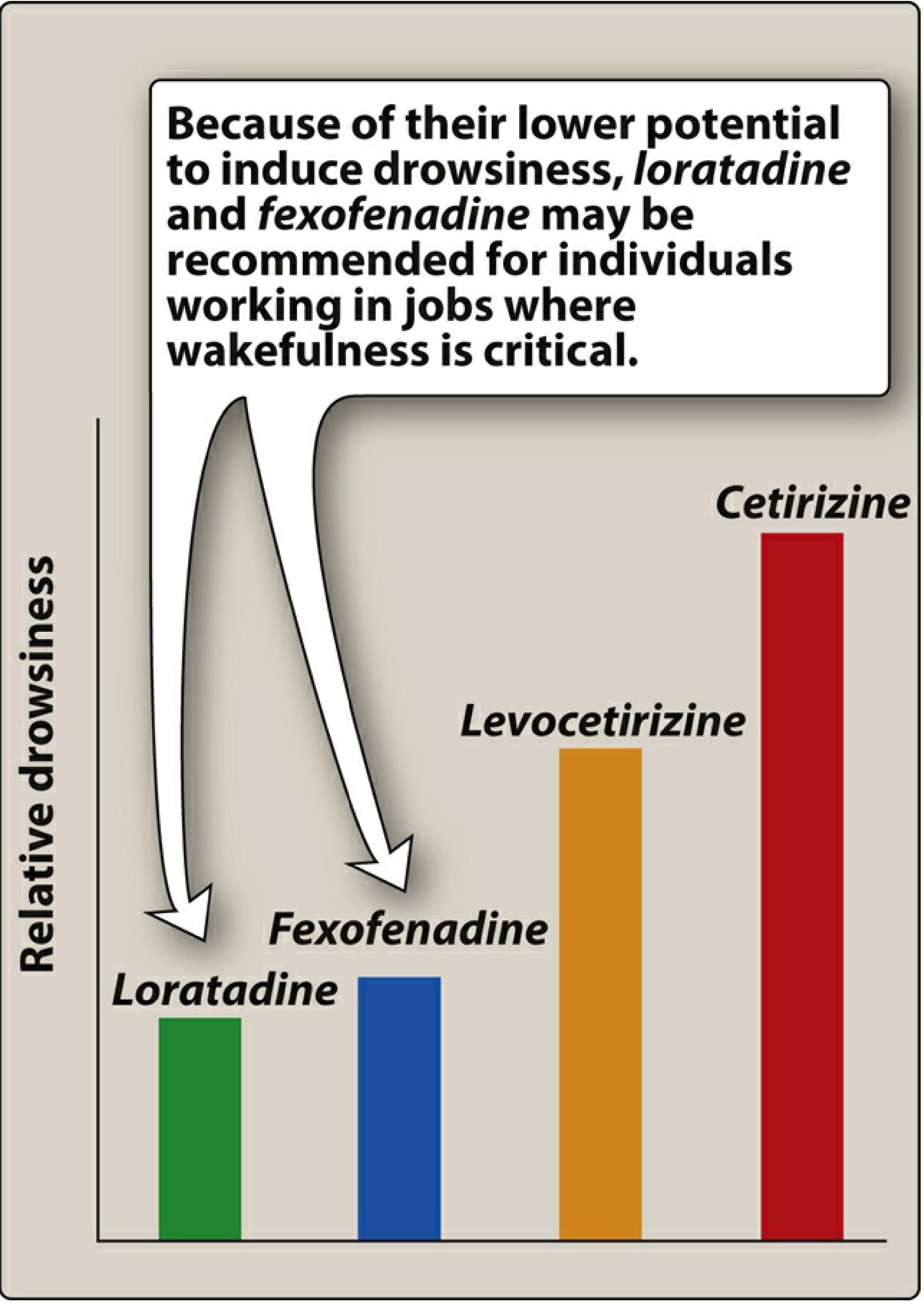
Relative Drowsiness Among Second-Generation Agents:
Clinical Uses
| Indication | Preferred Agent(s) |
|---|---|
| Allergic rhinitis | 2nd-gen oral (loratadine, cetirizine, fexofenadine) |
| Urticaria (hives) | 2nd-gen H1 antihistamine - first-line |
| Allergic conjunctivitis | Azelastine, olopatadine, ketotifen (eye drops) |
| Motion sickness | Diphenhydramine, dimenhydrinate, meclizine, promethazine |
| Nausea/vomiting | Promethazine, diphenhydramine |
| Insomnia (short-term) | Diphenhydramine, doxylamine |
| Pruritus (itch) | Hydroxyzine (sedating, useful at night), cetirizine |
| Anaphylaxis (adjunct) | Diphenhydramine IV/IM (adjunct only - epinephrine is the drug of choice) |
| Appetite stimulation | Cyproheptadine |
| Serotonin syndrome | Cyproheptadine |
| Cough suppression | Diphenhydramine |
Important: H1 blockers are NOT effective for bronchial asthma - histamine is only one of many mediators in asthma. Epinephrine (not antihistamines) is the drug of choice for systemic anaphylaxis.
Side Effects Comparison
| Effect | 1st Generation | 2nd Generation |
|---|---|---|
| Sedation | ++++ | - to + |
| Anticholinergic (dry mouth, urinary retention) | +++ | - |
| Cognitive impairment | +++ | - |
| Cardiac arrhythmia | + (some) | Rare (terfenadine/astemizole withdrawn for QT prolongation) |
| Appetite stimulation | + | - |
| Motion sickness use | Yes | No |
Intranasal Antihistamines (Topical)
- Azelastine nasal spray - rapid action; can combine with intranasal steroid (Dymista = azelastine + fluticasone)
- Twice-daily dosing; bitter taste is the main complaint
- Useful when quick symptom control is needed
Practical Pearls
- Take before allergen exposure for best effect (preventive > reactive)
- Loratadine or fexofenadine are best for daytime use (pilots, drivers, exam students)
- Cetirizine is slightly sedating but more effective for itching in some patients
- Hydroxyzine at bedtime is useful for chronic pruritus with insomnia
- Antihistamines alone do not relieve nasal blockage - add a decongestant or intranasal steroid for that