

1000378699.jpg
Interpret the Botox injections why they give what are the underlying causes.
analyse_media
Reading File
Now let me search the medical library for detailed information on post-stroke spasticity and Botox (botulinum toxin) treatment, while also searching for relevant medical images.
Finding Sources
Finding Sources
Searching Images
post-stroke spasticity botulinum toxin injection sites upper limb
Reading File
Finding Sources
Finding Sources
Reading File
Reading File
I now have all the information needed for a thorough, well-sourced interpretation. Here is the full analysis:
Document Interpretation: Mr. K.V.H. Gupta's Medical Records
What the Documents Say
Document 1 - "To Whomsoever It May Concern" Letter (Suryamukhi Hospital, Srikakulam)
Mr. K.V.H. Gupta; 61-year-old Male; Known history of Status Post residual Left ICH (Intracranial Hemorrhage) / brain stroke with residual Right Upper Limb & Lower Limb severe spasticity & gait dysfunction & currently requiring interval Botox injections for post-stroke spasticity. Further he needs continuous physiotherapy & further Botox injection optimization based on degree of spasticity & gait dysfunction.
Document 2 - Follow-up and Treatment Plan dated 30/7/24 (July 30, 2024), Dr. Raja Kumar SR
- BP: 120/80 mmHg (normal)
- SpO2: 98%
- PR: 73 bpm
- Weight: 85 kg
- Currently administering 10(?) units Botox injection for post-stroke spasticity
- Total dose: 400 U
- UL & EM (Upper Limb & Electromyography guidance, or likely "UL & EM muscles")
Clinical Interpretation: Why Botox is Being Given
1. The Underlying Cause - Left ICH (Left-Sided Intracranial Hemorrhage / Brain Stroke)
Mr. Gupta suffered a left-sided intracranial hemorrhage (ICH) - a type of hemorrhagic stroke where bleeding occurs within the brain tissue itself. Because the brain is organized contralaterally (each hemisphere controls the opposite side of the body), a left-sided bleed causes right-sided deficits. This explains why he has:
- Right Upper Limb (RUL) severe spasticity - the arm is likely held in a flexed, internally rotated posture
- Right Lower Limb (RLL) severe spasticity - typically causing an equinus foot/foot drop posture and a stiff-legged gait
- Gait dysfunction - difficulty walking due to the spastic lower limb
2. What is Post-Stroke Spasticity?
After a stroke damages the brain's upper motor neurons (UMN), the normal inhibitory control over spinal cord reflexes is lost. This results in:
- Hyperreflexia - exaggerated tendon reflexes
- Hypertonia - increased muscle tone with velocity-dependent resistance to passive movement (classic spasticity)
- Clasp-knife phenomenon - initial resistance then sudden "give" on passive stretch
- Flexor posturing in the arm, extensor posturing in the leg
- Clonus - rhythmic involuntary muscle contractions
- Contractures over time - permanent shortening of muscles and tendons
The muscles most affected in post-stroke hemiplegia are:
- Upper limb: shoulder internal rotators, elbow flexors (biceps), wrist/finger flexors
- Lower limb: hip adductors, knee flexors (hamstrings), plantar flexors (calf/gastrocnemius - causing equinus/foot drop)
This spasticity directly impairs rehabilitation, walking ability, dressing, and quality of life.
3. Why Botox (Botulinum Toxin Type A)?
Botulinum toxin type A (brand name Botox) is a focal neuromuscular blocking agent. It works by:
- Blocking the release of acetylcholine at the neuromuscular junction of the injected muscle
- This causes temporary, reversible paralysis/weakening of the overactive spastic muscle
- The effect typically lasts 3-4 months, which is why "interval" injections are needed (as stated in this document)
- The dose of 400 units total noted in the second document is consistent with standard multi-muscle dosing protocols for post-stroke spasticity
Botox injections for spasticity are most effective for the wrist and finger flexors and plantar flexors of the ankle, as stated in Bradley and Daroff's Neurology in Clinical Practice - exactly the muscles affected in Mr. Gupta's condition.
The goals of Botox in his case are:
- Reduce muscle tone in the right arm and leg
- Improve passive and active range of motion
- Improve gait - reduce equinus foot posture, improve walking pattern
- Facilitate physiotherapy - a looser limb can be more effectively exercised
- Prevent contractures - reduce the risk of permanent joint deformity
- Improve ease of care - easier to dress, bathe, and manage the limb
4. Why Interval Injections?
Because Botox effects are temporary (lasting approximately 3 months), repeated injections are needed on a regular schedule. The first document specifically states "interval Botox injections" and the treating team wants to optimize the dose and injection sites based on:
- The degree of spasticity (measured on the Modified Ashworth Scale or Tardieu Scale)
- Gait dysfunction severity (via clinical observation or gait analysis)
Per Miller's Review of Orthopaedics, when functional joint ranging is insufficient to control deformity, interventional modalities including botulinum toxin type A are indicated alongside orthotic prescription and serial casting.
5. The Role of Physiotherapy
The letter specifically states the patient needs continuous physiotherapy alongside Botox. This is because:
- Botox alone does not restore function - it only reduces the spastic resistance
- Physiotherapy during the "window" of reduced spasticity helps retrain movement patterns, strengthen weak muscles, and improve walking
- Without exercise, the benefit of Botox is largely limited to ease of passive movement rather than functional gain
Summary Table
| Finding | Explanation |
|---|---|
| Left ICH / Brain Stroke | Left-sided hemorrhagic stroke damaging upper motor neurons |
| Right-sided hemiplegia | Contralateral control: left brain controls right body |
| Severe UL & LL spasticity | Loss of UMN inhibition → uninhibited stretch reflexes |
| Gait dysfunction | Spastic equinus foot, stiff knee, scissor gait pattern |
| Botox 400 U (interval) | Temporarily blocks ACh release at neuromuscular junction → reduces tone |
| Physiotherapy required | To capitalize on Botox window for functional retraining |
| Dose optimization | Titrate to degree of spasticity and gait response on follow-up |
Sources: Bradley and Daroff's Neurology in Clinical Practice (UMN spasticity management, botulinum toxin evidence); Miller's Review of Orthopaedics 9th Edition (interventional modalities for post-stroke spasticity)
Create a detailed guide on Botox injection site anatomy for post-stroke upper/lower limb spasticity
For post-stroke spasticity, Botox targets the overactive spastic muscles in predictable patterns - flexors dominate in the upper limb, while extensors/plantar flexors dominate in the lower limb. Standard injection sites follow established anatomical landmarks, typically guided by EMG or ultrasound.
Searching sources...
Finding Sources
Finding Sources
Searching Images
upper limb spasticity flexor posture stroke hemiplegia anatomy
Finding Sources
Finding Sources
Finding Sources
Reading File
Finding Sources
Finding Sources
Reading File
Searching the Web
post-stroke spasticity botulinum toxin injection sites anatomy upper lower limb clinical guide 2024
Loading Image
Reading Web Page
https://pmc.ncbi.nlm.nih.gov/articles/PMC12116053
Finding Sources
I now have comprehensive material from textbooks, clinical guidelines, and recent 2024 literature. Here is the full guide:
Botox Injection Site Anatomy Guide: Post-Stroke Upper & Lower Limb Spasticity
Background: Why These Specific Muscles?
After a stroke, loss of descending cortical inhibition creates a stereotyped spastic posture pattern. In the upper limb, flexors dominate (the arm "curls in"). In the lower limb, extensors and plantar flexors dominate (the leg "stiffens out" but the foot plantarflexes/inverts). Botox targets the overactive muscles in each pattern to reduce tone, improve range of motion, and allow physiotherapy to achieve functional gains.
The standard of care is now ultrasound-guided or EMG-guided injection to the zone of maximum muscle thickness and motor nerve arborization (motor end-plate zone), as international consensus guidelines confirm this improves accuracy over manual landmark-only techniques.
PART 1: UPPER LIMB
The classic post-stroke upper limb posture is:
Shoulder adducted + internally rotated → elbow flexed → forearm pronated → wrist flexed → fingers flexed → thumb-in-palm
Each component has a responsible muscle group that is the injection target.
1.1 Shoulder - Internal Rotation & Adduction
| Muscle | Anatomical Location | Injection Landmark |
|---|---|---|
| Pectoralis major | Anterior chest, inserts on greater tubercle | 2-3 injection points along the muscle belly, mid-clavicular to axillary fold |
| Subscapularis | Deep, anterior surface of scapula | Posterior axilla approach or ultrasound-guided anterior approach through pectoralis minor - requires US guidance due to depth and proximity to brachial plexus |
| Teres major | Posterior axillary fold, inferior angle of scapula to lesser tubercle | Posterior approach, mid-muscle belly, lateral to scapular border |
Caution: The axillary nerve and brachial plexus run close to subscapularis. Ultrasound guidance is mandatory.
1.2 Elbow - Flexion
| Muscle | Anatomical Location | Injection Landmark |
|---|---|---|
| Biceps brachii | Anterior arm, 2-headed (long = supraglenoid, short = coracoid) | Mid-anterior arm, 2-3 injection points along the long axis of the muscle belly. Easily palpated |
| Brachialis | Deep to biceps, distal anterior humerus | Distal one-third of anterior arm, lateral to biceps tendon. US helpful to distinguish from biceps |
| Brachioradialis | Lateral forearm, radial border | Proximal forearm, lateral aspect, along the brachioradialis ridge |
Tip: Biceps and brachialis are the primary targets. The musculocutaneous nerve runs between biceps and brachialis - keep injections lateral and away from the medial neurovascular bundle.
1.3 Forearm - Pronation
| Muscle | Anatomical Location | Injection Landmark |
|---|---|---|
| Pronator teres | Medial epicondyle to radial shaft (obliquely) | Just distal to medial epicondyle, follow the oblique muscle belly toward the radial shaft. 1-2 injection points |
| Pronator quadratus | Deep, distal forearm, crosses radius to ulna | Distal volar forearm - deep injection required; US guidance preferred to avoid median nerve |
1.4 Wrist - Flexion (Primary Botox Targets)
These are the most responsive muscles to Botox in the upper limb, per Bradley and Daroff's Neurology in Clinical Practice.
| Muscle | Anatomical Location | Injection Landmark |
|---|---|---|
| Flexor carpi radialis (FCR) | Medial epicondyle → base of 2nd metacarpal | Proximal third of volar forearm, 1-2 cm lateral to midline; palpate its tendon at the wrist and trace proximally |
| Flexor carpi ulnaris (FCU) | Medial epicondyle + olecranon → pisiform | Medial border of volar forearm, along the ulnar border; palpate the ulna and inject just medial to it in the muscle belly |
| Palmaris longus | Medial epicondyle → palmar aponeurosis | Central volar forearm - thin muscle, 1 injection at mid-belly |
1.5 Fingers - Flexion
| Muscle | Anatomical Location | Injection Landmark |
|---|---|---|
| Flexor digitorum superficialis (FDS) | Medial epicondyle → middle phalanges | Mid-volar forearm, between FCR and FCU; wide muscle requiring 2-3 injection points. EMG confirmation helps confirm placement |
| Flexor digitorum profundus (FDP) | Ulna → distal phalanges | Deep to FDS, mid-forearm ulnar half; EMG or US essential as depth makes it harder to target precisely |
| Lumbricals / Interossei | Within the palm/hand | Small muscles; direct intrapalmar injection if finger curling persists despite forearm injections - technically demanding |
1.6 Thumb - Thumb-in-Palm Deformity
| Muscle | Anatomical Location | Injection Landmark |
|---|---|---|
| Flexor pollicis longus (FPL) | Radius → distal phalanx of thumb | Radial half of volar forearm, deep compartment; US-guided |
| Adductor pollicis | Oblique + transverse heads, within palm | 1st webspace, deep injection into the adductor muscle between thumb and index metacarpal |
| Thenar muscles (FPB, OpP) | Thenar eminence | Thenar eminence, small volumes (5-10 U each) |
PART 2: LOWER LIMB
The classic post-stroke lower limb posture is:
Hip adducted / scissors gait → knee stiff or hyperextended → foot plantarflexed + inverted (equinovarus)
The equinovarus foot is the most common and disabling deformity.
2.1 Hip - Adduction ("Scissors Gait")
| Muscle | Anatomical Location | Injection Landmark |
|---|---|---|
| Adductor longus | Pubic body → medial femur (linea aspera) | Medial thigh, 2-4 cm below groin crease; largest adductor, most superficial, easily palpated |
| Adductor magnus | Ischiopubic ramus → medial femoral condyle | Deep to adductor longus, mid-medial thigh; 2 injection points; US guides depth to avoid obturator nerve branches |
| Gracilis | Pubic symphysis → medial tibia (pes anserinus) | Medial thigh, just posterior to adductor longus; thin strap muscle, 1 injection point |
The Elias University Hospital visual guide (PMC 2025) recommends US-guided injection at maximum muscle thickness for each adductor, noting the obturator nerve branches run deep to adductor longus.
2.2 Hip - Flexion (contributing to stiff gait)
| Muscle | Anatomical Location | Injection Landmark |
|---|---|---|
| Iliopsoas | Iliac fossa + T12-L5 vertebrae → lesser trochanter | Approached via femoral triangle (just lateral to femoral artery, distal to inguinal ligament) or anterior US-guided approach. Highly sensitive location - femoral nerve lies lateral, femoral artery medial |
| Rectus femoris | AIIS → patella via quadriceps tendon | Anterior thigh, central; palpable as the central quadriceps muscle. 2-3 injection points at proximal and mid-belly |
Critical landmark for iliopsoas: Femoral nerve (lateral), femoral artery (medial). Inject in the muscle belly under fluoroscopic or US guidance. Never inject blind.
2.3 Knee - Stiff Knee Gait (failure to flex in swing phase)
| Muscle | Anatomical Location | Injection Landmark |
|---|---|---|
| Rectus femoris (also listed above) | Anterior thigh - the only 2-joint quadriceps | Primary target for stiff-knee gait. Inject at proximal third and mid-belly anteriorly |
2.4 Knee - Excessive Flexion ("crouched gait")
| Muscle | Anatomical Location | Injection Landmark |
|---|---|---|
| Semimembranosus | Ischial tuberosity → medial tibial condyle | Medial posterior thigh; inject at proximal third (25-40% of ischial tuberosity-to-condyle line) and distal third (60-80%) per neural arborization mapping |
| Semitendinosus | Ischial tuberosity → medial tibia (pes anserinus) | Medial posterior thigh, slightly more superficial than semimembranosus; same landmarks |
| Biceps femoris (long + short head) | Ischial tuberosity / femur → fibular head | Posterior lateral thigh; short head at distal third, long head at mid-thigh posterolaterally |
2.5 Ankle/Foot - Equinovarus (Most Common Lower Limb Target)
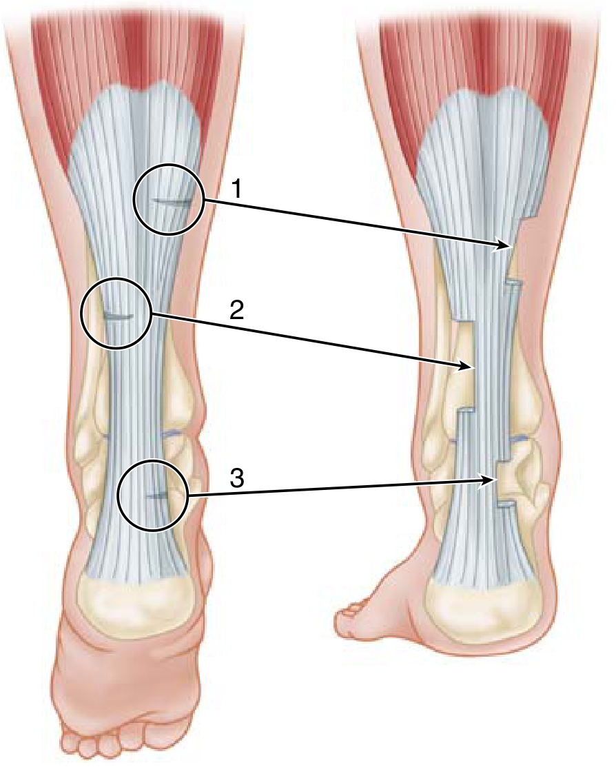
This is the primary target in Mr. Gupta's case. Per Miller's Review of Orthopaedics: the equinus is caused by overactivity of the gastrocnemius-soleus complex, and the varus by tibialis posterior and tibialis anterior.
| Muscle | Anatomical Location | Injection Landmark | Dose Range (onabotulinumtoxinA) |
|---|---|---|---|
| Gastrocnemius (medial head) | Medial femoral condyle → calcaneus via Achilles | Medial calf, upper third; 2 injection points in the muscle belly | 50-100 U |
| Gastrocnemius (lateral head) | Lateral femoral condyle → calcaneus | Lateral calf, upper third | 50-100 U |
| Soleus | Posterior tibia + fibula → calcaneus (deep to gastrocnemius) | Mid-calf, just distal to the gastrocnemius belly junction; inject deeper than gastrocnemius, palpate the firm muscle below the softer superficial calf | 50-75 U |
| Tibialis posterior | Posterior interosseous membrane → navicular + tarsals | Deep posterior compartment - requires US guidance; accessed medially between FDL and FHL, or from the posteromedial approach | 50-75 U |
| Tibialis anterior | Lateral tibia → medial cuneiform + 1st metatarsal | Anterior compartment, lateral to the tibial crest; easily palpated in the upper third of the leg | 50-75 U (if overactive) |
| Flexor digitorum longus / Flexor hallucis longus | Posterior tibia/fibula → toe phalanges | Deep posterior compartment, posteromedial approach; US essential | 25-50 U each |
Varus foot: Primary targets are tibialis posterior (main varus driver) and tibialis anterior if contributing. The 2021 study by Bensmail et al. (Ann Phys Rehabil Med) confirmed incobotulinumtoxinA efficacy for pes equinovarus in post-stroke lower limb spasticity.
PART 3: Dosing Summary and Practical Notes
Standard OnabotulinumtoxinA (Botox) Dose Ranges
| Region | Muscle Group | Typical Dose Range |
|---|---|---|
| Upper limb total | All muscles combined | 200-400 U per session |
| Shoulder | Pectoralis major + subscapularis | 50-100 U combined |
| Elbow flexors | Biceps + brachialis | 100-200 U combined |
| Wrist flexors | FCR + FCU | 50-100 U combined |
| Finger flexors | FDS + FDP | 50-100 U combined |
| Lower limb total | All muscles combined | 200-400 U per session |
| Calf (equinus) | Gastrocnemius + soleus | 150-300 U combined |
| Adductors | Adductor longus + magnus | 100-200 U combined |
| Hamstrings | Semimembranosus + semitendinosus + biceps femoris | 100-200 U combined |
| Total body maximum | Upper + lower | Up to 800 U (TOWER study; safe and effective) |
Mr. Gupta's documented dose of 400 U is therefore consistent with moderate multi-muscle treatment of either one limb fully or combined upper + lower limb at conservative doses.
Injection Guidance Techniques (in order of accuracy)
- Ultrasound (US) guidance - Gold standard. Real-time visualization of muscle belly, depth, and avoidance of neurovascular structures. Preferred for deep muscles (tibialis posterior, iliopsoas, subscapularis, FDP)
- EMG guidance - Confirms needle is in actively contracting muscle by sound/waveform. Good for distinguishing adjacent muscles (e.g., FDS vs. FDP)
- Electrical stimulation (e-stim) - Needle tip stimulates muscle contraction at low current (0.5-1 mA), confirming placement. Practical for outpatient settings
- Anatomical landmark / palpation - Adequate for superficial, easily palpated muscles (biceps, gastrocnemius, adductor longus). Less accurate for deep targets
Repeat Injection Timing
- Effect onset: 3-7 days
- Peak effect: 4-6 weeks
- Duration: 3-4 months
- Re-injection interval: Typically every 3-4 months (minimum 3 months between sessions to reduce antibody formation risk)
- Dose titration at follow-up: Based on Modified Ashworth Scale (MAS) change and gait analysis - exactly as stated in Mr. Gupta's letter ("optimization based on degree of spasticity and gait dysfunction")
Contraindications and Precautions
- Known hypersensitivity to botulinum toxin
- Myasthenia gravis or Lambert-Eaton syndrome (absolute contraindication)
- Pregnancy
- Anticoagulation - relative; use thinnest needle possible and apply compression
- Avoid injecting into clinically weak muscles (may worsen function)
- Do not inject near neurovascular bundles without US guidance (femoral triangle, axilla, deep posterior compartment)
Role of Physiotherapy (Mandatory Adjunct)
Botox alone provides a window of reduced tone. To convert this into functional gains, the following must follow immediately:
- Passive + active range-of-motion exercises
- Serial casting if contracture is developing
- Gait retraining / treadmill therapy
- Constraint-induced movement therapy (CIMT) for upper limb
- Ankle-foot orthosis (AFO) for equinovarus foot during the injection period
Per Bradley and Daroff's Neurology in Clinical Practice: "Treatment with any of the commercial botulinum toxins should include passive or active range of motion and treatments for pain to try to maintain better range of motion." Without concurrent physiotherapy, functional benefit is minimal.
Sources: Miller's Review of Orthopaedics 9th Ed; Bradley and Daroff's Neurology in Clinical Practice; Campbell's Operative Orthopaedics 15th Ed (2026); Adams & Victor's Principles of Neurology 12th Ed; PMC Visual Guide to US-guided BoNT-A Lower Limb Injections (2025); JRM International Consensus Guide
This is a shared conversation. Sign in to Orris to start your own chat.