
Pcnl for kidney
percutaneous nephrolithotomy PCNL kidney stone procedure
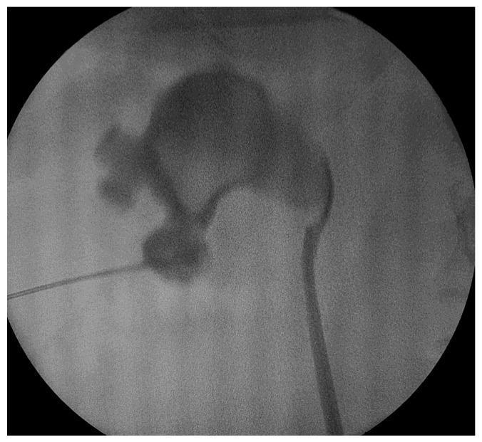
This diagnostic fluoroscopic image captures a percutaneous nephrolithotomy (PCNL) procedure. The image displays the renal collecting system opacified by contrast medium, highlighting the anatomy of the renal pelvis and several minor and major calyces. A radiopaque kidney stone (urolithiasis) is clearly visible within one of the lower pole calyces. A thin, linear hydrophilic guidewire is seen entering the kidney through a percutaneous access tract, directed toward the target calyx containing the stone. The image demonstrates the critical intraoperative step of achieving and maintaining percutaneous access to the intrarenal collecting system under fluoroscopic guidance. This material is pedagogically relevant for urology trainees and medical students to understand the radiological appearance of the pyelocaliceal system during minimally invasive surgical interventions for renal calculi.
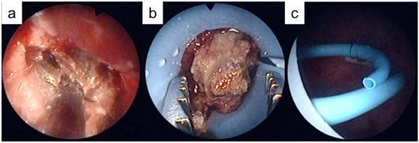
Endoscopic view of a percutaneous nephrolithotomy (PCNL) procedure, segmented into three clinical frames (a, b, and c). Frames a and b demonstrate the intra-renal management of a large renal calculus. The stone is light brown with a rough, crystalline texture, characteristic of calcium oxalate calculi. In frame a, a pneumatic lithotripter probe is applied directly to the stone surface for fragmentation. Frame b shows the use of metallic stone-grasping forceps to manipulate or extract fragmented stone pieces within the renal collecting system. Frame c depicts the placement of a flexible, light-blue ureteral stent or drainage tube, featuring dark graduation markings and a distinct distal lumen. The background shows the hyperemic mucosal lining of the renal pelvis. These images illustrate the key stages of PCNL: endoscopic stone identification, fragmentation, and post-procedural drainage in a patient with a complex renal anatomy, such as a horseshoe kidney.
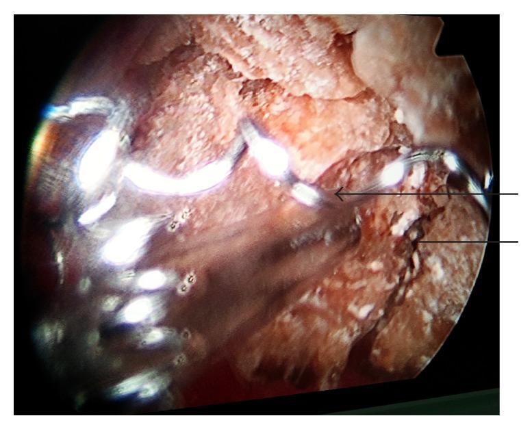
This intraoperative endoscopic image demonstrates a renal pelvic stone undergoing percutaneous nephrolithotomy (PCNL). The stone presents as a large, irregularly shaped, tan-to-brown mass with a rough, crystalline surface texture and focal white deposits. A key pathological feature is a migrated stainless steel embolization coil, visible as a bright, metallic, reflective spiral structure (indicated by an arrow) embedded within the stone matrix. The coil appears to have acted as a nidus for lithogenesis, with calculus material encrusting and engulfing the metallic loops within the pelvicalyceal system. The image illustrates a rare complication where a migrated vascular device facilitates secondary stone formation in the kidney. The surrounding field shows fragmented stone material and the moist internal mucosal environment typical of a urological endoscopic procedure.
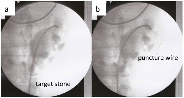
This diagnostic image pair displays retrograde pyelography of a human kidney, specifically within the context of a percutaneous nephrolithotomy (PCNL) procedure. The imaging modality uses a radiopaque contrast agent to opacify the renal collecting system, including the renal pelvis, major and minor calyces, and the ureter. In panel (a), a radiopaque 'target stone' is clearly visible in a lower calyx, and a flexible ureteroscope/catheter is seen traversing the ureter into the renal pelvis. In panel (b), the image demonstrates the deployment of a Lawson retrograde nephrostomy puncture wire, which appears as a thin radiopaque line extending from the upper calyx toward the skin surface. This illustrates a retrograde puncture technique for establishing percutaneous access in a kidney with complex anatomy (horseshoe kidney). The visualization highlights the relationship between medical instrumentation and the internal urinary tract architecture to guide surgical intervention for nephrolithiasis.
percutaneous nephrolithotomy outcomes complications
Percutaneous Nephrolithotomy (PCNL)
Indications
| Category | Details |
|---|---|
| Primary stones | Renal pelvic stones >2 cm, staghorn calculi |
| Lower pole stones | >1 cm (poor natural drainage) |
| Special anatomy | Stones in calyceal diverticula, horseshoe kidneys, poor drainage states |
| Stone composition | CT density >1000 HU (hard stones), cysteine content (relative indications) |
| Less common | Resection of TCC of renal pelvis, balloon dilatation/incision of PUJ obstruction, retrieval of foreign bodies |
Technique Overview
Positioning
- Prone access is most common
- Supine PCNL is also feasible and increasingly used
- Guidance: purely fluoroscopic OR combined ultrasound + fluoroscopy
Key Steps
- A high-quality pyelogram or 3D CT pyelogram is essential to map intrarenal anatomy and precise stone location
- The tract must be planned to allow maximal/complete stone clearance
- Posterior calyces allow better access to anterior calyces
- Anterior calyceal entry gives poorer intrarenal navigation
- Upper pole entry allows deep access to PUJ/upper ureter, but risks puncturing the posterior division artery or pleura
- Some interpolar calyces may be difficult to reach from either upper or lower entry
- Tract size: 16 to 32 F (30 F standard)
- Two guidewires are ideal: a stiff working wire + a safety wire
- Three dilatation systems:
- Balloon-mounted sheath system
- Serial plastic dilators
- Concentric/telescopic metal dilators
- Rigid nephroscope with pneumatic lithotripter or holmium laser
- Flexible nephroscope used for mid/upper calyceal stones
- Holmium laser debris washes out through the sheath or is retrieved with rigid nephroscope + suction
Stone Clearance Strategy
- Aim for complete stone clearance
- If not possible:
- Clear the renal pelvis first (to relieve obstruction)
- Clear lower pole calyces (fragments here may not drain spontaneously)
- Residual upper/interpolar stones can be managed later with ESWL
- For complex staghorn calculi - a single puncture may not suffice; upper pole access is preferred for single-tract approach
Mini and Micro-PCNL
- Mini-PCNL: sheath 16-24 F
- Micro-PCNL: even smaller
- Lower complication rates due to smaller tracts, though the principles of access and dilatation are identical
Special Populations
Morbid Obesity
- Multiple studies show no increase in overall morbidity compared to normal-weight patients (Pearle et al., 1998)
- However: higher BMI patients have more comorbidities (e.g., chronic anticoagulation) and a lower stone-free rate (Fuller et al., 2012)
- Practical challenges: two surgical tables may be needed; risk of rhabdomyolysis in lateral approach; extra-long Amplatz sheaths (≥20 cm) and nephroscopes may be required
Staghorn Calculi (Complex Branching Stones)
- Upper pole access preferred for single-tract management
- Allows coverage of upper pole, renal pelvis, and lower pole with rigid nephroscope
- Mid-calyceal stones addressed with flexible nephroscope + holmium laser
Complications
- Major complications: up to 7% of patients
- Minor complications: up to 25% of patients
| Complication | Details |
|---|---|
| Hemorrhage | Most significant; risk increases with stone burden and operative duration |
| AV fistula / pseudoaneurysm | Requires angiographic embolization; occurs in <1% of patients |
| Sepsis | Postoperative temp >38.5°C; requires blood cultures + broad-spectrum antibiotics |
| Pleural injury | Risk with upper pole access; may require chest tube |
| Adjacent organ injury | Bowel, spleen, liver |
| Ischemic stricture | Late complication - stricture of renal pelvis/infundibula post-PCNL (can be treated with balloon dilatation) |
| Retained fragments | May require secondary ESWL or re-look nephroscopy |
Post-Procedure Management
- Nephrostomy tube left in situ after standard PCNL
- In morbidly obese patients: balloon-type or re-entry Malecot catheters preferred (to prevent displacement)
- "Tubeless PCNL" (no nephrostomy) is used in selected uncomplicated cases
- Nephrostomy check / antegrade nephrostogram typically performed at 24-48 hours
- Residual fragments can be managed with ESWL or second-look flexible nephroscopy via the mature tract
PCNL vs. Alternatives
| Feature | PCNL | ESWL | URS (Ureteroscopy) |
|---|---|---|---|
| Stone size best suited | >2 cm | <1 cm | 1-2 cm |
| Stone-free rate (large stones) | Highest | Low | Moderate |
| Invasiveness | Moderate | Least | Moderate |
| Lower pole stones >1 cm | First-line | Poor clearance | Alternative |
| Staghorn calculi | First-line | Adjunct | Not recommended |
Recent Evidence (2025)
- A 2025 systematic review on outpatient PCNL (PMID 40684274) found PCNL can be safely performed as a day-case procedure in selected patients, with comparable outcomes to inpatient PCNL.
- A 2025 meta-analysis on PCNL bleeding risk factors (PMID 40760643) identified stone burden, access number, and operative time as the main predictors of severe post-PCNL hemorrhage.
- A 2025 meta-analysis on anesthesia type for PCNL (PMID 40252831) found no significant difference in stone-free rates between general and regional anesthesia, though regional anesthesia was associated with shorter hospital stay in some analyses.
- Grainger & Allison's Diagnostic Radiology, Chapter 85 (Interventional Uroradiology)
- Campbell-Walsh-Wein Urology, Chapter on Nephrolithiasis/PCNL