
How to write case history and priscription paper of physiotherapy
- Case history covers: patient demographics, chief complaint, history of present illness, past medical/surgical history, social/occupational history, and a systems review.
- Subjective examination (S): what the patient tells you - pain, onset, aggravating/relieving factors.
- Objective examination (O): posture, ROM, muscle strength, special tests, neurological tests.
- Assessment (A): diagnosis/clinical impression and problem list.
- Plan (P): treatment goals, modalities, exercises, and frequency/duration.
- The prescription paper summarizes modality settings, exercise prescription, and re-assessment schedule.
how to write physiotherapy case history and prescription paper format
https://www.apta.org/your-practice/documentation/defensible-…
How to Write a Physiotherapy Case History and Prescription Paper
PART 1: PHYSIOTHERAPY CASE HISTORY
SECTION A - Patient Demographics (Top of the Form)
| Field | Details |
|---|---|
| Name | Full name of the patient |
| Age / Date of Birth | |
| Gender | |
| Address & Contact | Phone number, email |
| Occupation | Important for ergonomic and functional context |
| Referred by | Doctor's name, specialty, date of referral |
| Date of first visit |
SECTION B - Chief Complaint (CC)
"I have pain in my lower back that started 3 weeks ago after lifting a heavy box."
- Location of complaint (use a body diagram if available)
- Onset: sudden or gradual
- Duration: how long it has been present
SECTION C - History of Present Illness (HPI)
| Letter | Meaning | Example |
|---|---|---|
| O | Onset | When did it start? What caused it? |
| L | Location | Exact site and radiation |
| D | Duration | Constant or intermittent? |
| C | Character | Sharp, dull, burning, throbbing, aching |
| A | Aggravating factors | Activity, posture, time of day |
| R | Relieving factors | Rest, ice, heat, medication |
| T | Timing | Morning stiffness, night pain, after activity |
| S | Severity | Pain scale 0-10 (VAS or NRS) |
- Behavior of symptoms 24 hours (24-hour pattern)
- Any previous episodes of the same complaint
- Treatment received so far and its outcome
SECTION D - Past Medical and Surgical History (PMH/PSH)
- Previous illnesses (diabetes, hypertension, osteoporosis, rheumatoid arthritis, cancer)
- Previous surgeries and dates
- Previous physiotherapy and outcome
- Hospitalizations
SECTION E - Drug/Medication History
- Current medications (painkillers, steroids, anticoagulants - important before applying electrotherapy)
- Allergies
SECTION F - Family and Social History
- Family history of relevant conditions
- Smoking, alcohol use
- Living situation (stairs at home, caregiver support)
- Recreational activities and hobbies
SECTION G - Occupational History
- Type of work (sedentary, manual, repetitive movements)
- Work hours, posture demands
- Any work-related injury (for medico-legal records)
SECTION H - Systems Review (Red/Yellow Flag Screening)
- Unexplained weight loss
- History of cancer
- Night sweats, fever
- Bilateral neurological symptoms
- Bowel/bladder dysfunction
- Constant, unremitting pain not affected by position
- Fear avoidance behavior
- Catastrophizing
- Low mood, anxiety, depression
- Poor workplace support
SECTION I - OBJECTIVE EXAMINATION
1. Observation / Inspection
- Posture (standing, sitting, lying)
- Gait analysis
- Swelling, bruising, muscle wasting, deformity, scar
2. Palpation
- Tenderness (point, diffuse)
- Temperature (warm = inflammation)
- Muscle spasm, trigger points
- Bony landmarks
3. Range of Motion (ROM)
- Active ROM (AROM) - what the patient can do
- Passive ROM (PROM) - what you can produce
- Record in degrees using a goniometer
- Note end-feel: soft, firm, hard, empty
4. Muscle Strength Testing
- Manual Muscle Testing (MMT) using the Oxford Scale 0-5
- Or dynamometer readings
5. Neurological Examination (if applicable)
- Dermatomes (sensation - light touch, pin prick)
- Myotomes (muscle power by nerve root)
- Reflexes (deep tendon reflexes - graded 0 to 4+)
- Upper/lower motor neuron signs
6. Special Tests
- Specific to the region (e.g., Straight Leg Raise for lumbar, Spurling's for cervical, McMurray's for knee, Hawkins-Kennedy for shoulder)
- Record: positive or negative with brief description of finding
7. Functional Assessment
- Activities of Daily Living (ADLs): dressing, walking, climbing stairs
- Standardized tools: Oswestry Disability Index, DASH Score, Barthel Index, Berg Balance Scale
8. Girth/Volume Measurements
- For swelling or muscle wasting
9. Postural/Biomechanical Analysis
- Spinal curvatures, limb length discrepancy, foot pronation
SECTION J - ASSESSMENT / CLINICAL IMPRESSION
- Physiotherapy Diagnosis - the impairment-based diagnosis (e.g., "Mechanical Low Back Pain with L4-L5 nerve root irritation")
- Problem List - list all identified impairments:
- Pain (severity, location)
- Reduced ROM
- Muscle weakness
- Functional limitation
- Prognosis - expected recovery timeline and influencing factors
- Short-term goals (1-2 weeks): e.g., reduce pain from 7/10 to 4/10
- Long-term goals (4-6 weeks): e.g., return to full work duties
PART 2: PHYSIOTHERAPY PRESCRIPTION PAPER
FORMAT OF THE PHYSIOTHERAPY PRESCRIPTION
PHYSIOTHERAPY PRESCRIPTION

Patient Name: _____________________ Date: __________
Age: ________ Gender: _____________ MRD/File No: ____
Referring Physician: _______________

DIAGNOSIS: ____________________________________________

PRECAUTIONS / CONTRAINDICATIONS:
_______________________________________________________

PHYSIOTHERAPY TREATMENT PRESCRIBED:

1. ELECTROTHERAPY MODALITIES:
   □ TENS         Settings: _______ Duration: _______ mins
   □ IFT          Frequency: _____ Duration: _______ mins
   □ Ultrasound   Intensity: __ W/cm²  Mode: CW/Pulsed  Duration: ___ mins
   □ LASER        Dose: _______  Duration: _______ mins
   □ SWD/MWD      Settings: _______ Duration: _______ mins
   □ Hot pack / Ice pack      Duration: _______ mins
   □ Traction     Mode: ________ Wt: _____ Duration: ___ mins

2. EXERCISES:
   □ Active ROM exercises        Repetitions: _____ Sets: _____
   □ Passive stretching          Hold: _____ sec   Sets: _____
   □ Strengthening exercises     Load: ______      Reps/Sets: _____
   □ Proprioception/Balance training
   □ Gait training / Ambulation
   □ Breathing exercises
   □ Home Exercise Program (HEP): ___________________________

3. MANUAL THERAPY:
   □ Joint mobilization (Grade: I / II / III / IV)
   □ Soft tissue massage
   □ Neural mobilization
   □ PNF stretching

4. FUNCTIONAL TRAINING:
   □ Transfer training
   □ ADL training
   □ Ergonomic advice / Postural correction

FREQUENCY: _____ sessions per week
DURATION:  _____ weeks (Total: _____ sessions)

RE-ASSESSMENT DATE: ____________________________________

Physiotherapist Signature: __________ Reg. No.: _______
Date: _____________
KEY TIPS FOR DOCUMENTATION
- Write legibly - or use printed/digital forms where possible.
- Use objective, measurable terms - avoid vague words like "improved slightly." Write "knee flexion improved from 90° to 120°."
- Date and sign every entry - include your registration number.
- Use standardized outcome measures - these make progress trackable and defensible (VAS, Oswestry, DASH, FIM, etc.).
- Record informed consent - note that the patient understood and agreed to the treatment.
- Document red flags - if present, note referral made.
- SOAP notes for every follow-up visit - brief S (any change in symptoms), O (objective findings), A (response to treatment), P (plan for next session).
- Contraindications must be documented - especially before using electrotherapy (cardiac pacemaker, pregnancy, metal implants, malignancy, open wounds).
FOLLOW-UP / PROGRESS NOTE FORMAT
- S: "Patient reports pain reduced to 4/10, able to walk to the bathroom without support."
- O: Knee flexion 110° (was 90°). No swelling. SLR negative.
- A: Improving. On track with goals.
- P: Continue current program, add closed-chain strengthening today.
Frozen shoulder with tenderness physiotherapy intervention
"adhesive capsulitis" AND "physical therapy"
frozen shoulder adhesive capsulitis physiotherapy exercises
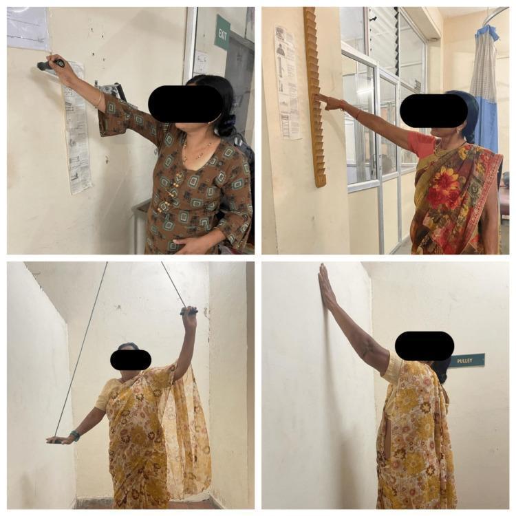
A composite of four clinical photographs demonstrating conventional physical therapy exercises for shoulder rehabilitation and range of motion (ROM) improvement. Top left: A patient performs 'wall washing' exercises, moving their hand in functional patterns against a wall surface to facilitate shoulder flexion and abduction. Top right: A patient uses a 'finger ladder' mounted on a wall, utilizing finger movements to incrementally increase shoulder elevation and abduction. Bottom left: A patient engages in shoulder pulley exercises using a suspended rope and handle system to perform assisted ROM activities, where the healthy arm helps lift the affected limb. Bottom right: A patient demonstrates a wall-climbing or stretching exercise to maximize vertical reach. These interventions are typically used in the management of adhesive capsulitis (frozen shoulder) or post-operative recovery to improve shoulder mobility, specifically targeting flexion, abduction, and rotation. The images represent standard physiotherapy protocols often compared with modern interventions like virtual reality-based therapy.
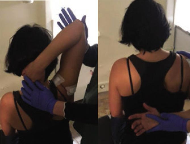
This clinical photograph consists of two panels illustrating post-procedural passive mobilization techniques for adhesive capsulitis (frozen shoulder) in a seated patient. The images demonstrate the application of physical therapy or orthopedic manipulation following glenohumeral joint hydrodistension. In the left panel, a practitioner wearing blue medical gloves performs passive abduction and external rotation; one hand supports the cervical-thoracic junction while the other guides the patient's flexed elbow and upper arm. Two white square adhesive patches, likely providing transcutaneous electrical nerve stimulation (TENS) or monitoring, are visible on the posterior-lateral deltoid region. In the right panel, the practitioner performs passive internal rotation by guiding the patient's arm into a 'hand-behind-back' position, emphasizing posterior capsule stretching. Both panels highlight the therapeutic range of motion exercises used to restore joint mobility and prevent adhesion recurrence in the shoulder complex. This clinical resource is relevant for physical medicine, rehabilitation, and orthopedic education regarding joint capsule pathologies.
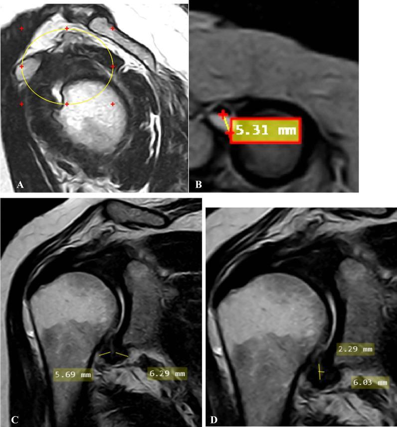
A multi-panel MRI study of the shoulder demonstrating radiological signs of adhesive capsulitis (frozen shoulder). Image A (oblique sagittal T2-weighted) shows the complete obliteration of the subcoracoid fat triangle within the rotator interval (encircled). Image B (oblique axial fat-suppressed PD-weighted) identifies a significant effusion (5.31 mm) within the long head of the biceps tendon sheath, exceeding the normal threshold. Images C and D (oblique coronal T2-weighted) focus on the axillary recess. Image C measures the pathological thickening of the joint capsule at the humeral (5.69 mm) and glenoid (6.29 mm) aspects. Image D provides measurements for the maximal height (2.29 mm) and width (6.03 mm) of the axillary recess space. This composite image illustrates key diagnostic criteria including rotator interval fat replacement, synovial effusion, and capsular thickening, which are critical for the radiological staging of adhesive capsulitis in a clinical setting.
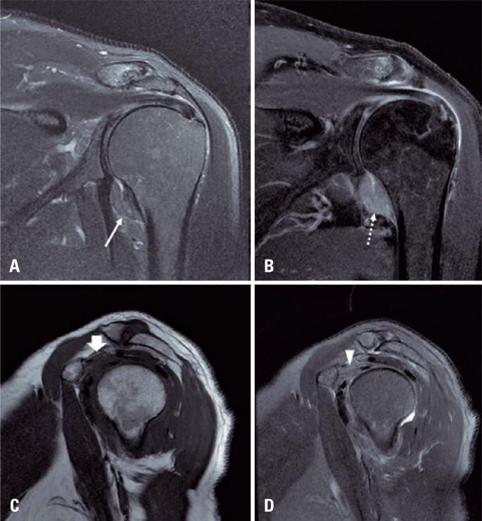
This diagnostic image set consists of four MRI views of a human shoulder illustrating the classic findings of adhesive capsulitis (frozen shoulder). Image A (coronal T2-weighted with fat suppression) and Image B (coronal T1-weighted post-gadolinium with fat suppression) demonstrate significant thickening of the glenohumeral joint capsule and pericapsular edema, particularly within the axillary recess. The post-contrast image (B) shows intense enhancement of the thickened capsule, indicating active synovitis and hypervascularity. Images C (sagittal T1-weighted) and D (sagittal T2-weighted with fat suppression) focus on the rotator interval. They reveal the characteristic obliteration of the normal subcoracoid fat pad by hypointense inflammatory tissue on T1 (arrow in C) and hyperintense edema on T2 (arrowhead in D). These imaging features are essential for confirming adhesive capsulitis in patients presenting with clinical shoulder stiffness and pain, allowing for the differentiation from other pathology such as rotator cuff tears or labral injuries.
PMID: 36861780
frozen shoulder with tenderness physiotherapy intervention stage based management 2024
Frozen Shoulder (Adhesive Capsulitis) - Physiotherapy Intervention
Understanding the Condition
| Stage | Name | Duration | Key Features |
|---|---|---|---|
| Stage 1 | Freezing (Painful) | 3-9 months | Severe pain, insidious onset, night pain, tenderness - minimal restriction |
| Stage 2 | Frozen (Adhesive) | 4-20 months | Pain decreasing, severe global stiffness dominates, compensatory scapular motion |
| Stage 3 | Thawing (Resolution) | 5-26 months | Pain settling, stiffness gradually resolving, ROM slowly returning |
Key clinical finding with tenderness: Tenderness is most prominent in Stage 1 (freezing phase). It is typically felt over the anterior capsule/rotator interval, biceps tendon groove, and deltoid insertion. This indicates active synovial inflammation - treatment must be pain-first before any aggressive mobilization.
STAGE 1 - FREEZING / PAINFUL STAGE (Tenderness Predominant)
1. Electrotherapy Modalities (Pain Relief)
| Modality | Parameters | Purpose |
|---|---|---|
| TENS | Burst/Conventional mode, 80-100 Hz, 30-45 mins | Immediate analgesia via gate control |
| IFT (Interferential Therapy) | 80-120 Hz, carrier frequency 4000 Hz, 15-20 mins | Deeper analgesia, muscle spasm reduction |
| Ultrasound Therapy | 1 MHz, 0.5-1.0 W/cm², pulsed mode (1:4), 5-7 mins over anterior capsule | Anti-inflammatory, reduces synovial thickening |
| LASER (Low Level Laser Therapy) | 5-10 J/cm², over tender points | Reduces local inflammation and tenderness |
| Cryotherapy (Ice pack) | 15-20 mins, 2-3 times/day | Acute pain and swelling control |
| Short Wave Diathermy (SWD) | Pulsed mode only (to avoid heating inflamed tissue) | Caution - avoid in acute stage |
Tenderness-specific note: Over tender points, use pulsed ultrasound (not continuous), TENS electrode placement around (not directly over) the most tender area.
2. Manual Therapy (Gentle, Low Grade)
- Grade I-II Maitland joint mobilization - oscillations at beginning of range, purely for pain relief, no stretch
- Gentle soft tissue massage - to trapezius, deltoid, periscapular muscles
- Pendulum (Codman's) exercises - gravity-assisted, gravity distraction reduces pain
3. Exercises
- Pendulum/Codman's exercises - forward, sideways, circular - small circles, pain free
- Active-assisted ROM - with a stick/wand, within pain-free range only
- Shoulder shrugs and scapular retraction - maintain scapulothoracic motion
- Elbow, wrist, hand exercises - prevent disuse atrophy distally
4. Education and Positioning
- Advice: avoid lying on the affected side at night (use a pillow under the arm)
- Posture correction - correct rounded shoulder/forward head posture
- Activity modification - avoid overhead reaching, heavy lifting, overhead work
- Explain the three-stage natural history - reduces fear and anxiety
STAGE 2 - FROZEN / ADHESIVE STAGE
1. Thermal/Physical Modalities
| Modality | Parameters | Purpose |
|---|---|---|
| Hot pack/Moist heat | 15-20 mins before exercises | Increase tissue extensibility before stretching |
| Continuous ultrasound | 1 MHz, 1.0-1.5 W/cm², 5-7 mins | Thermal effect - loosens capsule for stretching |
| SWD (Continuous mode) | 20 mins before mobilization | Deep heating of joint capsule |
2. Manual Therapy (Progressive, Grade III-IV)
- Maitland Grade III-IV mobilizations - sustained end-range techniques for stiffness
- Inferior glide (caudal distraction) - primary technique to restore abduction
- Posterior glide - restores internal rotation and flexion
- Anterior glide - restores external rotation (most restricted movement in frozen shoulder)
- PNF stretching (Contract-Relax) - post-isometric relaxation to gain range
3. Stretching Exercises
- Cross-body stretch (posterior capsule) - hold 30 seconds, 3-5 reps
- External rotation stretch with stick - supine, door frame or wand stretch
- Sleeper stretch - side-lying internal rotation stretch
- Wall-climbing / Finger ladder exercises - active-assisted elevation
- Pulley exercises - overhead pulley for assisted elevation
STAGE 3 - THAWING / RESOLUTION STAGE
1. Strengthening Exercises
- Rotator cuff strengthening - theraband external/internal rotation
- Scapular stabilizers - serratus anterior (wall push-up plus), lower trapezius (Y-T-W exercises)
- Deltoid strengthening - progressive resistance, side raises
- Closed-chain exercises - wall push-ups, quadruped weight-bearing
2. Proprioception and Neuromuscular Training
- Ball rolling on wall
- Perturbation training (unstable surface)
- Rhythmic stabilization techniques
3. Functional and Occupational Training
- Return to overhead reaching, ADL training
- Work-specific tasks
- Sport-specific training if applicable
JOINT MOBILIZATION GRADE GUIDE (Maitland)
| Grade | Technique | Used In | Purpose |
|---|---|---|---|
| I | Small amplitude, beginning of range | Stage 1 (tenderness) | Pain relief |
| II | Large amplitude, within range, no resistance | Stage 1 | Pain relief |
| III | Large amplitude into resistance | Stage 2 | Stretch, increase ROM |
| IV | Small amplitude at end of range, into resistance | Stage 2-3 | Break adhesions, ROM |
| V | Thrust (manipulation) | Selected cases only | Capsular release |
TENDERNESS MANAGEMENT - SPECIFIC PROTOCOL
- Identify tender points - biceps tendon, rotator interval, acromioclavicular joint, greater tuberosity
- Do NOT apply deep pressure directly over tender areas - use surrounding soft tissue massage instead
- Pulsed ultrasound is preferred over continuous when there is active tenderness
- TENS - electrode placement around (not on) the most tender area
- Ice before treatment (10-15 mins) to reduce acute tenderness before handling
- Mobilization direction - use pain-free accessory glides first; avoid painful arcs
- Corticosteroid injection co-management: when tenderness is severe, intra-articular injection (ideally ultrasound-guided) significantly reduces pain and allows physiotherapy to be more effective - as confirmed by the Rheumatology textbook (Elsevier 2022) and current clinical guidelines
CONTRAINDICATIONS TO PHYSIOTHERAPY IN FROZEN SHOULDER
- Active infection or septic arthritis
- Malignancy of the shoulder region
- Fracture or recent surgical fixation
- Severe osteoporosis (avoid high-grade manipulation)
- Uncontrolled diabetes (alters healing and response)
- Metal implant in the field (for SWD, ultrasound - adjust accordingly)
HOME EXERCISE PROGRAM (HEP)
- Stage 1: Pendulum exercises, gentle active ROM within pain-free range, ice after activity
- Stage 2: Pulley, wall climbing, cross-body stretch, external rotation with stick (10 reps x 3 sets, hold 30 sec per stretch)
- Stage 3: Theraband strengthening, full overhead activities
RECENT EVIDENCE NOTE
SAMPLE PHYSIOTHERAPY PRESCRIPTION FOR FROZEN SHOULDER (Stage 1 with Tenderness)
Diagnosis: Left Adhesive Capsulitis (Freezing Stage) with tenderness

MODALITIES:
- TENS: Conventional mode, 80 Hz, 200 µs, 30 mins
- Pulsed Ultrasound: 1 MHz, 0.8 W/cm², 1:4 duty cycle, 6 mins
- Ice pack: 15 mins post-session

MANUAL THERAPY:
- Soft tissue massage: periscapular muscles, 10 mins
- Maitland Grade I-II inferior glide and anterior glide mobilization

EXERCISES:
- Pendulum exercises: 10 circles each direction, 3x/day
- Active-assisted ROM with wand: Flexion, Abduction within pain-free range
- Scapular shrugs and retraction: 3 x 10 reps

FREQUENCY: 5 days/week
DURATION: 3 weeks, then re-assess

PRECAUTIONS:
- No forceful stretch in painful range
- Monitor for increased night pain (sign of flare)
- Refer for ultrasound-guided intra-articular injection if no improvement in 2 weeks